h1.qusth1 { display: none !important; }

QUESTION OF THE WEEK

Dr. Donovan's Articles

QUESTION OF HAIR BLOGS

Filtering by Category: Terms


Lichen planopilaris: Classic trichoscopic findings

Classic trichoscopic findings of LPP

Classic trichoscopy of active lichen planopilaris, an immune mediated scarring alopecia is shown below.

Classic trichoscopic image from a patient with active lichen planopilaris (LPP)

Classic trichoscopic image from a patient with active lichen planopilaris (LPP)

There is redness and scale around hairs (called perifollicular erythema and perifollicular scale). Some hairs are twisted (called pili torti). The areas of scalp devoid of hairs are no longer red as the immune system has destroyed hairs in that area and has since left the area. Treatments discussed in other posts as in the following link.

Treatments for LPP: What is available?


This article was written by Dr. Jeff Donovan, a Canadian and US board certified dermatologist specializing exclusively in hair loss.
Share This
No Comments

Scalp Itching in Psoriasis: Don't Forget about "Psoriasitch"

Scalp Itching in Psoriasis Affects Quality of Life

Psoriasis is an immune disease that affects multiple organs and structures. Dermatologists treat skin psoriasis on a daily basis. Rheumatologists treat psoriatic arthritis on a daily basis. 
The scalp is involved a large proportion of patients with psoriasis. In fact, nearly two thirds of patients with psoriasis have scalp psoriasis and it is often the first area where the disease shows up. 

Having scalp psoriasis affects people’s quality of life. Scalp itching is often present and the itching can be troublesome for many. In fact, when I teach other physicians about scalp psoriasis I always encourage them to remember the word “psoriasitch.” It’s a completely made up and completely invented word but it helps clinicians remember the often debilitating effects of the itching that comes with having psoriasis. Not only do we need to remember the flaking and redness and sometimes hair loss that goes with it - but we absolutely need to address the itching these patients live with. And so I call it “psoriasitch.”

The mechanisms that lead to itching are actually quite complex for patients with psoriasis and involve parts of the immune system, nervous system, endocrine system and blood vessels. The mast cell, a key cell normally involved in “allergic responses” in the body, seems to be a key cell type involved in generating itching in patients with psoriasis.

Trichoscopic image from a patient with psoriasis. The patient was extremely itchy. Scalp itching impacts qualify of life and must be addressed by practitioners.

Trichoscopic image from a patient with psoriasis. The patient was extremely itchy. Scalp itching impacts qualify of life and must be addressed by practitioners.


A wide variety of traditional anti-psoriasis agents can reduce itching. Topical steroids, vitamin D analogues, methotrexate, retinoids, and the newer biologics can reduce inflammation and reduce itching. But whether other non traditional strategies that affect the nervous system, endocrine system, blood vessels can also help stop itching is the subject of intense research interest worldwide.

The image here is a trichoscopic image from a patient with scalp psoriasis. The scale is white in contrast to the yellow of seborrheic dermatitis. There is scale around hairs (“perifollicular scale”) and certainly other conditions such as lichen planopilaris could be considered.


This article was written by Dr. Jeff Donovan, a Canadian and US board certified dermatologist specializing exclusively in hair loss.
Share This
No Comments

Secondary Hair Casts: Psoriasis

What are hair casts?

hair casts.png

Hair casts are thin, elongated, cylindrical concretions that encircle the hair shaft. Hair casts range in size from 2-7 mm and can be easily dislodged. The term was coined by Kligman in 1957.

Hair casts (sometimes called “pseudonits”) can be easily differentiated from true “knits" because they slide along hairs when grabbed with the fingers. They are usually asymptomatic and particularly common in young women.

Hair casts are said to be "primary" in nature when not associated with an underlying scalp disorder and "secondary" when associated with an underlying disorder. Common secondary causes include psoriasis, seborrheic dermatitis, pemphigus and traction alopecia and scarring alopecia. Many other causes are possible too including hair sprays and deodorants.

Hair casts are thought to represent material from both the internal root sheath and the external root sheath.

The photo here shows casts in a patient with psoriasis.


This article was written by Dr. Jeff Donovan, a Canadian and US board certified dermatologist specializing exclusively in hair loss.
Share This
No Comments

Smelly Scalp (Hair) Syndrome

Diagnosing and Treating an Unpleasant Scalp Smell

Every now and then, a patient reports that they have a problem with scalp odour. The story typically goes that their scalp smells bad and no matter what they do, they just can’t get rid of the smell.  The specific issue goes by a variety of names, although the most common names are smelly scalp syndrome (SSS) and smelly hair syndrome (SHS).


What causes SSS?

There is no one cause of smelly hair syndrome. There are many causes. The first thing that I tell my patients is that I need to use my eyes and ears to help determine the cause - use of my nose actually plays little role. 


Step 1: Hear the Story

To start with, I need to hear their story. I need to understand when the odor started and what things in the patient’s life changed around this time. I also need to understand several other issues including:

Were any new medications started?

Were any new hair products started?

Any new hair dyes started?

Were any new shampoos started?

How often does the patient shampoo?

What shampoos are used?

Did a child in the house start school?

Is their a new pet in the house?

Were any new hobbies started?

Is it just the scalp odor or does odour come from the body (armpits/groin?)

Has their been any changes in the patients health?

Are their any dental concerns?

Does the scalp itch?

Is their burning in the scalp?

Is the scalp tender?

Is their hair loss accompanying the odor ... or just odor?

Does the patient wake up with blood or secretions on the pillow?

Has the patient’s level of fatigue changed?

What is the patient’s stress level?

Has the patient travelled?

Has the patient’s diet changed?

Does the patient report any skin rashes?

Is their any concerns regarding sexually transmitted diseases?


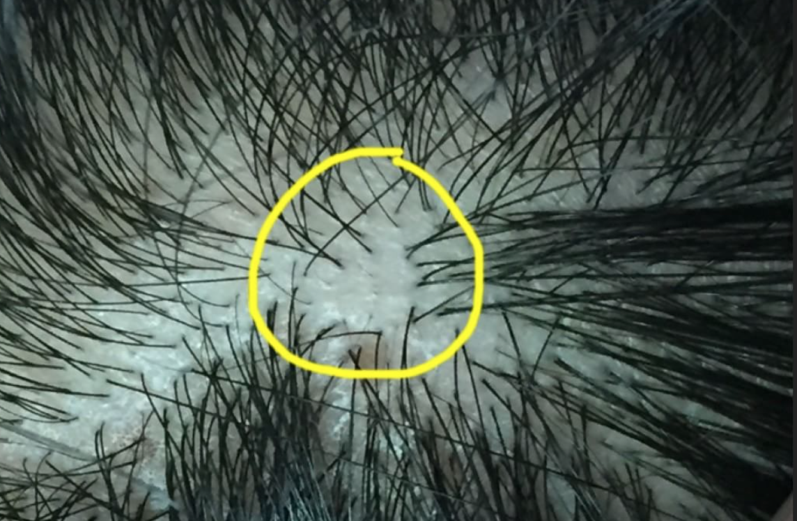
Step 2: See the Scalp

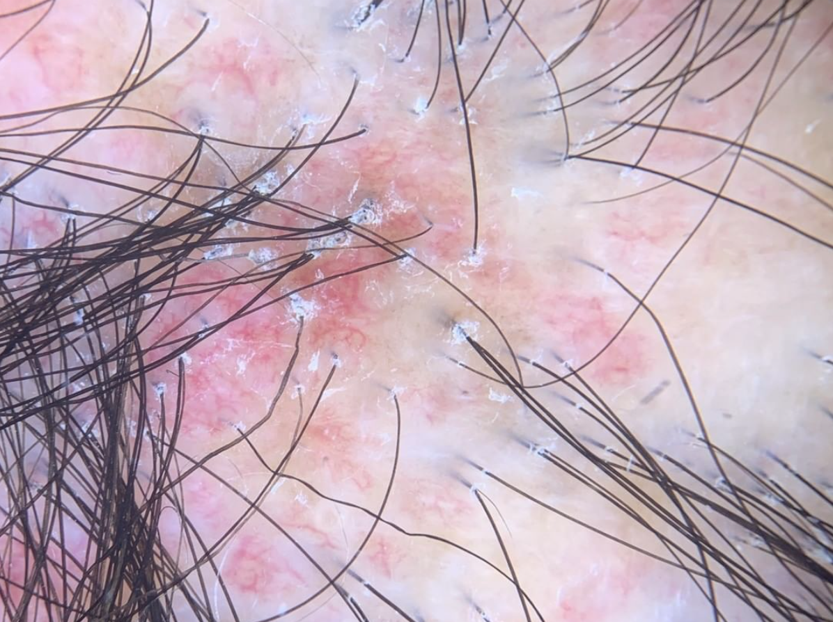
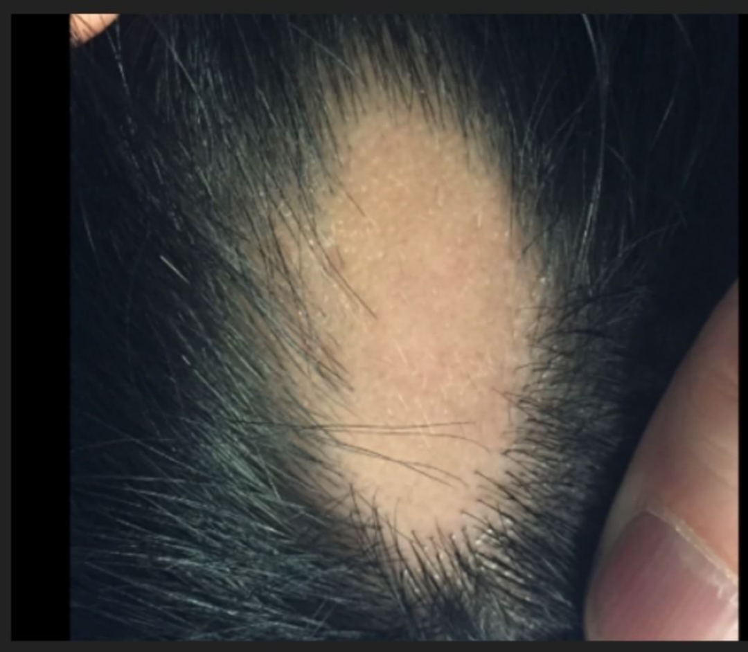
After getting a sense about the patient’s story, I need to see the scalp, eyebrows, eyelashes and body hair. I also need to see the nails and may even examine parts of the skin.

The scalp examination is important to determine if there are any abnormalities in the skin of the scalp and/or the hair follicles themselves. It is important to determine if there is redness, scale, pustules (pimples), openings in the skin, sores, breakdown. Of course, determining whether or not the smell is actually associated with hair loss is critically important.


Causes of SSS

There are many potential cause of SSS. The most common causes include:

  1. Seborrheic dermatitis 
  2. Psoriasis
  3. Fungal Infections
  4. Allergic contact dermatitis 
  5. Irritant contact dermatitis
  6. Scarring Alopecias
  7. Apocrine/Eccrine Gland Overactivity
  8. Metabolic Disturbances
  9. Infections
  10. Skin Cancers
  11. Hormonal disorders
  12. Infrequent Washing 


Tests for SSS

For individuals with smelly scalp, there are no specific tests per se. However, a number of tests may be performed including:
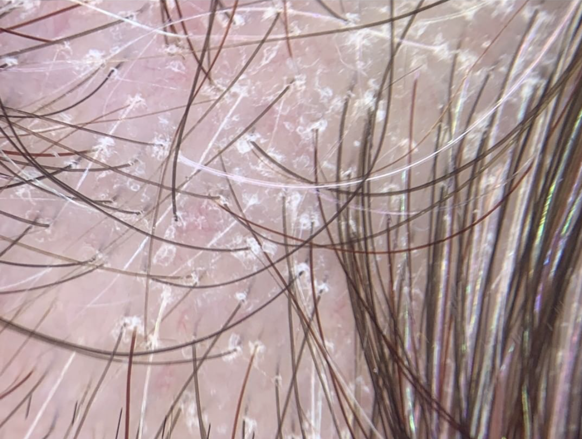
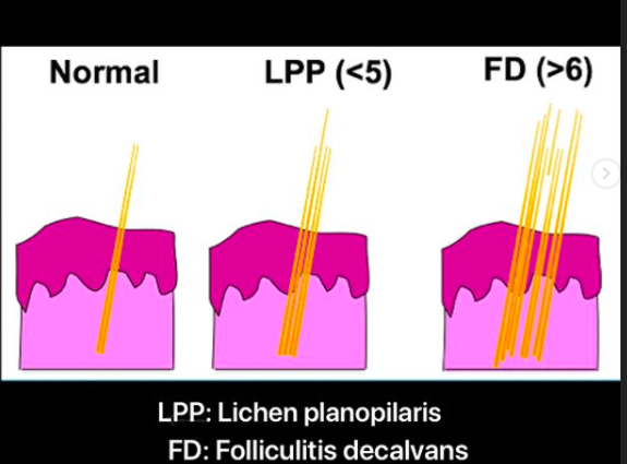
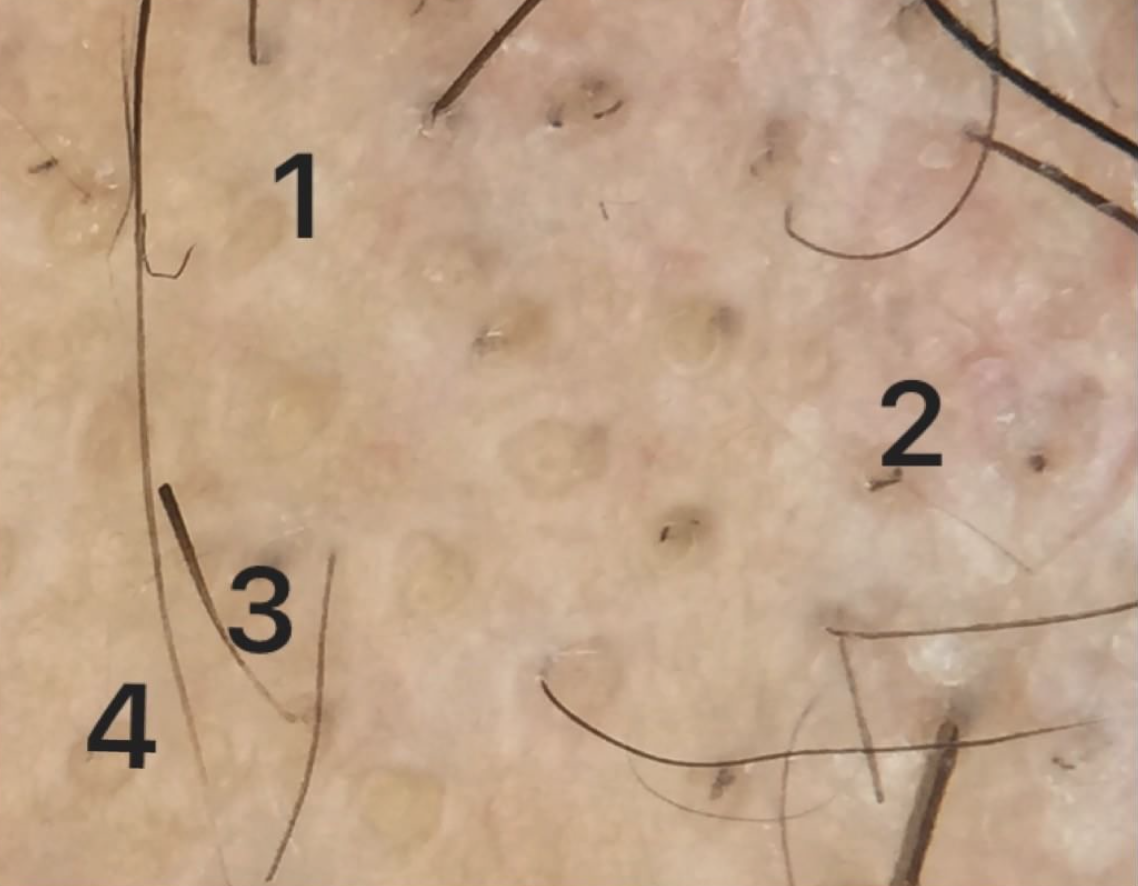
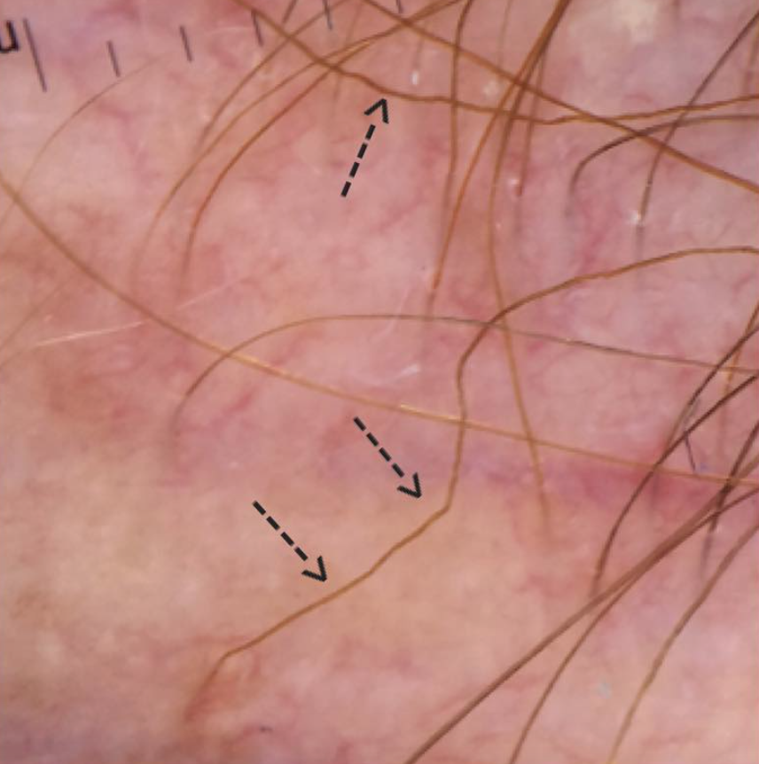
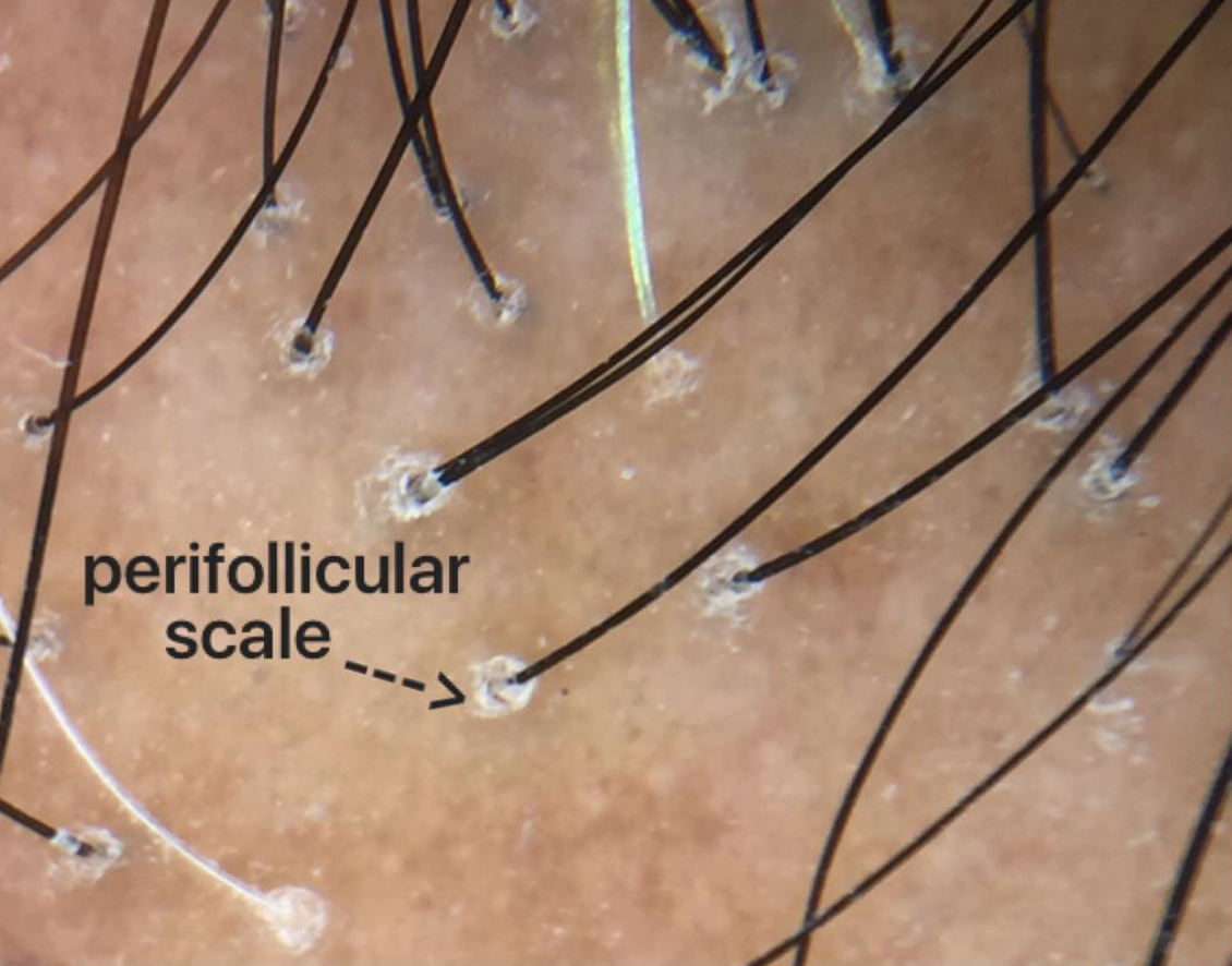
 

1. Dermoscopy

Dermoscopy is a technique that involves examination of the hair and scalp with a specialized instrument known as a dermatoscope. Dermoscopy can help physicians identify special features that point to a specific disease including seborrheic dermatitis, psoriasis, tinea capitis, scarring alopecias, folliculitis.

2. Swabs of Pustules

Any pustules on the scalp need to be swabbed. The presence of yellow pustules does not necessarily mean their is an infection in the follicle, but careful assessment is needed. A swab will help not only to identify bacteria that might be present on the scalp but also to determine the precise antibiotics that kill those bacteria. This information is helpful if a decision is made at any point to treat.

3. Skin scrapings

Skin scrapings are important if a diagnosis of tinea capitis (scalp ringworm) is being considered. The scrapings can be examined under the microscope using a standard KOH preparation or sent off to a laboratory for analysis.

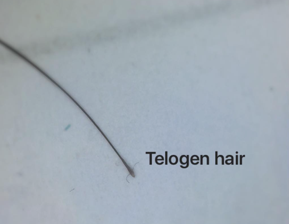
4. Examination of Hairs

Hairs themselves should be analyzed. A gentle pull of hairs may reveal secretions and crusts attached to hairs. These should be examined for bacteria and fungi as in the paragraph above.

5. Scalp Punch Biopsy

A scalp biopsy is rarely needed for patients with smelly scalp syndrome but should always be considered. Patients with scalp odor who have concerns about severe burning or pain or who have concerns about bleeding, weeping or oozing are most likely to benefit from a biopsy.

6. Blood Tests

Blood tests are usually normal in patients with scalp odor. However there are situations where I will consider blood tests for blood sugars, thyroid disease and hormone abnormalities.

7. Patch Testing

Patch testing is a method of determining whether a patient has a true allergy. It’s a very involved test and not appropriate for everyone but may be appropriate for a minority of patients with SSS who have marked scalp itching and tenderness especially those with rashes elsewhere on the body.


Treating SSS

The best treatment for SSS is to treat the underlying cause - if it can be identified. If the odor is coming from a scarring alopecia (like dissecting cellulitis) treatment for this condition is what is needed. If the smell is coming from a weeping skin cancer, the only definitive way to get rid of the smell is to treat that cancer. In other words, treatment for scalp odor is first and foremost centred around trying to lock in a diagnosis.

One needs to keep in mind that a cause is not always readily apparent. In such cases a variety of approaches can be taken. 


Treatments to Consider When No Cause Can be Found. 

In cases where the precise cause can not be identified, my general approach is to target scalp fungi, bacteria and inflammation. As I often say to physicians who visit with me in my clinic a helpful “memory tool” to treat scalp smell is to remember that dealing with challenging cases of scalp smell requires us to call in “smelly undercover FBI agents on a daily basis to deal with the slippery cover up.” 

This 'memory tool' reminds us we need to

a) uncover any scale

b) kill bacteria (letter B in FBI)

c) kill fungi (letter F in FBI)

d) reduce inflammation (letter I in FBI)

e) shampoo daily

f) slippery (reduce oils)

f) cover up any smell


The three letter FBI acronym stands for agents that target fungi (letter F), bacteria (letter B) and inflammation (letter I)

A variety of approaches are possible and there is no one treatment approach that fixed everything. But with a logical approach we can often combat challenging cases of scalp smell.

I generally start patients with a once daily to twice daily shampooing regimen which includes antibacterial cleansers alternating with ketoconazole shampoos and alternating with a sulphur shampoo containing salicylic acid. These are done in 3 day cycles (see below) amd are left on the scalp for three minutes each application. Depending on the specific situation, the patient may use a topical clindamycin/corticosteroid solution at night for a few weeks.


Initial Starting Routine:

Day 1 morning: Antibacterial cleanser 3 min

After morning shampoo: apply 3 drops peppermint oil in jojoba to scalp

Day 1 evening: Clindamycin/steroid solution 


Day 2 morning: Ketoconazole shampoo 3 min

After morning shampoo: apply 3 drops peppermint oil in jojoba to scalp

After morning shampoo: apply 3 drops peppermint oil in jojoba to scalp

Day 2 evening: Clindamycin/steroid solution 


Day 3 morning: Sulphur/SA shampoo

After morning shampoo: apply 3 drops peppermint oil in jojoba to scalp

Day 3 evening: Clindamycin/steroid solution 


Repeat cycle...


Let’s look at each of these steps in a bit more detail.


1. Uncover

In many of these smelly scalp syndromes, it is important to get rid of any scale or crust that might be harbouring bacteria. For this reason, I frequently recommend use of a shampoo containing salicylic acid. Salicylic acid helps remove scale. There are many ways to bring salicylic acid into a treatment program but shampoos are frequently the easiest. Neutrogena T sal is a common sulphur based shampoo which contains salicylic acid. Many patients with SHS like both components, but particularly the sulphur component.


2. Fungi

Yeast are a common culprit in scalp smell for many patients. Yeast known as Malassezia are thought to be the key underlying cause of seborrheic dermatitis. Any patient with scalp smell needs to eradicate yeast as a fundamental step in treatment. Ketoconazole is a good first step in eradicating yeast in patients with SSS. However other options can be considered including

a) zinc pyrithione shampoo 

b) ciclopirox shampoo

c) selenium sulphide

d) oral itraconazole

e) tea tree oil shampoo 

f) 1 teaspoon tea tree oil in 1 cup water


3. Bacteria

Bacterial load on the scalp should be reduced as part of a basic regimen to stop scalp smell. Antibacterial soaps containing triclosan were common antibacterial agents we used in the past. These are less commonly used given the ban on triclosan in many countries. However, I am a fan of  antibacterial washes for smelly scalp syndrome  -including harsh ones. Normally, we don't use soaps on the scalp as they tend to be harsh and tend to leave a residue. In many patients with SSS/SHS, this actually proves beneficial.

The use of antibacterial agents should be left on for 3 minutes.

Depending on the patient, bacterial load can also be controlled with

a) Topical clindamycin

b) Topical or oral metronidazole (Flagyl)

c) Oral doxycycline

d) Benzoyl peroxide body washes (10%) used as scalp cleanser

e) topical chlorhexidine 2-4 % applied with wash cloth (must be kept out of eyes and ears)

e) topical triclosan and phisohex - no longer used.


4. Inflammation

Inflammation in the skin of the scalp can change the types of oils that are produced and the types of free fatty acids that are released. In the early weeks of treating scalp smell, I recommend use of a mid potency topical steroid such as topical betamethasone valerate to help reduce inflammation. This can be used nightly with topical clindamycin compounded into it.


5. Reducing oils

A variety of options are available if it is discovered that the patient is producing too much oil. One can first see if shampoos like ketoconazole shampoo discussed above can help dry things out. The other option is to use various dry shampoos throughout the day. These can be washed out at night with one of the shampoos discussed above so that the clindamycin/betamethasone can be applied.

In rare cases, scalp smell can be improved by drastically and dramatically affecting the size of the oil glands with oral medications such as isotretinoin. In patients with bacterial scalp issues, reduction in oil glands makes bacteria less likely to remain on the scalp - a second benefit.


6. Cover up

There are a variety of other options to cover up smell. Use of essential oils such as peppermint oil, rosemary or lavender can be applied to the scalp. 3 drops of peppermint oil for every teaspoon of jojoba oil is a great start. This can be applied after the morning shampoo.

Rinses with baking soda help some. 1 teaspoon of baking soda is added to 1 cup of water along with a few drops rosemary. This is allowed to sit on the scalp 20 minutes.

For others, mixing 1/4 cup apple cider vinegar 3/4 cup water with 10 drops rosemary essential oil and 1/8 cup lemon juice forms a tonic than can be left on the scalp 30-40 minutes before rinsing off


Conclusion

Managing scalp door can be challenging. Too often it is forgotten that the fundamental step in treating scalp door is to treat the underlying cause of the door. That may require an examination by a dermatologist to determine that.  The use of suphur based shampoos with salicylic acid together with various antibacterial washes and anti-seborrheic shampoos is a good starting point. More advanced options should be discussed with a dermatologist including use of isotretinoin or metronidazole.


This article was written by Dr. Jeff Donovan, a Canadian and US board certified dermatologist specializing exclusively in hair loss.
Share This
28 Comments

Normal vs Compound Follicles

What is the difference?

lpp-vs-fd.png

Hairs emerge from the scalp through pores or hair follicle openings. Some pores have just one hair, but most normally have two or three hairs emerging through a single hair follicle opening. This is completely normal. 
It’s important to be able to quickly spot when something is not quite right. Most hair loss conditions lead to a reduction in the number of hairs coming out of each pore. Instead of seeing the plentiful bundles of two and three hairs one starts to see pores with either no hairs at all or just a single hair. 
Some scarring alopecias are associated an unusual feature- and that is an increase in the number of hairs coming out of the pores. When six or more hairs come out of a single opening we refer to this as a “compound” follicle. The scarring alopecias which frequently show compound follicles include folliculitis decalvans (tufted folliculitis) and sometimes acne keloidalis. It tends to be the scarring alopecias associated with neutrophils that are associated with formation of compound follicles.

Compound follicles occur because of the destructive enzymes released from the inflammatory process. These enzymes destroy tissue and promote fusion of follicles together. The photos here show compound follicles in folliculitis decalvans and single haired follicles in lichen planopilaris. 

SINGLE HAIRS IN LICHEN PLANOPILARIS 

SINGLE HAIRS IN LICHEN PLANOPILARIS


COMPOUND HAIRS IN FOLLICULITIS DECALVANS

COMPOUND HAIRS IN FOLLICULITIS DECALVANS


This article was written by Dr. Jeff Donovan, a Canadian and US board certified dermatologist specializing exclusively in hair loss.
Share This
2 Comments

Alopecia areata: Up close

Dermatoscopic (Trichoscopic) Features of Alopecia Areata

AA

Typical dermatoscopic findings in alopecia areata: 1) yellow dot 2) black dot 3) exclamation mark hair and 4) tapered hair. 


This article was written by Dr. Jeff Donovan, a Canadian and US board certified dermatologist specializing exclusively in hair loss.
Share This
No Comments

Telogen Effluvium: What are upright regrowing hairs (URH)?

URH in TE

urh

Telogen effluvium refers to a type of hair loss whereby the patient experiences increased daily shedding. Shedding typically occurs 2-3 months after a "trigger" such as weight loss, surgery, illness, low iron, crash diet, medication initiation or development of some internal illness.

Dermoscopy (shown here) does not have many specific findings in patients with telogen effluvium although many upright regrowing hairs (URH) may be seem along with hair follicles containing only a single hair follicle.  


This article was written by Dr. Jeff Donovan, a Canadian and US board certified dermatologist specializing exclusively in hair loss.
Share This
2 Comments

Focal Atrichia in AGA

Focal atrichia: What does it mean? 

Focal atrichia refers to small circular areas on the scalp that are devoid of hair. These areas are typically slightly larger than a pencil eraser. 

fa-in-aga


Focal atrichia is seen in both male and female androgenetic alopecia and more common in more advanced stages. They may contain a few tiny vellus hairs if one looks closely but eventually these tiny hairs disappear over time. Hair regrowth does not occur in these areas.

Studies by Olsen and Whiting (see references below) showed that focal atrichia was present in 44% of women with female pattern hair loss, including 67% of late onset vs 15% of early onset, compared to 3/146 (2%) of those with other hair disorders. Hu and colleagues showed that focal atrichia in men with balding was associated with more advanced stages.


Reference


Olsen EA, et al. Focal Atrichia: A Diagnostic Clue in Female Pattern Hair Loss. J Am Acad Dermatol. 2017.

Hu R, et al. Trichoscopic findings of androgenetic alopecia and their association with disease severity. J Dermatol. 2015.


This article was written by Dr. Jeff Donovan, a Canadian and US board certified dermatologist specializing exclusively in hair loss.
Share This
No Comments

Scarring alopecia: Acquired Pili torti

Acquired Pili torti

pt

Scarring alopecias are a group of diverse hair loss conditions that are associated with the presence of scar tissue in the scalp. This scar tissue can damage growing hair follicle and affect how they grow.

A common finding in many scarring alopecias is the twisting of hairs in a patient with otherwise straight hair. This “twisting” of hair is called pili torti and when it develops long after birth we call it “acquired pili torti.” This photos shows typical pili torti in a patient with frontal fibrosing alopecia. Some straight unaffected hairs can also be seen in the photo as well (bottom right). Dilated veins typical of FFA can also be seen.


This article was written by Dr. Jeff Donovan, a Canadian and US board certified dermatologist specializing exclusively in hair loss.
Share This
2 Comments

Lichen planopilaris up close: A look at "perifollcular scale"

Perifollicular scale: What does this term mean?

pfs

Lichen planopilaris ("LPP") is a scarring alopecia which causes permanent hair loss.

Affected individuals frequently develop hair shedding accompanied by scalp itching and sometimes scalp burning and scalp pain.

The accompanying photo shows the typical appearance by trichoscopy of lichen planopilaris (LPP). Single hairs are seen with white scale around these hairs. This whitish scale is known as perifollicular scale and sometimes also as follicular hyperkeratosis.

Treatments for LPP include topical steroids, topical calcineurin inhibitors, steroid injections, oral tetracyclines, oral hydroxychloroquine, oral methotrexate, oral mycophenolate, oral cyclosporine, oral low dose naltrexone. Some patients also respond to oral finasteride.


This article was written by Dr. Jeff Donovan, a Canadian and US board certified dermatologist specializing exclusively in hair loss.
Share This
No Comments

Telogen hairs: Not only in Telogen Effluvium

What are telogen hairs? When does one find them?


Telogen hairs are hairs that are shed from the scalp because they are done their growing. If you reach up and run your hands gently through your own hair, you’ll likely remove a telogen hair. (If you forcefully pluck hairs from the scalp you’ll remove a completely different type of hair called an anagen hair). Between 30-70 telogen hairs normally fall out of the scalp every day. On shampooing days (days that one shampoos the scalp), even more wiggle out so the number can reach well over 100 for some individuals. If one shampoos the scalp everyday, they remove telogen hairs on a consistent basis and so the loss each time remains much lower than if one shampoos once per week.


Telogen Hairs can be a Normal Finding

telogen

The finding of telogen hairs can be completely normal but an increased number of telogen hairs lost from the scalp compared to what one usually loses, is not normal. If one normally loses 40 hairs per day and now loses 80, this is not normal provided the frequency of shampooing remains the same. A common mistake that individuals make is assuming that the finding of increased shedding of telogen hairs confirms a diagnosis of “telogen effluvium.” This is not accurate. Individuals with a range of hair loss conditions will shed more telogen hairs than they normally do including the following: telogen effluvium, chronic telogen effluvium, androgenetic aloepcia, alopecia areata, as well as the many scarring alopecias and the inflammatory scalp diseases (such as seborrheich dermatitis). The finding of increased numbers of telogen hairs coming out from the scalp could mean the patient has a diagnosis of telogen effluvium - but does not necessarily mean this is the only diagnosis to consider.


This article was written by Dr. Jeff Donovan, a Canadian and US board certified dermatologist specializing exclusively in hair loss.
Share This
No Comments

Alopecia areata : Well demarcated

Common Clinical Features of AA

aa-welldemarcated

Alopecia areata is an autoimmune condition that affects 1.7 % of the world. Affected patient often develop well demarcated round or oval-shaped patches of hair loss.

Well-demarcated

The term well dermarcated means that one can often draw an imaginary line around the area of hair loss as opposed to other hair loss conditions like androgenetic alopecia where is can be challenging to determine where the hair loss starts and ends.


If one looks up close at a typical patch of alopecia areata, a number of findings are often present including (1) thin hairs or white hairs within the patch of hair loss and (2) “exclamation mark” hairs around the perimeter of the patch of hair loss. Spontaneous growth can occur in many patients. However as the size of the patch increases amd as the number of these patches increases, the chance of spontaneous regrowth diminishes. The most effective treatments for a single patch such as the one shown are topical steroids and steroid injections. I frequently add minoxidil if the patch is slower to respond to treatment.


This article was written by Dr. Jeff Donovan, a Canadian and US board certified dermatologist specializing exclusively in hair loss.
Share This
No Comments

Permanent Chemotherapy Induced Alopecia (PCIA)

A New type of Hair loss in Patients Undergoing Chemotherapy: PCIA

Every year about 650,000 patients undergo chemotherapy in the United States. Hair loss is a common side effect of chemotherapy and occurs in about 65 % of patients who receive chemotherapy. There are two main types of hair loss that can occur in patients undergoing chemotherapy. The first is hair loss that happens within weeks of starting the chemotherapy and then lasts several months before growing back.  This is known as temporary chemotherapy induced alopecia ("TCIA"). The second type is uncommon and occurs when patients fail to regroth their hair back to the level it was before undergoing chemotherapy. If hair has not grown back after chemotherapy by the 6 month after chemotherapy, we call this permanent chemotherapy induced alopecia (PCIA) and it is sometimes also called Chemotherapy Induced Permanent Alopecia (CIPAL).


Permanent Chemotherapy Induced Alopecia (PCIA) 

The failure of the hair to grow back fully 6 months post chemotherapy raises concerns about a phenomenon known as permanent chemotherapy induced alopecia (PCIA).     In recent years a number of studies have highlighted the possibility of PCIA in women with breast cancer treated with various chemotherapeutic agents, especially drugs known as taxanes. Docetaxel and paclitaxel are part of this group of drugs. The exact mechanisms are unclear although injury to the bulb as well as follicular stem cells are thought to be relevant. Adjuvant anti-estrogen hormonal therapy may be an important cofactor in many women with PCIA. A similar PCIA presentation has been reported in patients undergoing bone marrow transplantation. The scalp is predominantly affected in women with PCIA although a minority may have eyebrow, eyelash and body hair loss as well.


Different Clinical Presentations of PCIA

PCIA doesn't appear similar in all patients. In fact, three main types appear to exist including a diffuse type, a diffuse type with vertex accentuation (mimicking androgenetic alopecia) and a patchy type mimicking alopecia areata.   


Examination under the Microscope: Biopsies of PCIA

Histopathology of biopsy specimens shows a non-scarring alopecia with preservation of sebaceous glands, miniaturization, decreased anagen hairs, increased telogen hairs and end stage avascular fibrous tracts. There may be several histological presentations and the exact features remains to be defined although a high proportion show dysmorphic telogen germinal units. Some biopsies show peribulbar type inflammation.


How do we treat PCIA?

We don't really know yet how to best treat PCIA. The most common treatments described in the medical literature are oral and topical minoxidil. Both seem to provide benefit to at least a proportion of patients.  Other treatments are not known to provide benefit. 


Dr. Donovan's Articles for Further Reading

Preventing Hair Loss from Chemotherapy

Does hair always grow back after chemotherapy?


REFERENCES

Miteva M, Misciali C, Fanti PA et al. Permanent alopecia after systemic chemotherapy: a clinicopathological study of 10 cases. Am J Dermatopathol. 2011 Jun;33(4):345-50.  

Fonia A, Cota C, Setterfield JF et al. Permanent alopecia in patients with breast cancer after taxane chemotherapy and adjuvant hormonal therapy: Clinicopathologic findings in a cohort of 10 patients. J Am Acad Dermatol. 2017 May;76(5):948-957.

Rugo HS.  Real-world use of scalp cooling to reduce chemotherapy-related hair loss.  Clin Adv Hematol Oncol. 2017


This article was written by Dr. Jeff Donovan, a Canadian and US board certified dermatologist specializing exclusively in hair loss.
Share This
No Comments

Grieving and Hair Loss

Dr. Elisabeth Kubler-Ross was a pioneer in understanding how humans grieve. Further studies and observations over the years has shown that the way we grieve is similar regardless of what it is we are actually grieving.

As Dr. Kubler-Ross first described nearly 50 years ago, grieving commonly occurs through 5 stages that include:

  1. denial and isolation
  2. anger
  3. bargaining
  4. depression and
  5. acceptance.

Not all stages have the same length and don't necessarily occur one after the other like the chapters in a book. Not all patients reach the end - acceptance. There is tremendous variation in how people grieve but Kubler-Ross's model has served as a valuable model for decades.

There is little written about grieving and hair loss. You'll never hear about the topic at any hair meeting. It has become increasingly clear to me over the years that some forms of hair loss lead to such profound changes in a person's appearance that they trigger the same grieving responses as one might have with any illness or cosmetic alteration in appearance. Patients with rapid alopecia areata, scarring alopecias, hair shedding disorders as well as androgenetic alopecia often grieve the loss of an appearance they once had. For many, hair loss brings profound changes in one's self-identify and overall self confidence. Many affected patients also progress through the grieving stages of denial, anger, bargaining, depression and acceptance.

Reference

1. www.donovanmedical.com/hair-blog/grieving

2. Kubler-Ross. On Grief and Grieving. 2007.


This article was written by Dr. Jeff Donovan, a Canadian and US board certified dermatologist specializing exclusively in hair loss.
Share This
No Comments

Vellus Hairs on the Scalp

Vellus Hairs on the Scalp.png

Does One Find Vellus Hairs Normally?

Vellus hairs are tiny, short non-pigmented hairs. They are fine hairs with a caliber less than 30 micrometers by definition. It is not common to find vellus hairs on the scalp in an individual without hair loss. On a normal scalp only about 1 of every 25 hairs are vellus hairs. Most hairs on the scalp are large pigmented terminal hairs. During the course of male and female androgenetic alopecia, vellus hairs become more prevalent and may even become the dominant hair type (outnumbering terminal hairs) in advanced balding cases.

Reference

Ko JH et al. Hair counts from normal scalp biopsy in Taiwan. Dermatol Surg. 2012


This article was written by Dr. Jeff Donovan, a Canadian and US board certified dermatologist specializing exclusively in hair loss.
Share This
No Comments

Trichogram: An Older Less Commonly Used Technique

The "trichogram" is an older technique developed in 1957 for assessing hair loss. It involves quickly pulling out the roots of 50-70 hairs. The hairs are all placed on a glass slide and examined under the microscope.

By carefully comparing an affected area of hair loss to a presumed unaffected area alot of information can be ascertained. In androgenetic alopecia, there is a marked increase in the proportion of telogen hairs in the frontal scalp (as well as a variation in hair shaft diameter) compared to the occipital scalp. In a pure telogen effluvium the trichogram results are similar in the front and back of the scalp.

A trichogram can provide helpful information in assessing active alopecia area (where an increase in dystrophic hairs are seen) and hair loss from various toxins.

The technique is not commonly used these days as dermoscopy (trichoscopy) has provided a more sensitive and non invasive means of assessing complex scalp disorders.


This article was written by Dr. Jeff Donovan, a Canadian and US board certified dermatologist specializing exclusively in hair loss.
Share This
No Comments

Trichotillomania

Exclamation Mark Hairs

Exclamation mark hairs are short hairs that are thick at the top and thinner as they enter the scalp. These hairs are known to occur in the autoimmune condition alopecia areata but also can occur in trichotillomania (shown this picture), poisoning situations (ie thallium) and have also been reported in dissecting cellulitis.


This article was written by Dr. Jeff Donovan, a Canadian and US board certified dermatologist specializing exclusively in hair loss.
Share This
No Comments

How can one identify a telogen hair?

Hairs that are shed from the scalp are known as telogen hairs. These hairs lack pigment at the very end and do not have a root sheath around the ends either. Telogen hairs are known as "club hairs."

I always encourage my patients to bring in hairs they shed if they are worried about the type of hair they are seeing. 99.9 % of the time the hairs they see are telogen hairs. About 30-70 telogen hairs are released from the scalp each day. In some hair loss conditions (such as telogen effluvium), an increased number of telogen hairs are released every day


This article was written by Dr. Jeff Donovan, a Canadian and US board certified dermatologist specializing exclusively in hair loss.
Share This
No Comments

Psoriasis + Seborrheic Dermatitis

Sebopsoriasis

Sebopsoriasis is a scaly scalp condition with features of both psoriasis and seborrheic dermatitis.

Many patients with psoriasis have overgrowth of Malassezia yeast which are known to play a role in seborrheic dermatitis. Some evidence suggests that Malassazia yeast may even be involved in the pathogenesis of psoriasis. Patients with sebopsoriasis often have a family history of psoriasis or seborrheic dermatitis. Lesions are less silvery white than classic psoriasis bit more defined and deeply red than classic seborrheic dermatitis. Scale is often thicker in areas than seborrheic dermatitis.


This article was written by Dr. Jeff Donovan, a Canadian and US board certified dermatologist specializing exclusively in hair loss.
Share This
No Comments

Plan b: Is There a Plan B to Treating Hair Loss?

The first step in determining how to help someone with hair loss is figuring out his or her diagnosis. There is no bypassing this step.  The second step is determining a treatment plan that is based on the best medical evidence. 


Plan B: What is Plan B, Doc?

After reviewing a treatment plan with my patients, I'm often asked what treatment will be considered next. "What's plan B, doc?" Well, every treatment plan needs Plan B as well as a Plan C and Plan D.

Consider the 28 year old female with androgenetic alopecia. The best treatment option for her based on all her facts, review of her blood tests and scalp exam might be topical minoxidil. Plan B might be oral spironolactone with or without minoxidil. Plan C might be the addition of a laser comb or changing the anti androgen used. Plan D for her might be a trial of PRP. A solid treatment plan has an alphabet of plans. Not guesswork and not a random pull out of a hat option. But rather options based on a delicate combination of medical science and expert consensus, and personal experience.

What about the 53 year old female with frontal fibrosing alopecia? Plan A for her might be finasteride & steroid injections with hydroxychloroquine as Plan B. Doxycycline is reserved for her as Plan C. For another patient with FFA, Plan A might start with hydroxychloroquine & steroid injections. For her, finasteride is not on the list given the past history of breast cancer the patient had. Plan B is doxycycline and plan C is methotrexate.


Conclusion

Every treatment plan should have an alphabet of plans. That does not necessarily mean one will need to move down the list but the physician should have a clear plan for how to navigate.


This article was written by Dr. Jeff Donovan, a Canadian and US board certified dermatologist specializing exclusively in hair loss.
Share This
No Comments

Blogs by Topic


Share This
-->