h1.qusth1 { display: none !important; }

QUESTION OF THE WEEK

Dr. Donovan's Articles

QUESTION OF HAIR BLOGS

Filtering by Category: Diagnostic Tests


Standardized Hair Collections

Identifying AGA and TE

Telogen effluvium (TE) and androgenetic alopecia (AGA) are common, especially among women. There are many ways to differentiate a shedding disorder (TE) from AGA - and some women have both. 

A clinical examination of the scalp, a biopsy and a so called "hair collection" are three methods to evaluate a patient's diagnosis. Exactly which one I use depends on the specific clinical situation. Certainly not everyone with hair loss needs a biopsy and not everyone needs to perform a hair collection.

There are many different ways to perform a hair collection. Rebora studied the use of the 5 day hair collection, where shampooed hairs are trapped on a gauze 5 days after shampooing. The collected hairs are divided into three groups: telogen vellus hairs (less than 3 cm), intermediate hairs (3-5 cm) and long hairs (more than 5 cm). The presence of more than 10 % non broken hairs 3 cm or less is suggestive of the diagnosis of androgenetic alopecia (AGA).


Reference


Distinguishing androgenetic alopecia from chronic telogen effluvium when associated in the same patient: a simple noninvasive method.
Rebora A, et al. Arch Dermatol. 2005.


This article was written by Dr. Jeff Donovan, a Canadian and US board certified dermatologist specializing exclusively in hair loss.
Share This
No Comments

Which hairs should be taken during a scalp biopsy?

What is a scalp biopsy? Which hairs should be taken ?


A scalp biopsy is a 15-20 minute procedure that helps the dermatologist obtain a small piece of tissue for analysis.  The sample is processed in a history laboratory into thin sections and mounted on glass slides for review by a pathologist under the microscope.  A scalp biopsy is not required for most patients with hair loss. When the diagnosis is uncertain, it can be very helpful.

FOR DETAILS ON THE SCALP BIOPSY, CLICK HERE
 

Which hairs should be included?
 

First, an area of the scalp containing hair follicles should always be included in a biopsy. A scalp biopsy specimen from a completely bald area is not helpful!

Second, the hair follicles obtained should be taken from an area showing the specific abnormalities thought to be in keeping with the hair loss condition in question. If no such features are present, hair follicles might be obtained from an area of the scalp where the hairs are easily extractable (positive pull test), or from an area having the most symptoms (itching, burning or pain).

The key point is that obtained a scalp biopsy from a random area on the scalp does not usually yield useful information.


This article was written by Dr. Jeff Donovan, a Canadian and US board certified dermatologist specializing exclusively in hair loss.
Share This
No Comments

How do I diagnose hair loss?

Diagnosis comes from listening and looking

I'm often asked how I diagnose hair loss. Sometimes it's easy and sometimes it's challenging. What steps are taken? What is the basic approach?

This short clip provides a brief overview of the key steps of first listening to the patient's story of their hair loss and then examining the scalp up close.

Hope you enjoy


Jeff


This article was written by Dr. Jeff Donovan, a Canadian and US board certified dermatologist specializing exclusively in hair loss.
Share This
No Comments

What is the best test to do if I think I have celiac disease?

What is celiac disease?

Celiac disease is an autoimmune disease of the bowel. When individuals with celiac disease eat foods containing "gluten" (such as barley, rye, wheat), inflammation develops in the small bowel. This leads to damage to the small bowel which prevents it from properly absorbing food.  Celiac disease can develop at any age. It’s more common in Caucasians and those of European ancestry. Women are affected to a greater extent than men.

Patients with celiac disease may have many symptoms.  The most common symptom is diarrhea that lasts many weeks or months (termed "chronic diarrhea") as well as weight loss.   But a range of symptoms are possible, including abdominal pain, weight loss, bloating, gas, and constipation. In fact, celiac disease can sometime be challening to diagnose because it has many different ways of presenting.


Should patients with hair loss be tested for celiac disease?

Many patients with hair loss wonder if they should stop gluten or if they should be tested for celiac disease.  In most cases, the answer is "no."  However, testing for celiac disease may be recommended f the patient has abdominal symptoms or long standing weight loss. Sometimes I also check for celiac disease if a pateint has low iron levels that just don't seem to raise despite use of iron pills.  A very small percent of patients with autoimmune hair loss conditions (i.e. alopecia areata) do have celiac disease.  


The most common tests that are ordered to SCREEN if someone has celiac disease are:

1. Tissue transglutaminase Antibodies (tTG)

2. Endomysial Antibodies (EMA) 

3. Other tests "may" be ordered by the physician as well including IgA antibodies and specific genetic tests such as HLA-DQ2 and HLA-DQ8.

Overall, the tTG test is the best screening test. It's inexpensive, quantitative and a highly reproducible test.


What is done once a patient is diagnosed with celiac disease? 

Once diagnosed with celiac disease, a gluten free diet will be recommended. In some cases, a referral to a gastroenterologist may be recommended. Follow up blood tests may be ordered to assess how well an individual is doing with their gluten free diet.


This article was written by Dr. Jeff Donovan, a Canadian and US board certified dermatologist specializing exclusively in hair loss.
Share This
No Comments

More Benefits of Checking Iron Levels in Women with Hair Loss

Checking Iron Levels in Women with Hair Loss

As many of my patients know, iron metabolism is one of my favourite subjects. It also forms one of the components of the hair loss research that I do. 

I generally recommend that all women with concerns about hair loss have their iron levels checked using the simple blood tests called ferritin. Other blood tests may also be ordered for women with hair loss, such as thyroid tests and a complete blood test to measure the hemoglobin level.

Although it is somewhat contraversial among hair experts around the world, the iron level I like my patients to aim for is a ferritin level above 40-50 ug/L.  If the blood test shows less than this, then I recommend supplementation with iron pills.


New research outlines additional benefits of checking iron levels in women

In a recently published study, Swiss researchers studied 198 premenopausal women who had ferritin levels less than 50 ug/L and symptoms of fatigue. A proportion of women in the study received ferrous sulphate pills and another proprotion of women received placebo pills.


What were the results of the study?

At the end of the 12 week study, women who received iron noted a significant improvement in their overall level of fatigue compared to women receiving the placebo pills.  The ferritin level in women recieiving the iron pills increased by approximately 12 ug/L over the 12 weeks of the study.


Comment

Many premenopausal women have low iron levels. This study reminds us that there are many improtant benefits of iron, including helping improve the overall feeling of fatigue (if levels are low). Although I routinely follow iron levels in  my patients, this study reminds us that asking about improvement in fatigue levels may also be an important parameter to assess in making a decision about continuing iron supplements for the longer term.


Reference

Vaucher P et al. Effect of iron supplementation on fatigue in nonanemic 
menstruating women with low ferritin: A randomized controlled trial. CMAJ 
2012 Aug 7; 184:1247. (Click link for article)


This article was written by Dr. Jeff Donovan, a Canadian and US board certified dermatologist specializing exclusively in hair loss.
Share This
No Comments

Hair loss and lupus: Doc, do you think my hair loss could be from lupus?

Hair loss and lupus:

Anyone who is losing hair knows how frustrating it can be to find accurate information. It is often difficult to figure answers by searching the internet. Confiding in a friend about hair loss leads to one piece of advice, while a second friend offers completely different advice. It seems daunting to pinpoint which of the 100 causes of hair loss are responsible.

In the search for answers, it's not uncommon for a patient to ask me "Do you think my hair loss could be from lupus?" Are you going to test me for it?

What is lupus?

Lupus, also known by the medical term "systemic lupus erythematosus" is an autoimmune condition. It has the potential to affect nearly every body system from the skin to heart to lungs to brain to kidney (....and that is why the word "systemic" is used). It affects approximately 50 out of every 100, 000 people. Current estimates suggest there are 500,000 individuals in the USA and 50,000 individuals in Canada affected by lupus. Women are affected nearly 9 times more commonly than men. Black women are particularly affected.

What are the signs of lupus and should I get tested?

For every patient with hair loss that I see, I run through a series of simple "screening questions. " If the answers to all these questions are "NO" then I usually don't give any further thought to the patient in front of me having lupus. If the answer to one or more of the questions is "YES" it certainly does not mean the patient has lupus, but means that I might ask more "in depth" questions.

Typically, my "screening questions" for lupus that I ask patients with hair loss include the following 13 questions:

HAIR CLINIC SCREENING QUESTIONS FOR LUPUS:

1. Have you been experiencing extreme levels of fatigue lateley?
2. Do you experience headaches... and if so...how often do you get them?
3. Have you ever experienced a seizure in your life? how many?
4. Do you see or hear things that you think other people might not hear or see?
4. Do you have joint pains ...and if so ...which joints?
5. Do you have high blood pressure? Has it been difficult to control with medications?
6. Do you have pain in the chest when you take a deep breath?
7. Do you experience dry mouth or dry eyes?
8. Do you experience ulcers in the mouth, nose (and for women in the vagina) that you are aware of?
9. Do you develop rashes on the face or skin when you go out in the sun? Do you find you burn much easier than before?
10. Have you ever been told you have abnormal blood work results? ( especially, low hemoglobin, low white blood cells, low platelets)
11. Has anyone in your family been diagnosed with lupus in the past?
12. Have you ever had a blood clot?
13.(Women) Did you ever have a miscarriage and if so, how many?

Many, many individuals will answer " YES" to a question or two from the above list. It does not mean they have lupus. But if they answer "YES" to a few questions it points me down a path of very, very detailed questioning. The formal American College of Rheumatology criteria for diagnosing lupus can be found by clicking here.


Should I get a blood test for lupus?

The vast majority of patients with hair loss do NOT need to have a test for lupus. This can't be overstated enough. However, if the answers to a few of the screening questions above are " YES" then testing 'could' be at least considered. If I am even slightly suspicious, I usually order a blood test known as the ANA (anti-nuclear antibody test).   If the ANA blood test returns "positive", additional blood tests (Group 2) may be ordered. It takes alot of experience to interpret these tests and it can sometimes be challenging to diagnose someone with lupus.  Referral to a rheumatologist is often required.

Group 1 test for lupus

ANA (anti- nuclear antibodies)

  • 99 percent of people with lupus will have a positive test. However, many other conditions (and even normal healthy people) can have a positive ANA test. So having a positive ANA does not necessarily mean you have lupus.  Rarely, patients with lupus can even have a negative ANA test (especially early in the disease).


Group 2 tests for lupus (ordered if the ANA test is positive)

1. CBC (complete blood count)

  • Individuals with lupus may have low levels of red blood cells, white blood cells and platelets

2. Urinalysis

  • To check if there is protein or blood in the urine. This can be a sign of kidney damage.

3. Creatinine

  • Another measure of kidney health. Patients with kidney disease may have increased creatinine levels.

4. ESR ( erythrocyte sedimentation rate)

  • A measure of inflammation in the body. Many conditions can increase ESR, not just lupus.

5. C3 and C4 (complelment levels)

  • Complement levels may be lower in patients with autoimmune diseases and used to monitor activity of the disease


6. ENA (extractable nuclear antigens) which include the anti-Smith test

  • The ENA test measures many antibodies, including Jo, Sm, RNP, Ro, La, Scl-70. These antibodies can be positive in many different types of immunologic conditions. Patients with lupus may have a positive anti-Smith test (and sometimes other positive results from the ENA panel of tests too).

7. Antiphosphopilid antibodies

8. Anti-ds DNA test (anti-double stranded DNA test)


In summary, is the diagnosis of lupus should only be made by a medical professional. There are well over 100 causes of hair loss and certainly lupus is on that list and therefore needs to be at least considered. Simply having a positive ANA test doesn't automatically mean an individual has lupus. Obtaining a very detailed medical history and performing a detailed physical examination is very important in the overall evaluation of a patient suspected of having lupus.  Because lupus is a disease that affects many organs in the body, patients diagnosed with lupus are often treated by rheumatologists as well as other specialists (cardiologists, respirologists, neurologists, nephrologists, dermatologists). 


This article was written by Dr. Jeff Donovan, a Canadian and US board certified dermatologist specializing exclusively in hair loss.
Share This
No Comments

Preparing the Mind to Recognize the Many Forms of Hair Loss

In addition to being a hair transplant specialist and specialist in hair disorders, I do research in hair loss and devote time to teaching and lecturing medical students, interns, residents and physicians about hair loss. In fact, part of my time away from the office is spent teaching other doctors about hair loss and about hair transplant surgery. I’m lucky that my profession is not only my job but also a real joy.

I enjoy teaching others about the approximately 100 reasons for humans to have hair loss.

Androgentic alopecia, alopecia areata, telogen effluvium, lichen planopilaris, folliculitis decalvans, dissecting cellulitis, pseudopelade, morphea, ectodermal dysplasia. The list goes on and on.

Today, I gave a lecture about hair loss to medical students at the University of Toronto.  Hair loss is rarely covered in medical schools so it's a real privilege to have the chance to speak to a room full of bright students.  What I hope for after each lecture I give is that the learner goes home with an open mind to consider the many different kinds of hair loss that exist.

FD.jpg

Folliculitis decalvans affecting crown If someday they see a young 34 year old man with a bald crown that itches them like crazy will they instinctively think this is another case of “male balding” or is that doctor now open to consider that this man may instead have an unusual scarring hair loss condition called “folliculitis decalvans” ?

aad.jpg

Alopecia areata diffusa mimicking genetic hair lossor....When the young doctor finds themselves evaluating a 25 year old woman with hair loss in the centre of her scalp, low vitamin B12 blood levels and dozens of little dots in her nails will that doctor instinctively think this is an young woman with early “female balding” or will the doctor remember the lecture and consider that this could be an unusual form of alopecia areata (called “alopecia areata diffusa”)?

The French philosopher and Nobel Prize winner Henri Bergson once said that the human mind sees only what it’s prepared to understand. I consider it a great privilege to teach about hair loss and help others open their minds to the many different types of hair loss that they will likely encounter in their patients in the years to come.


This article was written by Dr. Jeff Donovan, a Canadian and US board certified dermatologist specializing exclusively in hair loss.
Share This
No Comments

Am I getting new hair growth or is it breaking off?

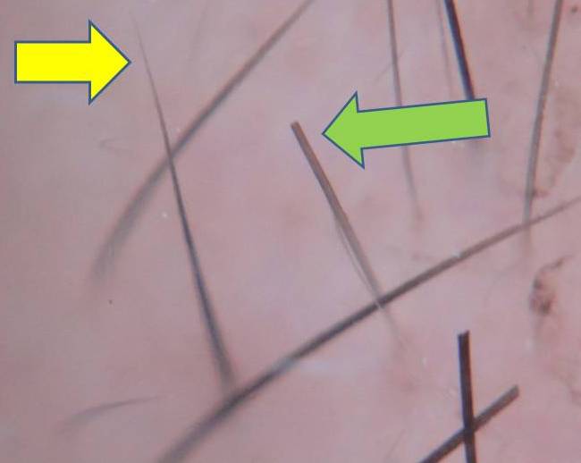
regrow vs broken.jpg

Whenever I examine the scalp, I methodically check for a number of different things. Essentially I have a "checklist" in my mind and all components need to be evaluated before I feel I have properly examined the scalp.

One essential part of the scalp examination is evaluating if patients have new growth - and if so - how much. I like to get a sense of how much hair the patient has grown in the last one month, the last three months and the last six months. Often when I remark to patients that they are experiencing a lot of new growth, I hear a reply such as

How do you know it's new growth?

How do you know my hair is not just breaking off?

The answer is straight forward. Newly growing hairs have 'pointy' ends whereas hairs that have broken off have 'blunt' ends. The photo on the right illustrates these differences. The yellow arrow highlights a newly growing hair with a pointy end and the green arrow identifies a broken hair with a blunt end.  The presence of broken hairs can be due to many causes, including excessive hair damage (heat damage, chemical processing, use of straightners).

Examining for newly growing and broken hairs is an important part of the scalp examination and should be done each time a thorough scalp exam is done.


This article was written by Dr. Jeff Donovan, a Canadian and US board certified dermatologist specializing exclusively in hair loss.
Share This
No Comments

How is a Scalp Biopsy Performed?

biopsy pearls.jpg

There are over 100 reasons for an individual to have hair loss. Sometimes the reason for a patient's hair loss is evident within seconds of meeting the patient. At other times, the diagnosis requires a small scalp biopsy.  A scalp biopsy is a short procedure, performed under local anesthesia, that allows a few hairs (and the skin surrounding those hairs) to be removed for future examination under the microscope.

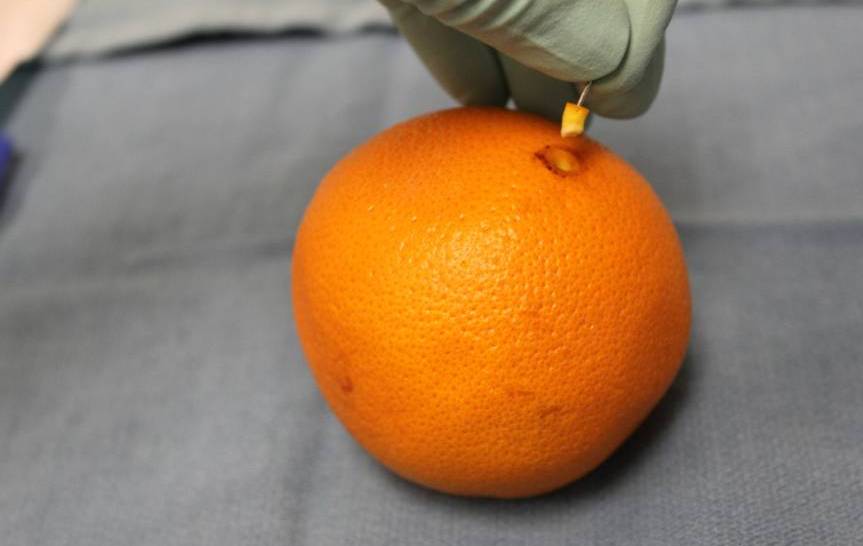
I routinely teach medical students, residents and fellows how to do proper scalp biopsies so that a good sample can be obtained.  It is absolutely essential to get a good sample - one that is deep enough, big enough and not damaged. Here, I outline the proper technique for obtaining a biopsy.  I often use an orange to demonstrate the procedure rather than showing the scalp. The orange background allows the key learning points to be very easily seen. Here, I'll use the orange as well to demonstrate the basic technique.

12 Steps to Performing a Scalp Biopsy

photo1.jpg

An orange is used to demonstrate the proper technique of performing a scalp biopsySTEP 1: Spend time finding an area of the scalp that best respesents the hair loss condition you think the patient has.

If the patient has an unusual pattern of hair loss, or I think that only some areas of the scalp are affected by the condition, I spend many minutes searching for the "perfect spot" to biopsy.  If I think the patient has a scarring alopecia such as lichen planopilaris, I look for an area with perifollicular erythema or scale. If I think the patient may have an unusual or atypical form of alopecia areata, I look for empty tracts or vellus like hairs.

photo2.jpg

STEP 2: Draw a circle around the area.

A blue Acculine Marking Pen can be used to outline the area. I like these pens because the marker does not wash off easily. Therefore, the circle will stay until it is wiped away at the end of the procedure.

photo3.jpg

STEP 3: Cut the hairs that are to be taken in the biopsy.

I routinely cut the hairs that I am going to sample. I clip the hairs to a distance of about 2-3 mm above the scalp. This way I can accurately see the angle that the hairs emerge from the scalp. This will be important in Step 6 (below).

photo4.jpg

STEP 4: Clean the skin.

The skin can be cleaned with many of the commercially available cleansing solutions. I typically use chlorhexidine.

photo5.jpg

STEP 5: Anesthetize the skin

The skin surrounding the hairs is frozen with a numbing solution such as 1% lidocaine with 1:200,000 epinephrine. After freezing the skin, I typically wait 10-15 minutes if possible. This allows the epinephrine to take full effect and drastically helps to reduce bleeding during the small procedure.

photo6.jpg

STEP 6: Use a 4 mm sized punch biopsy tool to puncture the skin.

The minimum size for a punch biopsy for scalp specimens is 4 mm.  These recommendations follow those of the North American Hair Research Society.

punch photo.jpg

The biopsy tool is placed directly over top of the hair - at the precise angle that the hairs are coming out of the scalp. The biopsy tool is then rotated back and forth quickly until the metal blade is completely submerged to the level of the subcutaneous fat. Then the biopsy tool is removed.

photo8.jpg

STEP 7: Delicately pull up on the specimen using the anesthetic needle.

Once the biopsy site has been made, it's important to treat the biopsy specimen very delicately. I use a 30 G needle (the same one used to freeze the skin) to "pull up" on the specimen.

photo9.jpg

STEP 8: Cut the biopsy specimen at the base.

Once the biopsy specimen has been pulled up, I cut it at the base in a horizontal manner using scissors. It is very important that this be done at the subcutaneous junction.

photo10.jpg

STEP 9: Remove the specimen.

After the specimen has been cut at the base, it can be easily removed from the scalp. It can be placed directly into 10% buffered formalin for transport to the histology lab.

empty.jpg

STEP 10: Suture the small opening.

The small opening can be closed with 3-0 or 4-0 nylon. Some physicians choose to leave it open without a suture or pack it with kaltostat.

photo13.jpg

STEP 11: Place petroleum jelly over the suture site to promote moist wound healing.

The biopsy site can be left uncovered. A bandage or dressing is not needed. A small amount of petroleum jelly can be placed over the incision site to keep it moist. Some physicians choose to use antibiotic ointment after the procedure and this is acceptable too. I typically advise patients to wash the area after 24 hours and to continue to apply petroleum jelly for an additional 5 days. It is well accepted that moist wounds heal better.

STEP 12: Remove the stitches in 10-14 days.

The small stitch is cut with scissors after 10-14 days.

All in all, performing a scalp biopsy is easy, and can be performed with little discomfort to the patient. The following represents a basic set up of a surgical tray:

tray set up.jpg

1. chlorhexidine solution 2. punch biopsy tool 3. needle driver 4. pick ups 5. scissors 6. q tips for applying petroleum jelly 7. suture 8. sterile drape 9. gauze


This article was written by Dr. Jeff Donovan, a Canadian and US board certified dermatologist specializing exclusively in hair loss.
Share This
18 Comments

What does a elevated "ferritin" blood test mean for someone with hair loss?

caution sign.jpg

Iron metabolism is one of my favorite subjects.  Iron has an important role in hair growth and it’s important to understand the intricacies of the many iron tests available. I like all of my patients, whether seeing me for medical treatment of hair loss or hair transplantation, to have iron levels in the normal range.

The “ferritin” blood test is a traditionally a measure of iron stores in the body. When I give lectures on hair loss, I often refer to ferritin as the ‘bank’ of iron – a measure of the amount of iron that is stored away in the body.  Hair follicles seem to be able to sense these iron stores – and if they are not high enough, the result can sometimes be hair shedding. Normally, I aim for patients, especially women, to have a ferritin of at least 50 ug/L.  If ferritin is lower than this I may perform additional blood tests to determine if there are any concerning reasons for the patient to have low iron. In most situations with a low ferritin, I will prescribe iron supplements.

If a low ferritin can sometimes be associated with hair loss, what does a high ferritin mean?

Although the upper range of normal for ferritin is 250-300, it's actually not very common to have a ferritin above 90.  If someone has hair loss, an elevated ferritin should always be thought of as a "caution sign." It might simply be a normal result, or a result of taking iron pills, but a bit more detective work needs to be done before coming to that conclusion. It is important to understand that the ferritin test can rise to levels above 90 ug/L for dozens and dozens of different reasons.  Sometimes it's completely normal finding, but other causes include inflammation, infections, cancers, liver disease, and certain other diseases. The disease known as hemochromatosis leads to markedly elevated ferritin levels.  Patients with various inflammatory scalp diseases (i.e. scarring alopecia, alopecia areata) can also sometimes have a high ferritin. These are just a few examples of conditions which are sometimes associated with a high ferritin.  The list is actually quite long and the cause can sometimes only be diagnosed with further detective work by the doctor. Therefore, the ferritin test is not the most specific test for figuring out iron problems, but it is certainly the best test to start with. 

If a patient has a low ferritin level, I can be pretty sure that the patient has iron deficiency.

However, if the ferritin is high (say, over 90), and the patient does not take iron supplements, I can't assume the patient simply has a normal iron status. In most of these situations, I ask many additional questions focusing on all the causes of the slightly high ferritin test.

In cases where the ferritin is high, but the patient is not taking iron, it’s important to look for other clues to determine if the patient (and the patient’s hair) might benefit from an iron supplement. All of the following tests need to be carefully interpreted together to make sure that iron is not given if the patients does not need it:

Hemoglobin (HGB).  Patients with significant iron deficiency can sometimes develop a low hemoglobin level or “anemia.”  However, in the “early” stages of iron deficiency, the body makes a normal number of red blood cells. The hemoglobin in such situations is normal.  However, hair shedding can still occur if the iron stores are low.

Red cell distribution width (RDW). The RDW gives a rough estimate of the size of the red blood cells that are being produced by the body. In cases of iron deficiency, the patient’s body may produce some normal size cells but also some very small cells. This leads to a greater variation in cell size.  Therefore, the RDW may be elevated in patients with iron deficiency.

Mean corpuscular volume (MCV). The MCV also gives another measure of the size of red blood cells. In cases of iron deficiency, the MCV is reduced because smaller red blood cells are produced.   This tests needs to be carefully interpreted by a physician as many other conditions can cause a lower MCV.

Transferrin saturation (Tr sat).  The transferrin saturation is calculated by taking into account two blood test results: the serum iron measurement (Fe) and the total iron binding capacity (TIBC) measurement. The Fe divided by the TIBC gives the transferrin saturation. The transferrin saturation can sometimes be reduced in individuals with iron deficiency.

Soluble transferring receptor (STfR). This can be an accurate measurement of iron deficiency and the test is completely independent of whether or not the patient has inflammation.  This is a very expensive test, and is usually not needed when all of the above tests are taken into account. It is rarely ordered. The soluble transfer receptor level is elevated in individuals with iron deficiency.

Taken together, a patient with elevated ferritin but low MCV, low hemoglobin, high RDW and high transferrin saturation has iron deficiency.  Further investigations would be needed to figure out why the patient is iron deficiency. But the investigations don't stop there - further tests would be needed to figure out why the patient has an elevated ferritin.


This article was written by Dr. Jeff Donovan, a Canadian and US board certified dermatologist specializing exclusively in hair loss.
Share This
9 Comments

What is the best lab test to see if thyroid problems are causing hair loss?

chart - thyroid tests.jpg

Are thyroid problems causing hair loss?

Thyroid hormones are produced by the  thyroid gland, which is a small gland located in the midline of the neck. Precise levels of thyroid hormones are important for normal hair growth. 

The TSH test

The release of thyroid hormones from the thyroid gland is an intricate process. A region of the brain known as the hypothalamus releases a hormone known as "TRH" or thyrotropin releasing hormone, which in turn triggers the tiny pituitary gland to release "TSH" or thyroid stimulating hormone.  In response to the release of TSH, the thyroid gland produces thyroid hormones, namely thyroxine (T4) and triiodothyronine (T3).

When the body produces too much or too little thyroid hormone this may lead to hair shedding. The single best test for thyroid problems is a blood test for “TSH” or thyroid stimulating hormone.  If the TSH is abnormal, several additional thyroid tests can be ordered, including a "free T4".

Hypothyroidism occurs in about 3 % of the population. Hypothyroidism occurs when the thyroid gland does not release enough thyroid hormone. Most patients who are hypothyroid have an abnormally high TSH level and a low free T4 level. This is known as "primary hypothyroidism"  Most patients who are hyperthyroid have the opposite pattern – a low TSH level and high free T4 levels.


This article was written by Dr. Jeff Donovan, a Canadian and US board certified dermatologist specializing exclusively in hair loss.
Share This
No Comments

Do I need any blood tests prior to a hair transplant?


The decision to order pre-operative blood tests depends on the hair transplant specialist. I recommend blood tests for CBC (complete blood count), PTT, INR (bleeding parameters) as well as Hepatitis B, Hepatitis C and HIV.   Patients with any previous heart problems may also be requested to obtain an electrocardiogram or “ECG.”


This article was written by Dr. Jeff Donovan, a Canadian and US board certified dermatologist specializing exclusively in hair loss.
Share This
No Comments

Miniaturization: The Clue to the Early Diagnosis of Androgenetic Alopecia

Miniauturization Follicles.jpg

One of the most common causes of hair thinning is androgenetic alopecia.  Men with androgenetic alopecia may notice hair loss at the top of the scalp as well progressive receding in the temples. Women with androgenetic alopecia notice thinning in the middle of the scalp.  The central hair part may become wider over time.  As hair thinning occurs the scalp becomes progressively more visible.   

I treat androgenetic alopecia with either 1) topical medications such minoxidil, 2) oral hormone blocking medications or 3) with hair transplantation. For some patients, I may recommend all three treatments. I encourage patients to consider using medical treatments in the early stages in order help maintain or improve the present hair density and prevent further loss over time.

Patients considering treatment for the very early stages of androgenetic alopecia often ask how I can absolutely sure they have androgenetic alopecia. Some of the doctors I teach ask the same question. How do you tell androgenetic alopecia is present if the patient does not actually have hair loss yet?What are the clues to the early diagnosis of androgenetic alopecia?

Androgenetic alopecia can be diagnosed based on the pattern of hair loss and by observing a process known as hair follicle “miniaturization.” When I lecture about androgenetic alopecia, I refer to miniaturization as the process by which hair follicles get skinnier over time.   It takes time for doctors to learn to identify hair follicle miniaturization, but I teach the following analogy to help others master this skill. 

Pretend that hair follicles are like tree trunks in the forest.  This analogy is kept in mind as the scalp is examined. If the size of the tree trunks is all the same – the patient does not have androgenetic alopecia.  If some of the tree trunks are fat and some of the tree trunks are skinny, the phenomenon of miniaturization is being observed. This is shown in the picture above. The most likely cause, by far, is androgenetic alopecia.

There are rarely other conditions that can be associated with miniaturized hairs besides androgenetic alopecia.  But this analogy is extremely important.  Many patients with concerns about hair loss end up receiving a diagnosis of androgenetic alopecia.  If there is no miniaturization, the diagnosis is wrong, and there is another reason for hair loss.


This article was written by Dr. Jeff Donovan, a Canadian and US board certified dermatologist specializing exclusively in hair loss.
Share This
No Comments

Blogs by Topic


Share This
-->