h1.qusth1 { display: none !important; }

QUESTION OF THE WEEK

Dr. Donovan's Articles

QUESTION OF HAIR BLOGS


Scalp pain, itching and burning: What are the drug-free options?

Practical non drug options for treating scalp symptoms

Scalp symptoms such as itching, burning and tenderness are common in a variety of conditions. Patients with seborrheic dermatitis, psorasis, scarring alopecia and telogen effluvium all can experience scalp symptoms. A variety of conditions (totalling well over 100) can cause patients to experience higher than normal levels of various scalp symptoms:

see The 6 D’s of Scalp Symptoms

Non drug options for scalp symptoms.

Treatments for scalp symptoms depend on the cause. If a patient has itching from psoriasis, the best option for the patient involves specific treatment for psoriasis. If the itching is due to an allergy, the best treatment is avoiding the suspected allergen altogether.

In some cases, the precise reason either can not be found and strategies are needed to reduce scalp symptoms. In other cases, the cause is known but treatments are not possible for the patient on account of side effects, cost, or ineffectiveness. A variety of non-specific drug-free treatment options can also be considered in such situations.

1. Ice packs or Cool water

Ice packs, frozen peas and cool towels are useful for many individuals with challenging scalp syndromes. These are safe to use provided they are not too cold and not left on too long.

2. Apple cider vinegar

Apple cider vinegar rinses are helpful for individuals with many different scalp syndromes including itching, burning and pain. Most often the apple cider vinegar is diluted 1:4 in water and applied to the scalp for 5-10 minutes before rinsing off.

3. Witch hazel

Several herbal ingredients are proposed to have an anti-irritant tendency and can be helpful in scalp pain syndromes. These include chamomile (Marticaria chamomilla), heart seed (Cardiospermum halicacabum), peony (Paeonia lactiflora), and the virginian witch hazel (Hamamelis virginiana). Witch hazel in particular has received great attention. We generally recommend application of pure witch hazel with a cotton ball for periods of 5-10 minutes before rinsing off. Many patients find relief from these agents.

See previous “Witch Hazel for Scalp Symptoms”

4. Allergen free shampoos

Although contact allergy must be considered in patients with scalp symptoms, a variety of allergen free shampoos can be considered even in the absence of any evidence of a true scalp allergy. A list of helpful low allergen shampoos is provided in the link below

LOW ALLERGEN SHAMPOOS

5. Vitamin C

Vitamin C or ascorbic acid occasionally helps some individuals with scalp nerve and pain syndromes. The dose is 500 mg daily.

6. Low level laser therapy.

Low level laser therapy (LLLT) involves the application of red light therapy to the scalp. The treatments were originally designed for use in androgneetic alopecia but have helped many patients with scalp dysesthesia. Some patients, however, find that the warmth of these devices makes their scalps feel worse. Therefore LLLT is not helpful for everyone.

Reference

Carr AC, et al. The role of vitamin C in the treatment of pain: new insights. Review article J Transl Med. 2017.


This article was written by Dr. Jeff Donovan, a Canadian and US board certified dermatologist specializing exclusively in hair loss.
Share This
1 Comment

Witch hazel for Scalp symptoms.

Witch hazel is commonly used to treat minor skin irritations such as insect bites. However, it has other potential uses in skin medicine including the treatment of various eczemas, acne, and blistered skin. Recent evidence would suggest that witch hazel may have benefits for red scalp.

Witch hazel (Hamamelis virginiana)  has been used for hundreds of years to treat superficial skin wounds and inflammatory skin conditions. Interestingly, history teaches us that native Americans produced witch hazel extract by boiling the stems of the shrubs. These extracts were then used to treat inflammatory conditions.

In 2014, Dr Trueb and colleagues reported outcomes of a witch hazel shampoo (Erol shampoo) in 1,373 patients. The shampoo is composed of extracts of H. virginiana and a shampoo base that is devoid of cocamidopropyl betaine and parabens.After a period of application of 4 weeks, the majority of patients reported an improvement of subjective manifestations of irritation, and rated the tolerance of both products as good to excellent. Overall, the majority of the patients were satisfied with the products.

The conclusion of the study was that this particular witch hazel shampoo should at least be considered in patients presenting with sensitive scalp and related conditions, such as the red scalp syndrome, and scalp burn-out.

Reference

Trueb et al. North American Virginian Witch Hazel (Hamamelis virginiana): Based Scalp Care and Protection for Sensitive Scalp, Red Scalp, and Scalp Burn-Out. Int J Trichology. 2014 Jul-Sep; 6(3): 100–103. 


This article was written by Dr. Jeff Donovan, a Canadian and US board certified dermatologist specializing exclusively in hair loss.
Share This
No Comments

Scalp Dysesthesia, Neurogenic Inflammation and Fibromyalgia

Fibromyalgia as a Cause of Scalp Dysesethesia


The “scalp dysesthesias” are a group of conditions whereby the patient has various types of symptoms in the scalp despite the scalp looking fairly normal. In other words, these are a group of disorders characterized by chronic cutaneous symptoms without objective findings. Patients complain of burning, stinging, or itching, which is often triggered or exacerbated by psychological or physical stress.

RECENT RELATED ARTICLES ON SCALP DYSESTHESIA

Scalp Exercises for the Scalp Dysesthesias

Approach to the Treatment of Scalp Dysesthesias

Scalp Dysestheisas: Misunderstood, Misdiagnosed and Poorly Managed

What causes scalp burning?

Drug-Free Options to Reduce Scalp Symptoms

Witch Hazel: What is it?

Scalp Dysesthesia Among Patients with Fibromyalgia

Breathing Exercises for Scalp Dysesthesias

Fibromyalgia and the Scalp

For anyone with scalp symptoms, a full examination is needed to determine the cause. Some are related to common conditions like seborrheic dermatitis, psoriasis, and other common dermatologic issues. However, sometimes the symptoms are due to a scalp “dysesthesia.” Symptoms from a dysesthesia can include burning, pain, itching, brushing, shoot pains and throbbing. The difference between a dysesthesia and a non-dysesthesia is that usually the scalp appears fairly normal in a dysesthesia and there is not much in the way or redness, scale, and other inflammatory systems

There are many causes to consider in patients with underlying scalp dysesthesia. Cervical spine disease and underlying depression/anxiety have received some attention in the research world. However, there are many causes to consider. The first of course is to ensure that the cause of the scalp symptoms are truly a dysesthesia and not some other explainable cause.

Today, we’ll spend time talking a bit about fibromyalgia as a potential cause of scalp dysesthesia. Fibromyalgia is a chronic pain disorder with a well-defined clinical phenotype which affects about 2-4 % of people in the United States. The condition has several key features including widespread pain and tenderness, high levels of sleep disturbance, fatigue, cognitive dysfunction and emotional distress. Research over the past decade has shown that abnormal processing of pain and other sensory input occurs in the brain, spinal cord and periphery and is related to the processes of central and peripheral sensitization. As such, fibromyalgia is deemed to be one of the central sensitivity syndromes.

Neurogenic Inflammation in Fibromyalgia

The method by which nerve signals communicate appear to be disturbed in fibromyalgia. We now know that the brain and spinal cord don’t process information about pain sensations in a normal manner. Patients with fibromyalgia often feel pain. Stimuli that might not be painful to most people are interpreted by the brain of someone with fibromyalgia as painful. For example, a scalp massage might be considered painful by someone with fibromyalgia.

A special type of inflammation known as “neurogenic inflammation” may contribute to the symptoms that patients with fibromyalgia experience. For example, there is now evidence of neurogenically-derived inflammatory mechanisms occurring in the peripheral tissues, spinal cord and brain in fibromyalgia. Neuropeptides, chemokines and cytokines are proposed to be the chemicals that drive this inflammation and are postulated to be the activators of both the innate and adaptive immune systems.  This contributes to the clinical features of fibromyalgia, such as swelling and dysesthesia, and may influence central symptoms, such as fatigue and changes in cognition. In turn, emotional and stress-related physiological mechanisms are seen as upstream drivers of ‘neurogenic inflammation’ in fibromyalgia. 

Scalp Dysesthesia in Fibromyalgia 

Individuals with fibromyalgia commonly experience a variety of scalp symptoms including itching, burning, pain, throbbing, shooting pains. For some it hurts to lay down on a pillow, brush the hair or even shampoo the scalp. These types of symptoms are much more common in fibromyalgia than we realized. Unfortunately, the symptoms are often ignored by the medical community or incorrectly diagnosed as dandruff, seborrheic dermatitis. All too common the symptoms are attributed to anxiety or depression. 

Despite being so common, scalp findings and symptoms in patients with fibromyalgia have not received much attention or study in the medical literature. We don’t really have a good understanding of the types of symptoms patients with fibromyalgia experience. Some of the pain has been attributed to the tightening of scalp muscles, but I think this is far too simplistic of a view. Now that we understand more about neurogenic inflammation in fibromyalgia it becomes clear that these cytokines, chemokine and neuropeptides are likely to be directly responsible for creating the pain, itching, and burning in fibromyalgia. A variety of other pain syndromes may be associated with the same neurogenic inflammation - including such entities as chronic fatigue syndrome. Many such entities have associated scalp conditions but have been poorly studied and documented. 

Treatment of Scalp Symptoms in Fibromyalgia

A variety of treatment options are available to address the scalp symptoms that occur in patients with fibromyalgia. The first of course is to determine precisely the cause. If the cause of scalp symptoms in a person with fibromyalgia is simply dandruff or seborrheic dermatitis and not related to a dysesthesia, then treating the dandruff or seborrheic dermatitis is what is needed. Patients with fibromyalgia still have the same set of conditions that anyone can have - it’s just that there is a whole set of conditions and considerations that can be different.

If symptoms are truly arising from a scalp dysesthesia, it’s important for the physician treating the scalp symptoms to work closely with the rheumatologist to formulate a plan for treating the scalp symptoms. When I work with patients who have scalp symptoms due to nerve related issues, I generally correspond with the main fibromyalgia treating team. 

Daily exercise and improving sleep as much as possible can help many people. Meditation techniques are helpful for some. There are hundreds of different types of meditation that are possible and there is no right or wrong way. Some of my patients with scalp pain simply find that turning on the TV helps take their mind off the symptoms (other patients of mind with fibromyalgia find the noise and light of the TV bothersome altogether so there is no one type of answer). Yoga, tai chi, prayer help many others. A variety of medications can be considered including gabapentinLyrica, and amitriptyline. Low dose naltrexone at 1.5 to 4.5 mg helps some patients with fibromyalgia-related scalp symptoms. Topical TKAL is proving helpful for some but not all individuals with fibromyalgia as sometimes rubbing creams into the scalp causes pain. We are also studying the role of N-acetyl cysteine

A variety of non-pharmacological options are available including apple cider vinegar rinses, use of witch hazel and use of essential oils. Ice packs and frozen peas placed on the scalp or neck provide relief to many patients. Cool water placed on scalp also helps many as well. 

scalp physiotherapy, neck massage, breathing exercises and daily exercise are also important.

Summary

A variety of scalp symptoms may be experienced by patients with fibromyalgia. A full examination is needed to determine the cause. Some are related to common conditions like seborrheic dermatitis, psoriasis, and other common dermatologic issues. However, sometimes the symptoms are due to a scalp dysesthesia. 

Symptoms from a dysesthesia can include burning, pain, itching, brushing, shoot pains and throbbing. The difference between a dysesthesia and a non-dysesthesia is that usually the scalp appears fairly normal in a dysesthesia and there is not much in the way or redness, scale, and other inflammatory systems. These types of symptoms are poorly understood. Unfortunately they are all too often dismissed by health care providers or attributed to other reasons. Treating is challenging but a multifaceted approach often provides some degree of help. 


This article was written by Dr. Jeff Donovan, a Canadian and US board certified dermatologist specializing exclusively in hair loss.
Share This
5 Comments

Good Hair Days for the Hair Specialist

The One Mandatory Requirement for Hair Specialists

Patients often comment about the quality and quantity of the hair in hair specialists they have seen in the past. I’m been storing and processing the information for a number of years now. I have a little storage closet in the back of my brain for comments and stories that patients share. The stories that patients share tend to be quite varied.


Stories and anecdotes that apply to the hair quality of the hair specialist typically go something like this:

“My doctor told me that I have way more hair than him and I just just be grateful for what I have.”

“I would never go to a bald hair specialist.”

“I prefer to go to a female specialist as they understand what hair means for a woman.”

“I don’t like going to her because her hair is perfect and she doesn’t understand what I’m going through.”

“A hair doctor should have good hair. Would you really want to go to a weight loss specialist who was overweight?”

“My hair doctor has experienced hair loss first hand. She knows what it’s like and that’s important to me.”

hair1

Hair and the Hair Specialist

The reality, at least in my mind, is that hair specialists need not themselves have good hair or be affected by hair loss to be a hair specialists - any more than kidney specialists have good kidneys or be affected by kidney disease or a heart specialist have a strong heart or be affected by heart disease. A mandatory requirement, however is a genuine desire to help one’s follow human with their hair loss concerns.

Practicing hair loss is about understanding what a patient experiences both emotionally and physically and applying one’s knowledge and skills to remedy the situation in the best way possible. A specialist with hair loss can be just as good as a specialist with good hair. A doctor with hair loss can become what society might call “a good doctor” or they can just as easily become “a bad doctor”. A doctor with with good hair can become either a good doctor or a bad doctor. The quality of a practitioner’s hair does not dictate the quality and sincerity of the care they provide.

It all comes down to a genuine desire to help. If the health care provider sets out with the goal to achieve the best possible outcome for his or her patient - the possibilities for the patient then open up considerably. If health provider sets out with the goal to achieve the best possible outcome for himself or herself, the possibilities for the patient likely remain quite limited.

hair2


This article was written by Dr. Jeff Donovan, a Canadian and US board certified dermatologist specializing exclusively in hair loss.
Share This
No Comments

Hair Loss Teaching Sessions: Hair Rounds Fall 2018

Teaching session fall 2018.png

Hair Rounds Fall 2018

Great afternoon working with our local dermatology residents and our amazing patient volunteers who gave up their time to help teach physicians about hair loss. We learned about scalp dysesthesias and the differential diagnosis of patients with marked scalp symptoms. We discussed the lymphocytic scarring alopecias including lichen planopilaris and frontal fibrosing alopecia and learned about the beneficial role of antiandrogens and retinoids in treating FFA. We finished with a discussion of female androgenetic alopecia and reviewed the definitions of miniaturized and vellus hairs. We finished the afternoon by reviewing what we know about the risk of cancer with antiandrogens such as finasteride and spironolactone.


This article was written by Dr. Jeff Donovan, a Canadian and US board certified dermatologist specializing exclusively in hair loss.
Share This
No Comments

Scarring Alopecia and Shedding Hair: Is Shedding Increased?

Is Shedding Increased?

SA and HS.png

Hair shedding is normal. Everyday humans lose hair from the scalp. For people who shampoo their hair every other day, typical shedding would involve 20-60 hairs on a non wash day and 40-140 on a shampooing day. These of course are averages and there is quite a bit of variation in these numbers across the population. People who shampoo daily notice less hairs and people who shampoo once weekly notice more hairs.

Increased hair shedding is said to occur when the individual feels they are losing more than they normally would lose. 
There are a variety of causes of increased hair shedding including telogen effluvium, chronic telogen effluvium, androgenetic alopecia, scarring alopecia and alopecia areata. 
Increased hair shedding is commonly seen in scarring alopecia. Hairs don’t grow particularly well with inflammation in the scalp and are triggered to shed. In contrast to telogen effluvium where only telogen hairs are shed, patients with scarring alopecia can shed telogen hairs (resting hairs) and anagen hairs (actively growing hairs). Shedding is important to recognize and document when evaluating a patient with scarring alopecia. This is especially true in follow up appointments after a treatment is started. For example, a patient with active lichen planopilaris who reports they once shed alot of hair every day and now after 4 months of hydroxychloroquine feels they are shedding less hair may be providing evidence that the treatment is helping. Of course, other information such as changes in scalp symptoms (itching, burning, pain) is also important to ascertain since reduction in symptoms may also indicate possible success of treatment. 
The prototypical hair shedding condition is telogen effluvium but that does not mean that all patients who report increased shedding have a diagnosis of telogen effluvium. Many different and distinct hair loss conditions are associated with increased daily shedding. Reduction in shedding for any type of condition generally signals the condition is improving to some degree.

Hair shedding is important to evaluate and monitor in all types of hair loss including scarring alopecia.


This article was written by Dr. Jeff Donovan, a Canadian and US board certified dermatologist specializing exclusively in hair loss.
Share This
2 Comments

Finasteride, Dutasteride and Breast Cancer in Women: What's the latest evidence?

Finasteride and Breast Cancer: No good evidence of Increased Risk

I just posted a new answer to our “Question of the Week.” I was asked to outline what is known at present about the risk of developing breast cancer in women who use finasteride and other 5 alpha reductase inhibitors like dutasteride. There is no good evidence that these drugs use increases the risk of developing breast cancer in men, and extrapolating from this data we don’t have good evidence that these drugs increase the risk of breast cancer in women at low risk.

The full answer to this week’s question can be read here:

Does Using Finasteride Increase the Risk of Breast Cancer?

To submit a new question for consideration of our Question of the Week, simply visit complete our online form


This article was written by Dr. Jeff Donovan, a Canadian and US board certified dermatologist specializing exclusively in hair loss.
Share This
No Comments

Hydroxychloroquine (Plaquenil) dosing: Are we following the new guidelines?

New Hydroxychloroquine Dosing


Hydroxychloroquine (Plaquenil) is an oral medication used int he treatment of several autoimmune diseases. In the field of hair loss, hydroxychloroquine is commonly used to treat lichen planopialris, frontal fibrosing alopecia, pseudopelade, discoid lupus and others.


What is the dose of Plaquenil?

Side effects of Hydroxychloroquine (Plaquenil) need to be very carefully reviewed before starting the drug. Taking the correct dose is important to reduce the overall chances of getting side effects. The optimal dose is 6.5 mg for every kg of lean body weight (not simply what the patient weighs but the lean body weight). "Lean body weight" is essentially the patients expected weight for their height and gender - it does not include the "extra" weight that some might carry. 

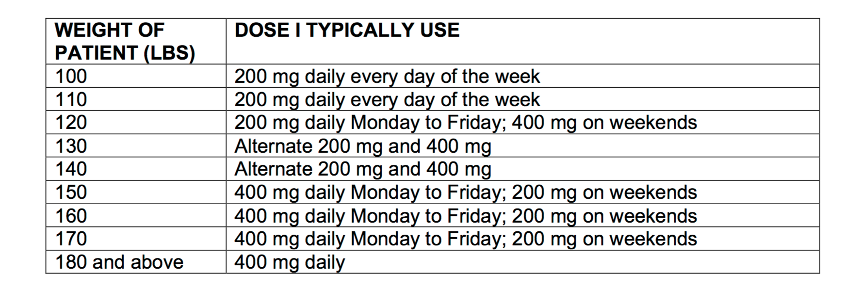
Instead of calculating lean body weight, newer guidelines advocate simply using the patient's true body weight and multiplying by 5 (instead of 6.5) to get the appropriate dose of Hydroxychloroquine (Plaquenil).  In our clinic we typically dose hydroxychloroquine according to the following grid:

plaquenil-dosing

SUMMARY and CONCLUSION

These new dosing suggestions are helpful to reduce side effects, especially the eye related side effects that can come from use of the drugs. My general feeling is that many patients I see are using slightly higher doses than in this chart.


This article was written by Dr. Jeff Donovan, a Canadian and US board certified dermatologist specializing exclusively in hair loss.
Share This
No Comments

End Of Day Scalp Symptoms: EODSS

EODSS

eodss.png

Scalp symptoms are common in everyday life. In addition to itching, a variety of symptoms are possible including burning, tenderness, pain, pins and needles, etc. Some patients find that symptoms seem to correlate with hair loss whereas some patients have concerns only about the symptoms and are not experiencing hair loss.

For some patients, symptoms are rare and occur every few months. For others, symptoms are present each and every day. Some patients wake up with symptoms whereas others ding their scalp feels fairly normal in the morning but worsens towards the end of the day. I refer to the second situation as “end of day scalp symptoms” (i.e. EODSS). There are millions of nerves on the scalp and these nerves all secrete chemical and respond to chemicals that come into the scalp from the blood stream and chemicals that are produced within the scalp itself. Some of these chemicals are part of the normal physiology of the scalp and some of these chemical are increased during states of inflammation, stress, or infection. All of these chemical have the potential to worsen scalp symptoms.

The exact reasons for EODSS is not clear but there are likely many reasons. The concentrations of dozens and dozens of different hormones, neurotransmitters and cytokines change from morning to bedtime. These all have the potential to impact the way our scalp feels. Cortisol and testosterone levels are high in the morning and drop in the afternoon. Melatonin levels increase in the evening. Even ‘core’ body temperature changes through the day - being lowest at 6 am and then peaking at round 6 pm to 9 pm. These changes all affect the events in the scalp.

EODSS are poorly researched and poorly studied. EODSS are not specific but may be more likely associated with certain conditions than others. A proportion of patients with inflammatory scalp disease in particular report EODSS. These include seborrheic dermatitis, psoriasis, scarring alopecia, scalp rosacea, burning scalp syndrome, and sensitive scalp syndrome. Some patients with depression and anxiety and some with shedding disorders also report EODSS.


This article was written by Dr. Jeff Donovan, a Canadian and US board certified dermatologist specializing exclusively in hair loss.
Share This
No Comments

PRP, Concentration and Volume: Analogy of Coffee vs Expresso

Analogy of Coffee vs Expresso

coffe vs expresso.png

I came across an interesting question today whereby a patient wanted to know specifically how much volume (in millilitres) of PRP various clinics used. As I’ll point out here, the question fails to take into account some of the finer details of the PRP procedure. To the hair follicle, volume does not matter as much as CONCENTRATION. What probably matters the most is the number of mL of HIGHLY CONCENTRATED PRP the clinic uses.

To explain the concepts of PRP volume and PRP concentration to patients and physicians, I generally use the analogy of Harry and Sam. 
Meet Sam. Sam works a busy job. Sam is feeling tired and thinks that some caffeine might help him feel more energized and so visits his local coffee shop. Sam is presented with two options for his beverage- a strong 60 mL (2 oz) espresso or a 350 mL (12 oz) large volume coffee/Americano. While making his decision Sam is advised that whichever beverage he chooses he is only allowed one sip and only one sip. This one sip rule is a strange rule but that’s how the coffee shop operates. Sam decides on the espresso. He estimates that one sip of espresso might have 30 mg of caffeine compared to one sip of the large volume coffee/Americano might have only 5 mg of caffeine. Sam considers the espresso as a better choice to wake him up!

Now meet Harry. Harry is a hair follicle. Harry is feeling tired and thinks that some PRP might help the follicle feel more energized and so Harry visits a local PRP clinic. Harry is presented with two options for PRP- 8 mL of PRP at 5 times above baseline volume or 15 mL of PRP at 2-3 times above baseline blood levels. 
While making his decision the PRP doctor advises hair follicle Harry that whichever PRP concentration he chooses his little hair follicle is only allowed to bathe in one tiny injection from the injecting needle.

Harry ultimately chooses the higher concentration of PRP estimates that one little injection of PRP at 5 times above baseline has a much higher concentration of growth factors than PRP at 2-3 times above baseline.

Hair follicle Harry considers the high concentration PRP as a better choice to wake up his hair follicle.


This article was written by Dr. Jeff Donovan, a Canadian and US board certified dermatologist specializing exclusively in hair loss.
Share This
No Comments

Placebo Effect: Pill Color Actually Matters

Pill Color Actually Matters

placebo.png

The mind is powerful. It can make us feel better or make us feel worse. The way we think has the potential to eradicate symptoms in certain diseases. The mind can also cause symptoms to occur and make us feel worse. 

The science of placebo pills or "sugar pills" is fascinating and reminded us of just how powerful the mind really is. Study after study has shown just how powerful placebo effects are in humans. Placebo pills have been shown to help a wide range of health conditions - which points to the powerful effect of the mind to heal disease. Nowadays, placebo pills can even be purchased from the internet.

One systematic review published in 1996 examined the importance of pill color. Blue and green colored pills were shown to be associated with a tranquilizing effect when yellow, red, orange were seen as having a stimulant effect. White colored pills have been shown to perform well as pain pills. 

The color of pills is important and something we often take for granted. Pharmaceutical companies put a great deal of time and effort into choosing the precise color of pills we buy.
A recent report in the International Journal of Biotechnology suggested that red and pink pills are increasingly popular. Pink pills in particular are seen as sweeter and prompt patients to take them in order to achieve the intended effects. In some studies, data suggests that middle-aged people preferred red tablets compared to younger adults and more women prefer red tablets than men. 

References 

AJ de Craen et al. Effect of color of drugs: systematic review of the perceived effect of drugs and their effectiveness. BMJ 1996. 

RK Srivastava, Aarti T. More. Some aesthetic considerations for over the counter (OTC) pharmaceutical products. International Journal of Biotechnology, 2010; 11 (3/4): 267


This article was written by Dr. Jeff Donovan, a Canadian and US board certified dermatologist specializing exclusively in hair loss.
Share This
No Comments

Dutasteride in Young Women: Not Usually.

Dutasteride Prescribing for Pre-menopausal Women Requires Caution

If any drug is to be prescribed, the prescriber needs to know everything about the drug. So does the pharmacist who is dispensing the drug. The prescriber also needs to figure out what information is important to convey to the patient. If there are 10,000 things that the patient ‘could’ be told, which of these 10,000 things really should be relayed to the patient? The answer is mainly those pieces of information that are important to know about and the side effects that are common.

Dutasteride is a 5 alpha reductase inhibitor that is not formally approved for use in women. It is FDA approved for treated prostate problems in men and approved in South Korea for treating male balding. Dutasteride is sometimes used off label in many countries for treating androgenetic alopecia in men. In fact, studies have shown that dutasteride is more effective than finasteride in treating male pattern balding. It may or may not be associated with a higher risk of side effects such as sexual dysfunction.

The question often arises - if it’s used off label in treating male balding, can dutasteride be used off label in treating female genetic hair loss (i.e. female pattern hair loss)?

That answer is maybe - but maybe not.

Dutasteride for Female Hair Loss: Top 10 things to know

The following are my professional opinions and recommendations about the use of dutasteride for treating hair loss in women.

1 . Dutasteride is not FDA approved for women. That doesn’t it can’t be used for women it just means that it was formally approved for another use. Anyone using it needs to understand that. Many hair loss treatments we use are off label, so that fact alone is not alarming in any way.

2. Dutasteride is an oral medication that can be used to treat several types of hair loss in women, including post-menopausal female androgneetic alopecia and post menopausal frontal fibrosing alopecia. There’s evidence to back up the claim that it helps these conditions, albeit not everyone with these conditions.

3. Side effects of dutasteride in women include mood changes (depression), fatigue, weight gain, decreased sex drive, breast tenderness, breast enlargement, muscle soreness and rarely changes in blood counts.

4. Dutasteride has a long half life of 4-5 weeks. This simply means it stays around in the body a long time. In fact, dutasteride can even be detected in the body 6 months after stopping the drug.

5. Because of the long half-life, some women benefit from use once, twice or three times weekly. Daily use may not be required for all patients.

6. Because of the long half-life, women with pre-existing depression should not start dutasteride without careful discussion with their physician. It may worsen depression and if the drug is stopped it may take months to fully get out of the system. Other treatment options should be considered first.

7. Because of the long half-life, women with pre-existing sexual dysfunction should not start dutasteride without careful discussion with their physician. It may worsen sexual dysfunction and if the drug is stopped it may take months to fully get out of the system. Other treatment options should be considered first.

8. Because of the long half-life, pre-menopausal women who are planning to become pregnant at any time in the future should not use dutasteirde. My personal view is that dutasteride is medication that can be considered in post menopausal women and can be considered in pre-menopausal women over 40 who do not plan to have additional children or do not plan to have children provided it is used with contraception. Dutasteride, like finasteride, blocks conversion of testosterone to dihydrotestosterone (DHT) and by reducing levels of DHT may cause abnormalities in the external genitalia of the male fetus. The importance of using contraception and preventing pregnancy while using a 5 alpha reductase inhibitory drug can not be overemphasized. Unfortunately, this message may not be getting out to the public appropriately as some studies (i.e. Teirchert et al, 2017) have suggested that an extremely large proportion of women using finsateride and dutasteride may not using appoprirate contraception.

9. Given the limited number of studies in women, I do not recommend dutasteride for women with previous history of breast cancer or for women who have an elevated high risk for breast cancer. I particularly find the NIH Breast Cancer Risk Assessment Tool helpful in that regard and use it to help patients understand their baseline risk of breast cancer. Although there is no evidence that anti-androgens like spironolactone or finasteride increase the risk of breast cancer in women at low risk for developing breast cancer, we do not know whether women at very high risk for developing breast cancer place themselves at increased risk by using these drugs. It is unlikely but these studies have simply not been done.

10. Pre-menopausal women considering anti-androgen therapy may wish to discuss the use of other anti-androgens with their physician as well as other treatments. This would include spironolactone (Aldactone) and finasteride. Both of these drugs are also off label and both of these mediations are not to be used during pregnancy or by women who are trying to become pregnant. However, the shorter half life means that the drug is cleared rapidly from the body if side effects develop and if pregnancy is being considered in the near future.

REFERENCES

Teirchert M et al. Contraindicated use of 5‐alpha‐reductase inhibitors in women. Br J Clin Pharmacol. 2017 Feb; 83(2): 429–431. 


This article was written by Dr. Jeff Donovan, a Canadian and US board certified dermatologist specializing exclusively in hair loss.
Share This
2 Comments

Does Using Spironolactone Increase Breast Cancer Risk?

Spironolactone and Breast Cancer: No evidence of Increased Risk

I just posted a new answer to our “Question of the Week.” I was asked to outline what is known at present about the risk of developing breast cancer in women who use spironolactone. Fortunately, there is no evidence that spironolactone use increases the risk of developing breast cancer.

The full answer to this week’s question can be read here:

Does Using Spironolactone Increase the Risk of Breast Cancer?

To submit a new question for consideration of our Question of the Week, simply visit complete our online form


This article was written by Dr. Jeff Donovan, a Canadian and US board certified dermatologist specializing exclusively in hair loss.
Share This
No Comments

Hormonal Changes in Late Onset Congenital Adrenal Hyperplasia

Hormonal Changed in Late Onset CAH Resemble PCOS

Late onset on “non-classic” congenital adrenal hyperplasia (LOCAH) is an uncommon genetic disorder that is most often (95 % of the time) caused by mutations in 21-hydroxylase gene. This mutation leads to reduced levels of the 21 hydroxylase enzyme.  Late onset CAH from deficiencies or mutations in other genes such as  11β-hydroxylase (CYP11B1) and 3β-hydroxysteroid dehydrogenase (HSD3B2) are extremely rare.

Patients with LOCAH have CYP21A2 mutations that lead to reduced levels on the 21 hydroxylase enzyme but not a complete absence.  The amount of functional 21-hydroxylase enzyme determines the severity of the disorder. Low levels of the enzyme result in low levels of hormones such as cortisol and/or aldosterone and high levels of androgens (male hormones such as testosterone and androstenedione).

As a result of low cortisol, patients may experience changes in energy levels, blood pressure, blood sugar levels, as well as impaired ability of the body to respond to stress, illness, and injury. Aldosterone plays a key role in helping the body maintain the proper level of sodium and water and helps maintain blood pressure.  


Diagnosis of LOCAH

The patient's signs and symptoms may point to a possible diagnosis.  Even though LOCAH has several endocrine issues (high androgens and low cortisol), generally speaking, the clinical features of LOCAH are due to the excess of the androgens. Taking a careful medical history from the patient may reveal premature pubarche (i.e. the development of pubic hair, axillary hair, and/or increased apocrine odor prior to age 8 years in girls and age 9 years in boys). Affected children may be tall and have accelerated linear growth velocity, and advanced skeletal maturation.

Although LOCAH is estimated to occur in less than 1:1000 individuals, overall about 2-9 % of all women with hyperandrogenism may have late onset CAH. It has been estimate that 5 % of all women with hirsutism have LOCAH. Other symptoms can closely resemble the constellation of symptoms seen in women with polycystic ovarian syndrome. For example, women with late onset CAH may develop a variety of symptoms including frontal baldness, hirsutism, acne,  and irregular periods. Other symptoms include a delay in the timing of the very first period, early onset of pubic hair, accelerated growth, reduced final height and infertility.  

In a 2000 study by Moran and colleagues, the three most common symptoms among adolescent and adult women with LOCAH were hirsutism (59%), oligomenorrhea (54%), and acne (33%). Studies in 2009 by Bidet and colleagues suggested that the initial presenting symptoms in 161 women with late onset CAH were hirsutism (78%), menstrual dysfunction (54.7%), and decreased fertility (12%).


Testing for LOCAH

Blood tests. Generally, additional testing is ordered to help confirm the diagnosis.  These tests may include a blood test to measure the concentration of 17-hydroxyprogesterone (17-OHP) on day 3-5 of the menstrual cycle. Levels of 170–300  ng/dL have been found to be an excellent screening tool. These should be obtained in the morning and during the follicular (preovulatory) phase of the menstrual cycle.

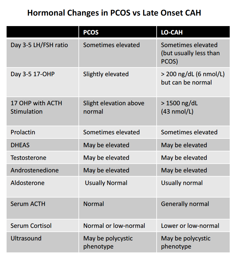
The clinical features of late onset CAH in postpubertal adults may be difficult to differentiate from those of the polycystic ovary syndrome (PCOS). The following table shows some of the key differences in women with PCOS vs LOCAH. Note that even 17 OHP concentrations may be within the normal range for individuals with late onset CAH.  LH levels can be elevated in LOCAH and the LH/FSH ratio above 3:1 can also sometimes be observed. Prolactin levels are sometimes modestly elevated and DHEAS, testosterone and androstenedione levels may or may not be elevated. Aldosterone and cortisol levels are often normal may be lower range . Low sodium or high potassium levels may be present in the blood and abnormalities in glucose levels might be present.

Comparison of Testing in PCOS vs Late Onset CAH. DOWNLOAD PDF VERSION

Comparison of Testing in PCOS vs Late Onset CAH. DOWNLOAD PDF VERSION

ACTH Stimulation Test. An adrenocorticotropic hormone (ACTH) stimulation test may also be ordered which involves measuring the concentration of 17-OHP in the blood before ACTH is administered to the patient and then again 60 min after ACTH is given. This test is typically conducted through an endocrinologist.  The acute ACTH stimulation test remains the gold standard to confirm decreased 21-hydroxylase activity.  

To perform the ACTH stimulation test, a blood sample is first collected to measure baseline hormone concentrations. Then, synthetic ACTH (Cortrosyn, 0.25 mg) is administered. A second blood sample is collected 30–60 minutes later.  When the ACTH-stimulated 17-OHP value exceeds 1500  ng/dL a mutation is likely.

Ultrasound of Ovaries. Ultrasounds of the ovaries may not necessarily help differentiate PCOS from LOCAH because ultrasounds in LOCAH may show an ovarian morphology similar to polycystic ovary syndrome (PCOS) in about 50 % of patients.


Other tests

In addition to performing blood tests for day 3-5 17 OHP, other tests may be recommended by the physician caring for the patient. They include cortisol, androstenedione, testosterone, free testosterone, DHEAS, progesterone, sodium, potassium, creatinine, glucose, hemoglobin A1C. LH and FSH may also be measured. Aldosterone may be tested. Blood pressure measurements will also be obtained.


REFERENCES


56. Azziz R, Dewailly D, Owerbach D. Nonclassic adrenal hyperplasia: current concepts. Journal of Clinical Endocrinology and Metabolism. 1994;78(4):810–815. 

Witchel et al. Nonclassic Congenital Adrenal Hyperplasia Int J Pediatr Endocrinol. 2010; 2010: 625105. 

Moran C, Azziz R, Carmina E, et al. 21-hydroxylase-deficient nonclassic adrenal hyperplasia is a progressive disorder: a multicenter study. American Journal of Obstetrics and Gynecology. 2000;183(6):1468–1474.

Bidet M, Bellanné-Chantelot C, Galand-Portier M-B, et al. Clinical and molecular characterization of a cohort of 161 unrelated women with nonclassical congenital adrenal hyperplasia due to 21-hydroxylase deficiency and 330 family members. Journal of Clinical Endocrinology and Metabolism. 2009;94(5):1570–1578.  

Scaroni C et al. HLA and hormonal studies in 5 patients with late-onset 21-hydroxylase deficiency syndrome (21 OHDS). Journal of Endocrinological Investigation. February 1986, Volume 9, Issue 1, pp 65–70


Carmina E et al. The endocrine pattern of late onset adrenal hyperplasia (21-hydroxylase deficiency). J Endocrinol Invest. 1984 Apr;7(2):89-92.


This article was written by Dr. Jeff Donovan, a Canadian and US board certified dermatologist specializing exclusively in hair loss.
Share This
No Comments

Androgenetic Alopecia (AGA): AGA Under the Microscope

AGA Under the Microscope

aGA under scope.png

There is often some degree of confusion as to what constitutes a typical biopsy from a patient with androgenetic alopecia (AGA). Here, I will review some of they key features of both horizontal and vertical sections (i.e. the two main ways a biopsy can be done). AGA is a non-scarring alopecia. In both horizontal and vertical sections sebaceous glands typically appear quite prominent. A reduction in sebaceous gland density should prompt one to consider that another diagnosis, such as scarring alopecia, might be present. 
Miniaturized hairs are a feature of AGA. In vertical sections, the miniaturized and vellus hairs simply appear as hairs which root themselves in the mid dermis rather than deep down in the subcutaneous fat. In horizontal sections, miniaturization can be appreciated by a shift in the terminal to vellus hair ratio (“T:V” ratio) from above 6:1 to well below 4:1 and possibly well below this.

Inflammation is very much a part of AGA. The legendary dermatologist Dr David Whiting showed that 40% of biopsies from patients with AGA showed perifollicular inflammation. This inflammation sits in the lower infundibulum and isthmus. The cells that comprise the inflammation are largely T cells. Perifollicular inflammation occurs early in the course of AGA and likely drives the development of perifollicular fibrosis and drives the progressive miniaturization of hairs.

Perifolliuclar fibrosis is also part of AGA and the concentric fibrosis can best be appreciated in horizontal sections. Perifollicular fibrosis can often be mistaken and misdiagnosed as scarring leading to a misdiagnosis of lichen planopilaris (LPP) and other scarring alopecias. However, the retention of sebaceous glands and absence of lichenoid change in the outer root sheath favours a diagnosis of AGA over LPP.

Cell death of keratinocytes in the outer root sheath is not a typical feature of AGA. Apoptosis is present in a proportion of dermal papilla cells and lymphocytes in the perifollicular inflammatory infiltrate - but not in the actual outer root sheath. Lichenoid change in the outer root sheath might cause one to consider a diagnosis of LPP rather than AGA.


This article was written by Dr. Jeff Donovan, a Canadian and US board certified dermatologist specializing exclusively in hair loss.
Share This
No Comments

Hair Extensions: The 2011 Spike

The 2011 Spike

hair extensions.png

Hair extensions continue to be very popular. This interest started to increase significantly in the late 1990s but increased even further over the last 7-8 years. Hair extensions were once an extremely private practice among women. That has changed in part due to the ever increasing acceptance of hair extensions, especially among celebrities. There are many types of extensions. including clip in, micro link, fusion and tape-in and weave-in extensions.

The last decade has witnessed a marked increase in the sale of hair extensions. In the United States alone there has been a 30 % increase in the number of salons offering hair extensions.

As one can see in this “Google Trends” search, interest in hair extensions saw a major peak in 2011 and this interest still remains strong to the present day.

China continues to be the number one supplier of human hair for wigs and extensions followed by India. The 2011 spike was not only in Google trends but in a reality in the hair industry. Between 2009 and 2011, China reportedly tripled its imports of Indian hair. The market for hair extensions is greatest in the United Kingdom, United States, Australia and South Africa.


This article was written by Dr. Jeff Donovan, a Canadian and US board certified dermatologist specializing exclusively in hair loss.
Share This
No Comments

Perifollicular Fibrosis and Perifollicular Inflammation in Androgenetic Alopecia

Perifollicular Inflammation and Perifolliuclar Fibrosis are Part of Balding

It had long been though that male and female patterned hair loss (‘balding') is a relatively non-inflammatory process. The last decade has repeatedly shown that this is not the case and that inflammation is likely very much a part of the process and likely contributes in some way to the balding process itself.

The current model of AGA suggests that ‘micro inflammation’ in AGA might trigger apoptosis and perifollicular fibrosis which in turn causes hairs to miniaturize and shed.

Inflammation in AGA: How common is it anyways?

Inflammation is commonly seen in androgenetic alopecia and is likely part of the condition itself. Studies by Whiting showed that perifollicular inflammation was present in 40 % of AGA biopsies. The inflammation present in AGA is different than the inflammation seen in the destructive scarring alopecias like lichen planopilaris. In 2000, Mahe used the term “microinflammation” to describe this types of inflammatory process.

Inflammation in AGA: Where is it found?

Inflammation in AGA is mainly seen in the upper parts of the hair follicle. Inflammation is commonly found in both the isthmus and isthmus which are the upper portions of the hair follicle. Minor amounts of inflammation around hair follicles can be seen in the normal scalp as well in in androgenetic alopecia. However, more marked degrees of inflammation are not normally seen in the scalp but can be a feature of androgenetic alopecia. Ramos in 2016 showed that inflammation is more common around miniaturizing hairs and this inflammation seems to be associated with a form of cell death known as apoptosis. In 1992, Jaworsky and colleagues showed that biopsies of males and females with androgenetic alopecia showed the presence of activated T-cell infiltrates about the lower portions of follicular infundibula. Inflammatory cells infiltrated the region of the follicular bulge, the putative source of stem cells in cycling follicles. It was postulated that the progressive fibrosis of the perifollicular sheath might begin with T-cell infiltration of follicular stem cell epithelium and that the perifollicular fibrosis actually impairs hair growth.

Cell death (Apoptosis) is AGA: Where does it occur?

Cell death via programmed cell death or ‘apoptosis. is also very much a part of AGA. It is known that apoptosis is dermal papilla cells is important in AGA and is proposed to reduce the cell mass of the dermal papilla and in turn lead to miniaturization.

In 2010, El-Domyati and colleagues showed that apoptosis (by bcl-2 immunostaining) was present in both the dermal papilla and in perifollicular lymphocytes (i.e. the white blood cells that comprise the perifollicular inflammation). The authors showed that the apoptosis in dermal papilla cells was associated with reduced cell activity and cell division (as measured by proliferating cell nuclear antigen staining PCNA).

Can we detect how much inflammation is present in AGA without a biopsy?

To date, there is no better way to detect inflammation than a biopsy. Of course, biopsies are seldom done in AGA because the diagnosis is largely clinical. However, careful examination of the scalp with dermosocpy may help pinpoint inflammation occurring beneath the scalp. In 2004, Deloche showed that peripilar signs (PPS) by dermoscopy were correlated with the presence of inflammation beneath the scalp.

Does inflammation and fibrosis really matter?

The inflammation and fibrosis seen in androgenetic alopecia probably does matter. The inflammation likely triggers abnormalities in how hair cycles (i.e. telogen effluvium) and likely contributes to the progressive ‘miniaturization’ of hairs over time.

Perifollicular inflammation in AGA appears to occur very early in the condition. In 2009, El-Domyati showed the perifollicular inflammation was present early the condition long before perifollicular fibrosis started to be seen. Over time, as perifollicular fibrosis became more commonly seen as perifollicular inflammation started to decrease. In other words, the two phenomena seemed to have an inverse correlation. Gonzalez showed in 2010 that perifollicular fibrosis was even a common findings in androgenetic alopecia occurring in teenagers.

Perfifollicular inflammation and fibrous probably affect how hairs grow. In 1993, Whiting performed some classic studies that have shaped how we think about inflammation in AGA. He showed that patients with perifollicular inflammation and fibrosis have poorer responses to minoxidil. Individuals with moderate or dense lymphocytic inflammation and perifollicular fibrosis may have poorer responses to minoxidil.

What causes the perifollicular fibrosis anyways?

It’s not entirely clear what causes the perifollicular fibrosis to occur. In 2006, Yoo and colleagues proposed that TGF-beta (transforming growth factor beta) seemed to play a role. They showed that testosterone treatment increased the expression of type I procollagen at mRNA and protein level and this was associated with a rise in TGF-beta protein levels by 81.9 % in dermal fibroblasts. Conversely, pretreatment of finasteride inhibited the ability of testosterone to make pro collagen RNA and protein and decreased the expression of TGF-bet by 30 %. Interestingly, pretreatment of follicles with a TGF-beta antibody inhibited pro collagen expression leading the authors of the study to conclude that testosterone triggers TGF-beta expression and perfiollciular fibrosis in AGA. They also postulate that one mechanisms of finasteride may be to reduce TGF-beta and therefore pro collagen expression.

Conclusion

Androgenetic alopecia is no longer viewed as a “non-inflammatory” condition. Inflammation is very much a part of AGA and this inflammation likely drives the development of perifollicular fibrosis and an inflammatory millieu that drives the apoptosis of dermal papilla cells and therefore the progression miniaturization of hair follicles.

So, do we need to treat the inflammation? That answer is ‘probably’ - it’s just we don’t quite know how to best do this yet. There are many different types of anti-inflammatory treatment including corticosteroids, doxycycline, tacrolimus, TNF-inhibitors, immunomodulatory and immunosuppressants. It’s a bit of a guess as to how best to address the inflammation in AGA and more research is needed. It’s extremely likely this will play a beneficial role, particularly the earlier such anti-inflammatory treatment is started. Treatment will likely be needed over an extended period rather than for a few days or weeks.

REFERENCES

Deloche C, et al. Histological features of peripilar signs associated with androgenetic alopecia. Arch Dermatol Res. 2004.

El-Domyati M, et al. Androgenetic alopecia in males: a histopathological and ultrastructural study.. J Cosmet Dermatol. 2009.

El-Domyati M, et al. Evaluation of apoptosis regulatory markers in androgenetic alopecia. J Cosmet Dermatol. 2010.

Gonzalez ME, et al. Androgenetic alopecia in the paediatric population: a retrospective review of 57 patients. Br J Dermatol. 2010.

Jaworsky C, et al. Characterization of inflammatory infiltrates in male pattern alopecia: implications for pathogenesis. Br J Dermatol. 1992.

Mahé YF, et al. Androgenetic alopecia and microinflammation. Int J Dermatol. 2000.

Nirmal B, et al. Evaluation of Perifollicular Inflammation of Donor Area during Hair Transplantation in Androgenetic Alopecia and its Comparison with Controls. Int J Trichology. 2013.

Ramos PM, et al. Apoptosis in follicles of individuals with female pattern hair loss is associated with perifollicular microinflammation. Int J Cosmet Sci. 2016.

Whiting DA. Diagnostic and predictive value of horizontal sections of scalp biopsy specimens in male pattern androgenetic alopecia. J Am Acad Dermatol. 1993.

Yoo HG, et al. Perifollicular fibrosis: pathogenetic role in androgenetic alopecia. Biol Pharm Bull. 2006.


This article was written by Dr. Jeff Donovan, a Canadian and US board certified dermatologist specializing exclusively in hair loss.
Share This
16 Comments

Coffee, Caffeine and Immunity: Caffeine is an Immunomodulator

Caffeine is an Immunomodulator

caffeine-immunity.png

Caffeine appears to have an anti-inflammatory effect in some situations and helps calm down an “overactive” immune system. In animal (rodent) models, it’s quite clear that caffeine can affect a variety of different types of inflammation. The effects on humans appear similar, with caffeine having an effect on multiple parts of the immune system - particularly immune cells known as lymphocytes.

To date, there is good evidence to suggest a suppressive effect of caffeine on the proliferation of “stimulated” lymphocytes. In other words, immune cells that have become too active can be quieted down with caffeine. Other immune cells such as natural killer cells and macrophages also exhibited a reduced activity in the presence of high doses of caffeine.

Caffeine may also be truly Immunosuppressive in some situations given the reduced levels of anti-inflammatory cytokines such as TNF-α, IL-2 and IL-6. Moreover, certain receptors, such as TLR1, TLR2, TLR4, and MHC class I-related chain B molecules are also decreased by caffeine.A recent study showed that caffeine inhibits STAT1 signaling and downregulates inflammatory pathways involved in autoimmunity.
identified. 


In actual human epidemiologic studies, coffee has mixed effects. Caffeine (coffee) consumption appeared to increase the risk of developing type 1 diabetes and rheumatoid arthritis. In contrast, coffee may help somewhat to prevent ulcerative colitis and multiple sclerosis. There does not appear to be an association ether way for other autoimmune diseases such as systemic lupus erythematosus, psoriasis and Crohn's disease.

In the world of hair loss, there are very few studies which have objectively examined the benefits of caffeine. Some studies have suggested a minor benefit for caffeine containing shampoos but these studies are small and have not yet been independently replicated to any significant degree.

It is fascinating to consider that caffeine has the potential to effect so many parts of our immune system. More studies are needed to understand whether or not caffeine can integrate into the treatment of various types of hair loss.


This article was written by Dr. Jeff Donovan, a Canadian and US board certified dermatologist specializing exclusively in hair loss.
Share This
No Comments

Alopecia Areata: Dermatoscopic (Trichoscopic) Features

AA Nov 05.png

Dermatoscopic (Trichoscopic) Features

Up close examination of the scalp via dermoscopy allows the physician the opportunity to confirm the suspected type of hair loss and also evaluate how active the condition is at the present time. There are many known “dermatoscopic” features of alopecia areata and I will review five common ones here:

Yellow dots (red arrow) represent empty hair follicle openings (pores) and are very common. The more severe of a disease a patient has, the more yellow dots are seen all over the scalp.

Vellus hairs (white arrow) are short and thin regrowing hairs and are also common in patients with alopecia areata.

Black dots (black arrow) represent hair follicles broken at the surface and indicate an active disease state.

Tapered hairs (blue arrow) are long hairs that become quite thin as the are about to enter the scalp. They too represent active disease.

Exclamation mark hairs (green arrow) are short 4 mm hairs that are actually broken hairs. They are wider at the top compared to the bottom and are found in patients with active disease.


This article was written by Dr. Jeff Donovan, a Canadian and US board certified dermatologist specializing exclusively in hair loss.
Share This
2 Comments

Blogs by Topic


Share This
-->