h1.qusth1 { display: none !important; }

QUESTION OF THE WEEK

Dr. Donovan's Articles

QUESTION OF HAIR BLOGS

Filtering by Category: Scarring Alopecia


Treatment of AKN with Long Pulsed Alexandrite Laser

AKN: Long pulsed Alexandrite as an option

AKN-image

Acne keloidalis nuchae is a scalp condition that commonly affects the back of the scalp. Patients develop what they frequently term 'bumps' at the back of the scalp. These frequently are associated with hair loss and the bumps themselves may stay and enlarge. In advanced cases the areas coalesce to form a large plaque. 

Treatments for AKN include topical steroids, antibiotics, retinoids, steroid injections. A variety of laser treatments may also be possible.  In previous studies the 810-nm diode laser and 1,064-nm Nd:YAG laser have been used for treating AKN with promising results.

Tafnik and colleagues set out to study the benefits of the 755-nm alexandrite laser in 16 male patients with AKN. Their study showed a significant decrease in the mean papule, pustule count, keloidal plaque size, and pliability at the fourth and sixth laser sessions when compared with baseline. The main complication was a temporary reduction in hair density in the treated area in 4 of 16 patients as a result of the laser treatment. This was accepted by the patients because of its reversible course.  No lesional recurrence was detected in the follow-up period.


STUDY CONCLUSION

This study provides evidence that the 755-nm alexandrite laser may provide options for treating AKN. The laser appears safe and effective in the condition and recurrence rates are fortunately low. 


REFERENCE

Tawfik A, et al. A Novel Treatment of Acne Keloidalis Nuchae by Long-Pulsed Alexandrite Laser. Dermatol Surg. 2018.


This article was written by Dr. Jeff Donovan, a Canadian and US board certified dermatologist specializing exclusively in hair loss.
Share This
No Comments

Pulsed Azithromycin for Folliculitis Decalvans

Pulsed therapy for Folliculitis decalvans

Folliculitis decalvans is a type of scarring alopecia and causes permanent hair loss. Affected individuals develop crops of papules, and pustules. The most effective treatment options are antibiotics and isotretinoin. 


Pulsed Therapy for FD

In an effort to reduce side effects from the daily use of a drug, "pulsed therapy" is frequently used for some medications. Pulsed therapy refers to delivery of a medication for short periods of time (i.e. the 'pulse') followed by periods of time whereby the patient does not receive any medications at all.  Pulsed therapy is common with oral steroids, oral anti-fungal medications as well as some antibiotics.

A new study has examined the possibility of using pulses of azithromycin to treat folliculitis decalvans.  The researchers studied 19 patients with mean age 27 years. Treatment was with azithromycin 500 mg per day for 3 consecutive days and repeated every 2 weeks. The severity of the disease was evaluated before treatment and after 1, 3 and 6 months.  

The study showed that azithromycin reduced the number of lesions as well as the disease activity. 


Conclusion

Pulsed azithromycin is among the antibiotic options for FD. Pulses of azithromycin are sometimes used as treatments for acne so this method of using azithromycin in a pulsed manner is not new. Side effects of azithromycin should be carefully review before starting. 

Download our Azithromycin Handout for Patients


REFERENCES

Andre MC et al. Effective Treatment of Folliculitis Decalvans: Azithromycin in Monotherapy. Hair Therapy and Transplantation. 

Antonio JR et al. Azithromycin pulses in the treatment of inflammatory and pustular acne: efficacy, tolerability and safety.J Dermal Treatment 2008;19(4):210-5. doi: 10.1080/09546630701881506.

Parsad D et al. Azithromycin monthly pulse vs daily doxycycline in the treatment of acne vulgaris.J Dermatol. 2001 Jan;28(1):1-4.


This article was written by Dr. Jeff Donovan, a Canadian and US board certified dermatologist specializing exclusively in hair loss.
Share This
No Comments

Trichoscopy of Central Centrifugal Cicatricial Alopecia

CCCA: Trichoscopy

ccca

Central centrifugal cicatricial alopecia (CCCA) is a scarring alopecia that commonly affects women with afrotextured hair.  It has a genetic basis in some women. The condition starts with central hair loss in most affected women and this is followed by expansion of the hair loss outwards. There may be symptoms such as itching, or pins and needles, but many women are asymptomatic. 

In an article earlier this year, I discussed some very interesting studies which showed a five fold increased risk of uterine fibroids among women diagnosed with CCCA.   


Dermatoscopic Features of CCCA
 

It is critically important to identify CCCA in the early stages in order to try to stop hair loss. Today I'd like to focus on the up close features of CCCA using a handheld dermatoscope.  We refer to this as trichoscopy. 

The trichoscopic features of CCCA are few. Miteva and Tosti in 2014 published the first real compressive overview of the trichoscopic features of CCCA. They retrospectively images obtained from 51 women with histologically proven CCCA and  compared to controls (which included 30 dermatoscopic images from histologically proven cases of scarring traction alopecia and discoid lupus erythematous).   


The Peripilar White Gray Halo

ccca

The so called "peripilar white gray halo" was found in 94% of patients and was highly specific and sensitive for CCCA. This halo was seen around the emergence of hair follicles.

The halo was shown to correspond on pathology to the lamellar fibrosis surrounding the hair follicle outer root sheath.


Reference

Miteva and Tosti. Dermatoscopic features of central centrifugal cicatricial alopecia. J Am Acad Dermatol. 2014.


This article was written by Dr. Jeff Donovan, a Canadian and US board certified dermatologist specializing exclusively in hair loss.
Share This
No Comments

Trichoscopy of Late DLE

Late DLE: Features


Screen Shot 2018-03-04 at 10.54.42 PM.png

Discoid lupus (DLE) is an autoimmune condition affecting the scalp and skin. It can cause permanent hair loss in affected individuals. About 5% develop systemic lupus erythematosus, an autoimmune condition with the potential to affect many organs of the body. Late scalp lesions of DLE show hyperpigmentation, white structureless areas and telangiectatic vessels,


This article was written by Dr. Jeff Donovan, a Canadian and US board certified dermatologist specializing exclusively in hair loss.
Share This
No Comments

Trichoscopy of Early DLE

DLE - Early Features

dle

Discoid lupus (DLE) is an autoimmune condition affecting the scalp and skin. It can cause permanent hair loss in affected individuals. About 5% develop systemic lupus erythematosus, an autoimmune condition with the potential to affect many organs of the body. Early scalp lesions show whitish scale, follicular plugging and a perifollicular white halo. Aggressive treatment of early DLE can prompt hair growth in some individuals


This article was written by Dr. Jeff Donovan, a Canadian and US board certified dermatologist specializing exclusively in hair loss.
Share This
No Comments

Normal vs Compound Follicles

What is the difference?

lpp-vs-fd.png

Hairs emerge from the scalp through pores or hair follicle openings. Some pores have just one hair, but most normally have two or three hairs emerging through a single hair follicle opening. This is completely normal. 
It’s important to be able to quickly spot when something is not quite right. Most hair loss conditions lead to a reduction in the number of hairs coming out of each pore. Instead of seeing the plentiful bundles of two and three hairs one starts to see pores with either no hairs at all or just a single hair. 
Some scarring alopecias are associated an unusual feature- and that is an increase in the number of hairs coming out of the pores. When six or more hairs come out of a single opening we refer to this as a “compound” follicle. The scarring alopecias which frequently show compound follicles include folliculitis decalvans (tufted folliculitis) and sometimes acne keloidalis. It tends to be the scarring alopecias associated with neutrophils that are associated with formation of compound follicles.

Compound follicles occur because of the destructive enzymes released from the inflammatory process. These enzymes destroy tissue and promote fusion of follicles together. The photos here show compound follicles in folliculitis decalvans and single haired follicles in lichen planopilaris. 

SINGLE HAIRS IN LICHEN PLANOPILARIS 

SINGLE HAIRS IN LICHEN PLANOPILARIS


COMPOUND HAIRS IN FOLLICULITIS DECALVANS

COMPOUND HAIRS IN FOLLICULITIS DECALVANS


This article was written by Dr. Jeff Donovan, a Canadian and US board certified dermatologist specializing exclusively in hair loss.
Share This
2 Comments

Isotretinoin for Folliculitis Decalvans: Good or Very Good?

Folliculitis decalvans: Isotretinoin among the top choices

Folliculitis decalvans is a less common type of scarring alopecia. Dermatologists who treat the condition often choose between antibtioics and isotretinoin when deciding on management strategies. Sometimes both are employed. 

Previous studies have shown the both can be helpful. In some studies, including those by Vano-Galvan the importance of antibiotics was emphasized. In other studies, the benefits of oral isotretinoin were emphasized. 

A new study from Turkey evaluated the benefits of isotretinoin in 39 male patients with folliculitis decalvans. The mean age was 38 years and all received oral isotretinoin for a range of 1-8 months. The dose range in the study was 0.1 to 1.02 mg/kg each day. The authors showed benefit with use of isotretinoin in 82 % of patients. Doses greater than 0.4 mg/kg daily were most helpful.


Conclusion

It's clear that both antibiotics as well as isotretinoin sit at the top of the list of effective treatments for folliculitis decalvans.  While earlier studies emphasized the importance of antibiotics, the benefits of isotretinoin are increasingly clear. 

Download our FD Handout for Patients

Download our Isotretinoin Handout for Patients


Reference

Aksoy B, et al. Isotretinoin treatment for folliculitis decalvans: a retrospective case-series study. Int J Dermatol. 2018.

Tietze JK et al. Oral isotretinoin as the most effective treatment in folliculitis decalvans: a retrospective comparison of different treatment regimens in 28 patients. J Eur Acad Dermatol Venereol. 2015 Feb 24. doi: 10.1111/jdv.13052. 

Vano-Galvan et al. Folliculitis decalvans: a multicentre review of 82 patients. J Eur Acad Dermatol Venereol. 2015 Feb 12. 


This article was written by Dr. Jeff Donovan, a Canadian and US board certified dermatologist specializing exclusively in hair loss.
Share This
1 Comment

Scarring alopecia: Acquired Pili torti

Acquired Pili torti

pt

Scarring alopecias are a group of diverse hair loss conditions that are associated with the presence of scar tissue in the scalp. This scar tissue can damage growing hair follicle and affect how they grow.

A common finding in many scarring alopecias is the twisting of hairs in a patient with otherwise straight hair. This “twisting” of hair is called pili torti and when it develops long after birth we call it “acquired pili torti.” This photos shows typical pili torti in a patient with frontal fibrosing alopecia. Some straight unaffected hairs can also be seen in the photo as well (bottom right). Dilated veins typical of FFA can also be seen.


This article was written by Dr. Jeff Donovan, a Canadian and US board certified dermatologist specializing exclusively in hair loss.
Share This
2 Comments

Scalp Burning: Many reasons but diagnosis is essential

Scalp Burning: Before talking treatment, talk diagnosis

In our clinic, many patients present with concerns about scalp burning. There are many reasons for scalp burning and the precise treatment depends entirely on the diagnosis of the burning. A carefully obtained history, along with an examination of the scalp is needed. Some patients with burning also have itching and some have pain. 


Causes of Scalp Burning


1.  Diseases/Disorders of the scalp

Individuals with scalp burning needs a thorough examination to evaluate for underlying scalp disease. A variety of inflammatory scalp disorders can trigger burning including scalp psoriasis, seborrheic dermatitis, scarring alopecia, dermatomyositis, tinea capitis, sunburns, irritant and allergic contact dermatitis. Common hair loss conditions such as androgenetic alopecia, telogen effluvium and scarring alopecia are also associated with burning in some cases.


2.  Dysesthesias

The scalp dysesthesias, as described by Hoss and Segal in 1998, are a group of conditions that give physical symptoms in the scalp without any other unusual findings at the time of examination (normal scalp examination). In addition to scalp burning, many patients with scalp dysesthesias have itching and pain.

The cause of scalp dysesthesias is not clear. One study (reference below) suggested that a high proportion of women with scalp dysesthesias had cervical spine disease. It seems that patients worsen with stress and improve with anti-depressants (venlafaxine, amitrytyline). Many respond to topical or oral gabapentin.

The burning scalp syndrome (similar to burning mouth syndrome) is a variant of scalp dysesthesia. Sensitive scalp syndrome may also be as well.


3. Depression and Other Psychological Issues. 

There is a well known relationship between the brain and the skin and this has been referred to as the 'brain-skin' axis. Stressful life events are a well known trigger to scalp burning. Burning is more common in patients with a host of psychological and psychiatric diagnoses including anxiety, depression,  post traumatic stress disorder, schizophrenia.


4. Drugs

Drugs can trigger scalp burning, both topical drugs and oral medications. Topical medications containing alcohol are frequent triggers or scalp burning. Topical calcipotriol, topical steroids, and a host of anti-dandruff shampoos can trigger burning. Oral medications, including cyclophosphamide can trigger scalp burning.


5.  Damaged Nerves and Small fiber neuropathies

Scalp burning may be a result of damage to nerves. As mentioned above, cervical spine disease may be one such condition. But diabetes, multiple sclerosis, and stroke can all give scalp symptoms.

Many issues affecting the tiny nerves of the scalp can cause scalp burning. This is seen in many of the autoimmune diseases including Sjogren’s syndrome.


6. Sleep Deprivation  

Sleep deprivation has been associated with many cutaneous symptoms including scalp burning.


Treatment for Burning Scalp

The treatment of burning scalp will depend on the diagnosis. For patients with scarring alopecia, treatments such as topical steroids, steroid injections and oral anti-inflammatory mediation such as doxycycline or hydroxychloroquine will frequently help stop the scarring alopecia itself as well as the burning. For burning due to psoriasis, a variety of topical steroids, topical vitamin D analogues can help.  The scalp dysesthesias are frequently more challenging to treat but options include topical steroids, oral gabapentin, topical gabapentin, oral amitryptyline, and topical capsaicin. Avoiding harsh shampoos is important. 

Breathing, exercise and scalp exercises are also important as outlined in prior posts.


Conclusion 

In summary, there are many reasons for a patient to present with concerns about burning scalp. A careful and detailed history along with a scalp examination is important. Many times, a scalp biopsy is needed.


This article was written by Dr. Jeff Donovan, a Canadian and US board certified dermatologist specializing exclusively in hair loss.
Share This
No Comments

Scarring Alopecia: What do all the colors mean?

Red and White in Scarring Alopecia: It often matters

Scarring alopecias are a group of hair conditions whereby scar tissue forms in the scalp and this scar tissue ultimately destroys hair follicle stem cells. There are dozens and dozens of different scarring alopecias but there are several that we see most commonly: lichen planopilaris, frontal fibrosinf aloepcia, folliculitis decalvans, central centrifugal cicatricial alopecia, pseudopelade, discoid lupus, dissecting cellulitis and acne keloidalis.

red-white-scar


Many of the scarring alopecias are associated with some type of inflammation present in the skin. This inflammation causes the skin around the hair follicles to take on various shades of red. Redness in the scalp in a patient with scarring alopecia should always be given attention because there is a chance it means the patient’s disease is active. (It does not always as sometimes scalps become red with chronic steroid use). This has been labelled “step 1” in the photo. Over time if the hair follicle is destroyed the inflammation disappears from the area as the immune system has nothing further to attack. What is left is a white scarred area that no longer has the original redness (step 2). Of course, if the disease is successfully treated inflammation may also be reduced from the area as well and redness will also disappear. In other words, it is sometimes possible to block step 1 from progressing to step 2 with appropriate treatment. All in all, it is important to understand the significance of various color changes on the scalp in the setting of scarring alopecia.


This article was written by Dr. Jeff Donovan, a Canadian and US board certified dermatologist specializing exclusively in hair loss.
Share This
2 Comments

Risk of Fibroids Increased in Women with CCCA

Five Fold increased Risk of Fibroids in Women with CCCA  

ccca

A new study, published in JAMA Dermatology, has given evidence that women with central centrifugal cicatricial alopecia (CCCA) are at increased risk of developing benign uterine tumors known as fibroids.  The medical terms for these are uterine leiomyomas.

CCCA is a type of scarring alopecia that occurs predominantly in women with afro-textured hairs. This new data suggests that a genetic predisposition to develop excessive scar tissue in other area of the body may be central to the underlying mechanisms that cause these two diseases.  

The researchers analyzed data from over 487,000 black women and examined the incidence of fibroids in women with CCCA and those without CCCA. Out of 486,000 women in the general population,  3.3 % had fibroids. However, 13.9 % of women with CCCA were found to have fibroids. Taken together, this works out to a five fold increased risk of fibroids in women with CCCA.


Conclusion

There is an increased risk of uterine fibroids in women with CCCA.  Whether there is an increased risk of other scarring related diseases of the body warrants further study.


REFERENCE

 
Dina et al. Association of Uterine Leiomyomas With Central Centrifugal Cicatricial Alopecia. JAMA Dermatology, 2017; DOI: 10.1001/jamadermatol.2017.5163


This article was written by Dr. Jeff Donovan, a Canadian and US board certified dermatologist specializing exclusively in hair loss.
Share This
No Comments

Hair Transplants for Folliculitis Decalvans: Is it even possible?

FOLLICULITIS DECALVANS

HAIR TRANSPLANT CANDIDACY CRITERIA


fd.jpg

The criteria we use in our clinic for evaluating folliculitis decalvans candidacy are among the most strict of all the scarring alopecia criteria. Hair transplants for folliculitis decalvans can be very challenging. Chances of success are low although successes to occur. In order to be a candidate for hair transplant surgery,  ALL FIVE of the following criteria MUST be met in a patient with folliculitis decalvans:


1.  The PATIENT should be off all hair-related medications.

Ideally the patient should be off all topical, oral and injection medications to truly know that the disease is "burned out (burnt out)". However, in some RARE cases, it may be possible to perform a transplant in someone using medications AND who meets criteria 2, 3 and 4 below.  This should only be done on a case by case basis and in rare circumstances. It is a 'last resort' in a well-informed patient. 


2. The PATIENT must not report symptoms related to the FD in the past 24 months. 

The patient must have no significant itching, burning or pain and no bleeding. One must always keep in mind that the absence of symptoms does not prove the disease is quiet.  Even the periodic development of itching or burning from time to time could indicate the disease has triggers that cause a flare and that the patient is not a candidate for surgery. The patient who dabs a bit of clobetasol now and then on the scalp to control a bit of itching may also have disease that is not completely quiet.  The patient with itching every now and then is also a worry. 


3. The PHYSICIAN must make note of no clinical evidence of active disease in the past 24 months. 

There must be no scalp clinical evidence of active FD such as perifollicular erythema, pustules, crusting, perifollicular scale (follicular hyperkeratosis). This assessment is best done with a patient who has not washed his or her hair for 48 hours.

The most important clinical features in our opinion are SCALP CRUSTING and REDNESS AROUND THE HAIRS. Some scalp redness may be persistent in patients with scarring alopecia even when the disease is quiet. Therefore scalp redness alone does not necessarily equate to a concerning finding. Perifollicular redness (redness around the hairs) however is more concerning for disease activity.  In addition, the pull test must be completely negative for anagen hairs and less than 4 for telogen hairs.  A positive pull test for anagen hairs indicates an active scarring alopecia regardless of any other criteria.


4. Both the PATIENT and PHYSICIAN must demonstrate no evidence of ongoing hair loss over the past 24 months.  

There must be no further hair loss over a period of 24 months of monitoring off the previous hair loss treatment medications. This general includes the patient's perceptions and physician's perception that there has been no further loss, physician's measurements showing no changes in the areas of hair loss, as well as serial photographs every 6-12 months showing no changes. 


5. The patient must have sufficient donor hair for the transplant. 

Not all patients with FD have sufficient donor hair even if their disease has become quiet.   

In situations where there is concern that the FD may be active or concern that the surgery may not be a success, strong consideration should be given to performing a 'test session' of 50-100 grafts and observing their survival over a period of 6-9 months. Less than 40 % uptake would intake a contraindication, although ideally one would hope for survival of more than 70% of the grafts.


For Further Reading


Lichen Planopilaris Transplant Candidacy

Frontal Fibrosing Alopecia Transplant Candidacy

Trichotillomania Candidacy: Can a patient with trichotillomania have a hair transplant?  


This article was written by Dr. Jeff Donovan, a Canadian and US board certified dermatologist specializing exclusively in hair loss.
Share This
No Comments

Are hair transplants possible for individuals with scarring alopecia?

Are hair transplants a good option for scarring alopecia?

LPP-HT

The answer to that questions is sometimes "yes" and sometimes "no". For many individuals who step into the office, the answer is frequently "no". A hair transplant is not a good option for them - at least right now. Not because we can't perform hair transplants in individuals with scarring alopecia but rather because the person sitting in front of me has a scarring alopecia that is currently active. They have ongoing hair loss and they report they have less hair than one year ago. Some have persistent itching, burning or tenderness in the scalp. These individuals are not candidates for a hair transplant any time soon.


A Balanced View of Hair Transplantation

It might sound surprisingly to have such a negative view of hair restoration for scarring alopecia. I would say that my view is balanced. The positive side of this topic is that a hair transplant can be a good option once the disease becomes quiet ... and stays quiet for a few years (ideally off medication). On previous blogs,  I have shared my personal views on the criteria we use when considering whether an individual is a good candidate for a hair transplant. These are mainly centered around lichen planopilaris (LPP) and frontal fibrosing alopecia (FFA) as these have been studied most extensively in our center.

CRITERIA FOR TRANSPLANTATION OF LPP

CRITERIA FOR TRANSPLANTATION OF FFA


This article was written by Dr. Jeff Donovan, a Canadian and US board certified dermatologist specializing exclusively in hair loss.
Share This
No Comments

Sinus tracts in Dissecting Cellulitis

Dissecting Cellulitis (DSC)

DSC-56

DSC is a rare scarring alopecia. It often affects young men. A key feature is boggy tender nodules that develop in the scalp, some of which drain pus. "Sinus tracts" are another feature and this refers to the presence of small tunnels that interconnect under the scalp.

This photo shows the appearance of one such "sinus tract" after it has entered a healing phase. This area will be permanently scarred with some degree of permanent hair loss in this area.

Treatment for DSC includes isotretinoin, antibiotics, TNF inhibitors. Second line agents include zinc, dapsone, colchicine. Surgical excision and laser therapies are also considerations. Some forms are challenging to treat.

For more information on DSC, see our Dissecting Cellulitis Handout for Patients


This article was written by Dr. Jeff Donovan, a Canadian and US board certified dermatologist specializing exclusively in hair loss.
Share This
No Comments

Frontal Fibrosing Alopecia (FFA) and "Baby Hairs"

FFA Destroys Vellus Hairs 

Many patients come to the office with worries that they might have frontal fibrosing alopecia (FFA). This autoimmune condition is becoming much more common and many patients are now aware of its existence. Given that alterations of the frontal hairline are so common in many hair loss conditions a great amount of confusion frequently arises.

Last week, we review some of the helpful and unhelpful pieces of information that patients relay when evaluating for FFA. 

HELPFUL and UNHELP INFORMATION WHEN CONSIDERING FFA

Today we'll take a closer look at one feature that is seen on examination - and that is the presence of absence of vellus hairs. 


The Frontal Hairline in FFA

FFA


Despite being a complex condition for which no cause is presently understood, FFA actually appears "simpler" than many conditions. The photo shows dermatoscopic images of FFA (right) and female pattern hair loss (FPHL, left). In FFA one can see that most of the hairs look fairly similar - all single hairs of similar caliber with no vellus hairs ("baby hairs" present. In contrast, the photo of FPHL looks much more complex. Thick hairs and thin hairs are seen and most importantly abundant "vellus" hairs are seen in the frontal hairline. There are other changes that help differentiate FFA from FPHL including redness around hairs, scaling, twisting of hairs (pili torti), atrophy or thinning of the skin and recession of the hairline itself. But the absence of vellus hairs is a fundamentally important difference that differentiates FFA from FPHL.


This article was written by Dr. Jeff Donovan, a Canadian and US board certified dermatologist specializing exclusively in hair loss.
Share This
No Comments

Low dose naltrexone for LPP

New Avenues for Treating Lichen Planopilaris

LPPLDN

Download PDF on LDN

Naltrexone is a medication that was approved in 1984 (at 50 mg) for treating addiction to opioids. Subsequently, it was shown that low doses rather than high doses sometimes have a remarkable effect on the immune systems. This opened the door to trying to better understand the benefits of low dose naltrexone (LDN). Studies have shown that LDN can help people respond better to many immunological conditions including HIV, cancer, and autoimmune diseases like lupus, Crohn’s disease, multiple sclerosis. It has also been used in chronic pain. New evidence suggested benefit in lichen planopilaris as well. A very small study in 4 patients suggested that LDN at a dose of 3 mg can reduce the signs of symptoms of this scarring alopecia. Side effects were not noticed. 


Low dose naltrexone: How does it work?


It is believed that our internal opioid and endorphins have an important effect on the immune system. It is now understood that various immune system cells also have opioid receptors on their surface. It is the ability to block opioid receptors in the body between 2 am and 4 am that is proposed to give the beneficial effects. Blockade in this manner lead to changes in the immune system and increase in the body’s endorphin and encephalin levels. These are powerful modulators of the immune system.

The typical dose of “low dose naltrexone” is 1.5 to 4.5 mg taken at bedtime. A compounding pharmacy generally takes the 50 mg pills and compounds in a topical solution. The perfect way of compounding LDN is not entirely clear, although use of calcium carbonate as a 'filler' is generally best avoided as it may interfere with absorption. 


Side effects of LDN


Many patients take LDN without side effects. However, the side effects include difficulty sleeping (one of most commonly seen in our practice), vivid dreams, and rare headaches. A full review of side effects is important for anyone starting LDN. 


Conclusion

More study is needed of LDN in various hair loss conditions. I have no doubt this study of LPP (referenced below) will open the floodgates to increasing use in patients with LPP in 2018. Good study is needed to monitor the short term and long term benefits.


Reference


Strazzulla LC, et al. Novel Treatment Using Low-Dose Naltrexone for Lichen Planopilaris. J Drugs Dermal 2017


This article was written by Dr. Jeff Donovan, a Canadian and US board certified dermatologist specializing exclusively in hair loss.
Share This
2 Comments

Advanced AGA: Often a Scarring alopecia

Androgenetic Alopecia: Advanced Stages


age-advanced

Advanced androgenetic alopecia (AGA) is sometimes associated with the presence of scar tissue beneath the scalp. This can sometimes cause an uneven and asymmetrical appearance of hair loss and even cause the physician to consider other diagnoses. Chronic sun damage (which is shown here in the photo) accelerates the development of this type of scar tissue in many men with male balding. Therefore advanced androgenetic alopecia can be thought of as a type of "scarring alopecia."


This article was written by Dr. Jeff Donovan, a Canadian and US board certified dermatologist specializing exclusively in hair loss.
Share This
No Comments

Do I have Frontal Fibrosing Alopecia?

Do I have FFA or not?

FFA of the frontal hairline. 

FFA of the frontal hairline. 

Frontal fibrosing alopecia is increasingly common. It's still a relatively rare condition overall but there is no doubt that the number of women being diagnosed is increasing. On account of the increase individuals in the general public are much more aware of this otherwise uncommon diagnosis. Patients often want to know if they have this condition. The short answer is that anyone wondering if they have FFA really should see a dermatologist for a comprehensive review and examination of the scalp. 

I enjoy helping patients understand why they do or do not have FFA. Many patients are convinced they have FFA when they clearly do not.  The following are some pieces of information that patients frequently feel points to a diagnosis of FFA when it fact it does not. Following this, I have outlined 5 pieces of information that actually is helpful (and increases the odds somewhat that a true diagnosis of FFA could be present). Exceptions of course exist and nothing replaces an in person review for an up close examination.


5 PATIENT COMMENTS THAT ARE NOT HELPFUL IN DIAGNOSING FFA

In my experience with countless numbers of patients who think they have FFA (but don't).... the following pieces of information are not helpful to making the diagnosis even though patients may think that it is!


Comment 1: "My hairline is changing"

Why is it not helpful? Hairlines change for many reasons including androgenetic alopecia, traction alopecia, alopecia areata and telogen effluvium. Frontal fibrosing alopecia is certainly on that list but there are too many reasons for frontal hairloss to make the observation of hairline changes a key feature for diagnosing FFA. A patient with hairline changes could have FFA but could have other conditions too.


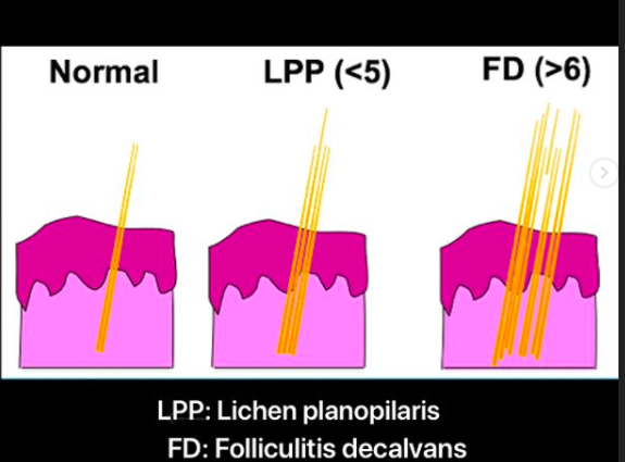
Comment 2: "I see 'lonely' hairs"

Not everyone knows what a lonely hair is. But a patient who understand what is meant by a "lonely hair" has generally done a lot of reading and are extremely knowledgeable about hair loss! The reality however is that most "lonely hairs" that most people find in the scalp are not what really constitutes the gist of what these hairs are all about. My feeling is that the presence of more than 6 hairs across the frontal hairline at a distance of greater than 1.5 cm from the main hairline probably does in fact raise suspicion for true 'lonely hair' phenomenon.


Comment 3: "I have redness and scarring in my scalp" 

Redness alone does not constitute a diagnosis of FFA. In fact, FFA tends to be more pinkish and subtly red than overtly red. There are simply too many scalp conditions that cause redness to give this observation any significance.

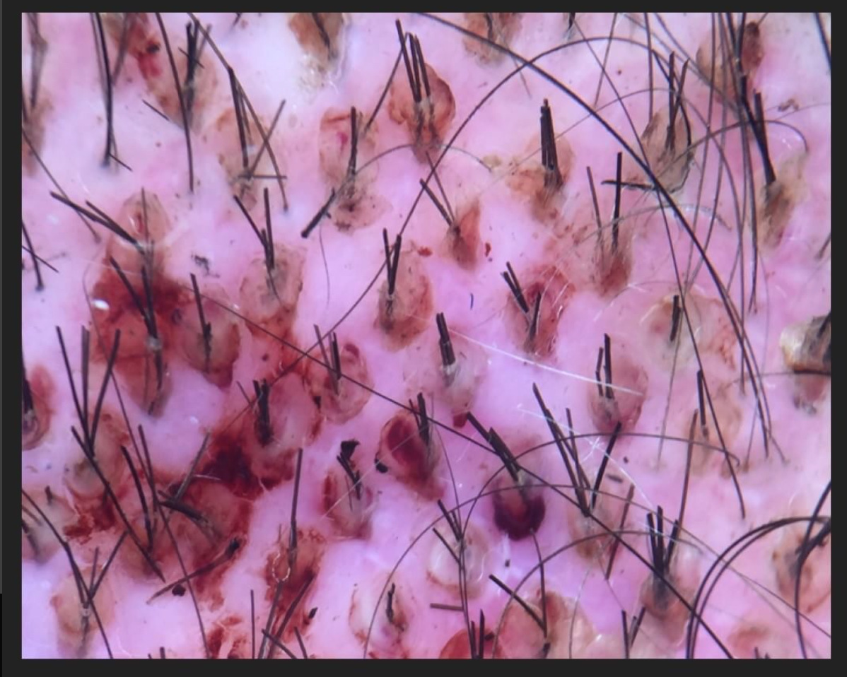
I'm always concerned when patients feel they can "see scarring" because one can't truly see scar tissue as it is mostly under the scalp. In advanced scarring alopecia, one certainly can see evidence of scarring but these cases are usually fairly advanced. In all fairness, one can however see scalp color changes (mostly to a white color) that would suggest the presence of scarring. However, one can't actually see scarring.


Comment 4: "I now have so many baby hairs"

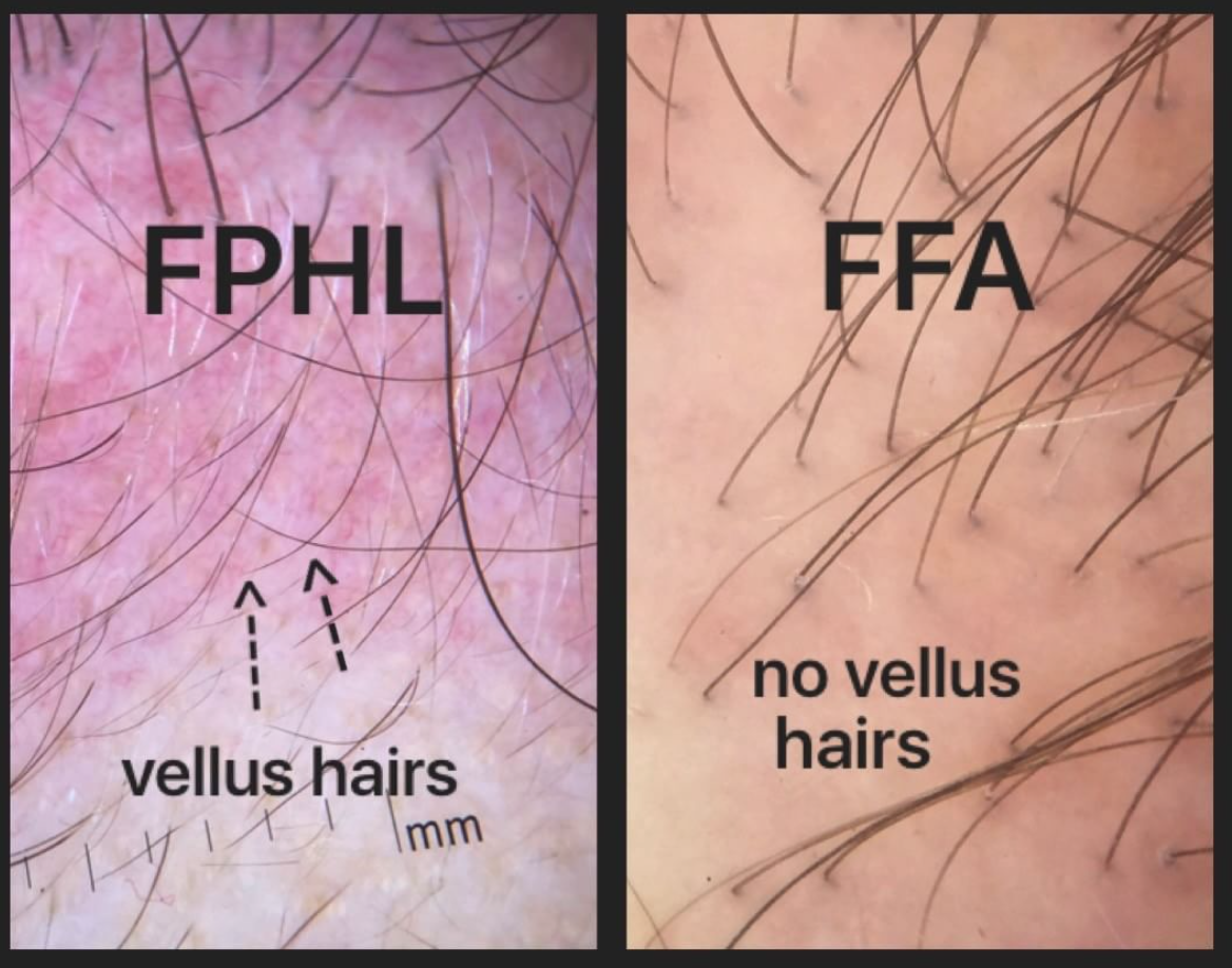
Baby hairs are not really a feature of FFA. In fact, the presence of baby hairs probably argues against the diagnosis of FFA for most patients than actually favours the diagnosis. This is because FFA tends to be a destructive process that involves the preferential destruction of baby hairs (medically termed vellus hairs). The presence of abundant baby hairs is not a feature of progressive FFA.


Comment 5: "I have no family history of balding so it only makes sense something else is going on"

The argument that an individual must have some other diagnosis other than genetic hair loss on account of the lack of genetic hair loss in the family is never a valid argument. There are women who come to be diagnosed with androgenetic alopecia despite a family history of men and women with thick hair. The patient's account of her family history is not very relevant to most diagnoses of hair loss. Sounds strange but this is true: I frequently diagnose androgenetic alopecia in women who stated their family is comprised of individuals with 'good hair.'


TOP 5 FINDINGS AND COMMENTS THAT ARE HELPFUL IN DIAGNOSING FFA

Nothing can substitute for a careful review of one's story and examination of their scalp with dermoscopy. The following pieces of information are helpful when considering a diagnosis. Not all patients have the following features, but they greatly increase the odds that what we are dealing with is FFA.


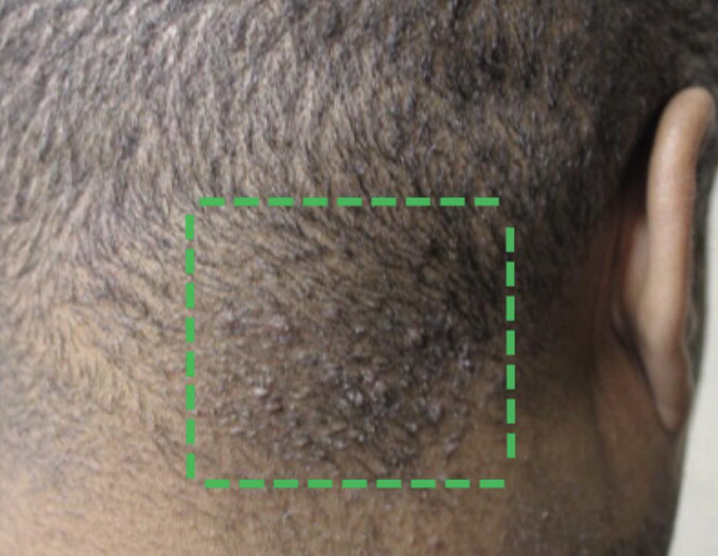
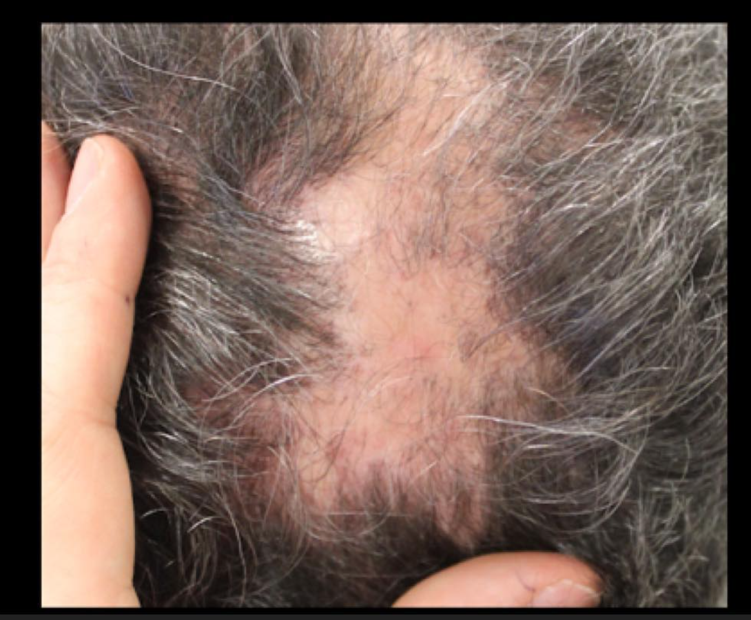
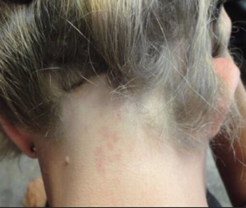
FINDING 1. Both sideburns are lost (above the ears). Not thinned but lost. The regions below where the spectacle of the glasses sit has been depleted of hairs.

Loss of the hair frim both sideburns is actually quite an important piece of information when it comes to diagnosing FFA.  This finding is mot present in everyone with FFA but is quite common if one looks.


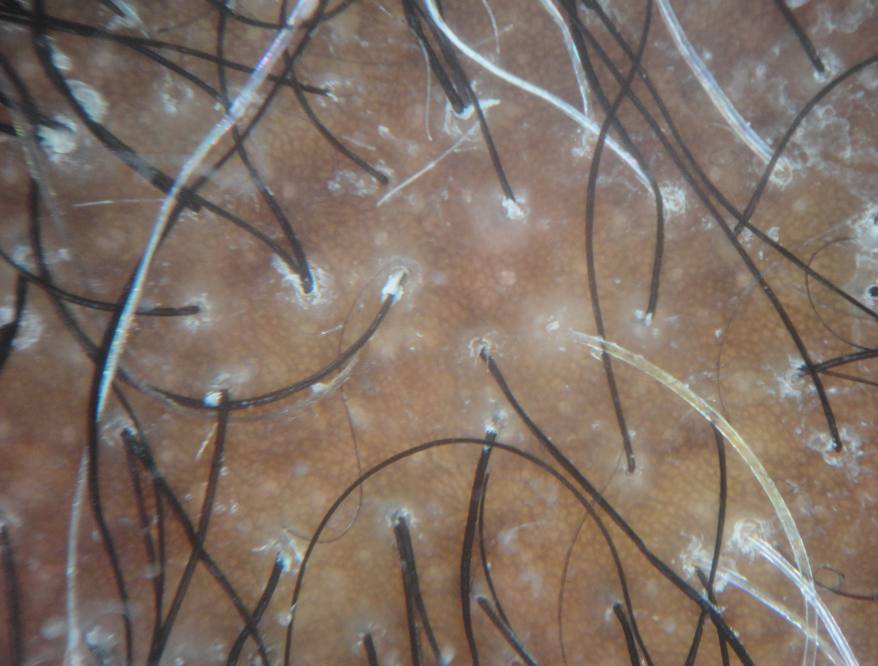
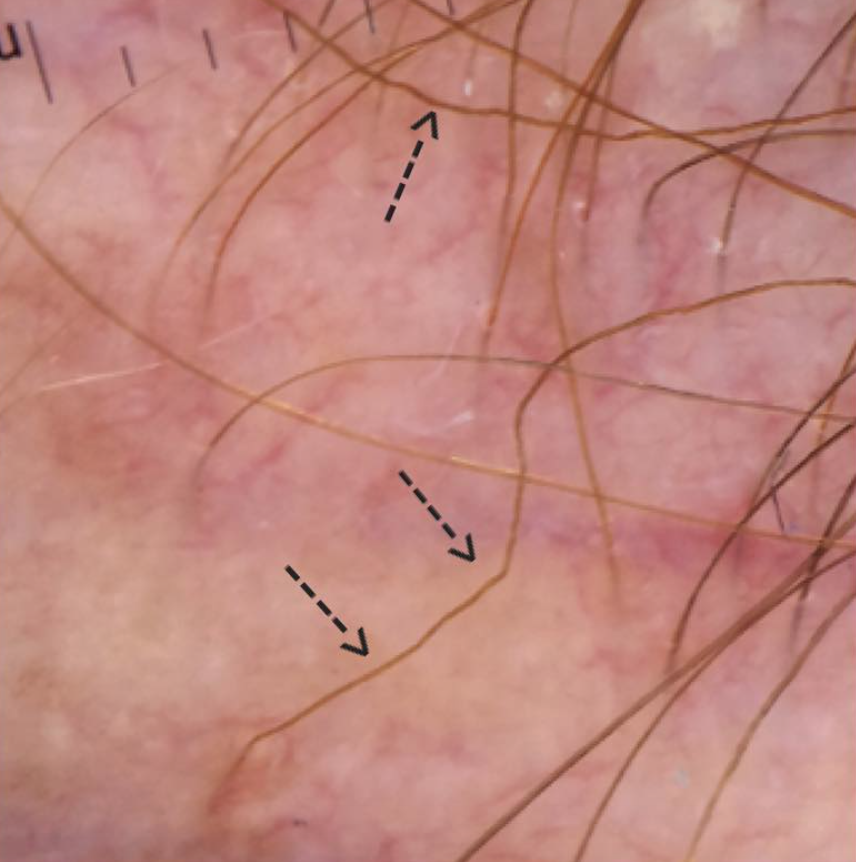
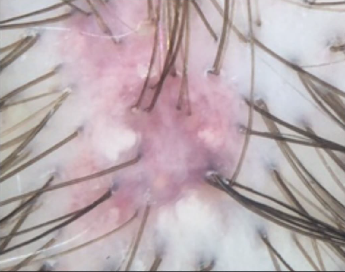
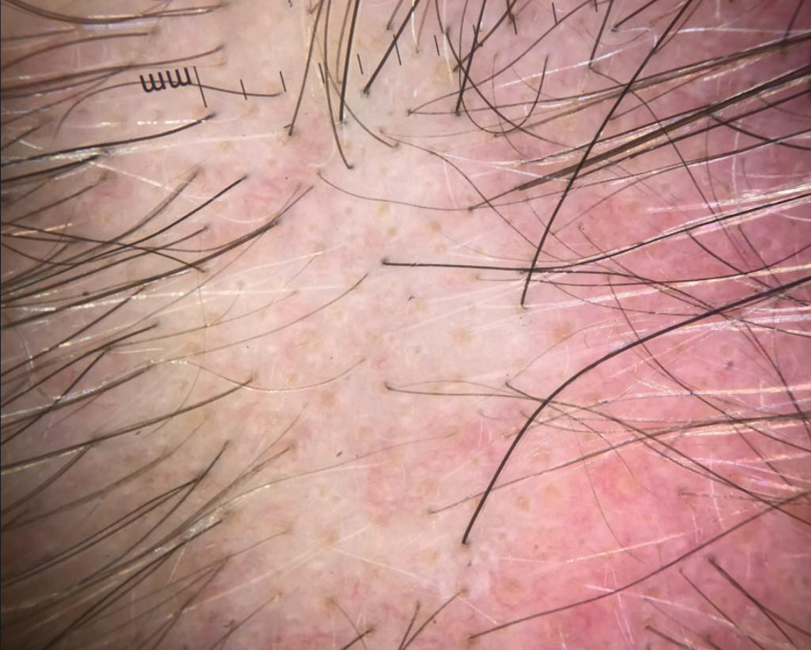
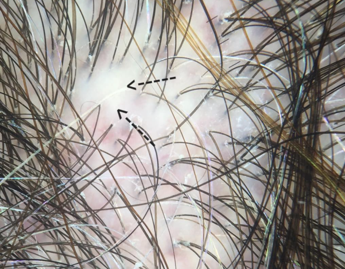
FINDING 2. Baby hairs are not seen in the frontal hairline but rather many single long hairs are seen. Not all the hairs have redness around them but many do have a faint redness

FFA is a process that destroys fine vellus hairs - but it does so with inflammation. The presence of small amount of inflammation (usually without symptoms) is a key finding.


FINDING 3. If the eyebrows are lost, they are significantly changed

Eyebrow loss is common in FFA and in a large number of individuals with FFA, they are the first finding. Eyebrow loss of course does not happen in all women with FFA and loss of eyebrows may even come after the loss of the frontal hair. However, significant eyebrow loss requiring tattooing, microblading does raise the odds a bit that the presentation fits with possible FFA.  Still, one needs a careful examination before concluding that anyone's eyebrow loss is FFA. Nevertheless, changes in the eyebrows are common to FFA.


FINDING 4. The patient is between 46 and 66

FFA is rare in the 20s and 30s. That's not to say it can't occur as we have patients in their teenage years affected. However, from a statistical point of view, women under 40 who come in with worries that they have FFA actually rarely end up having FFA. There are exceptions of course but it is not a common occurrence. In our clinic, 98 % of patients who receive a diagnosis of FFA are older than 46.


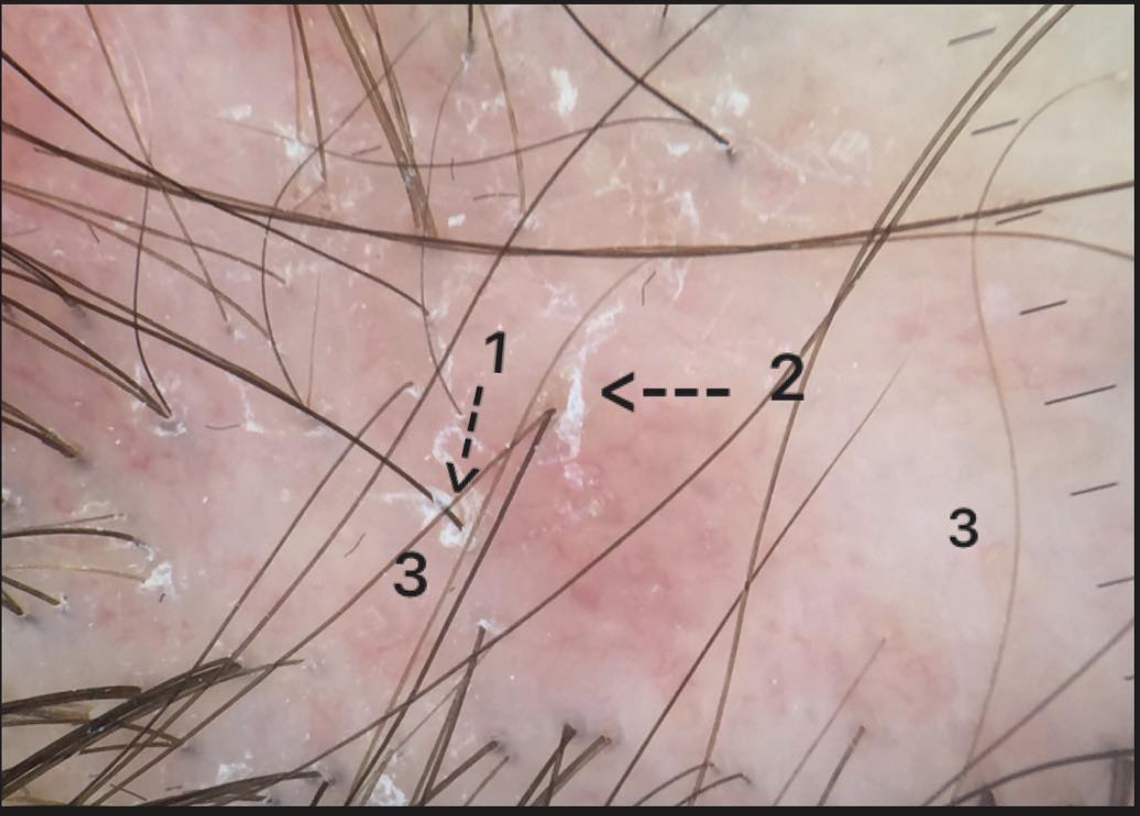
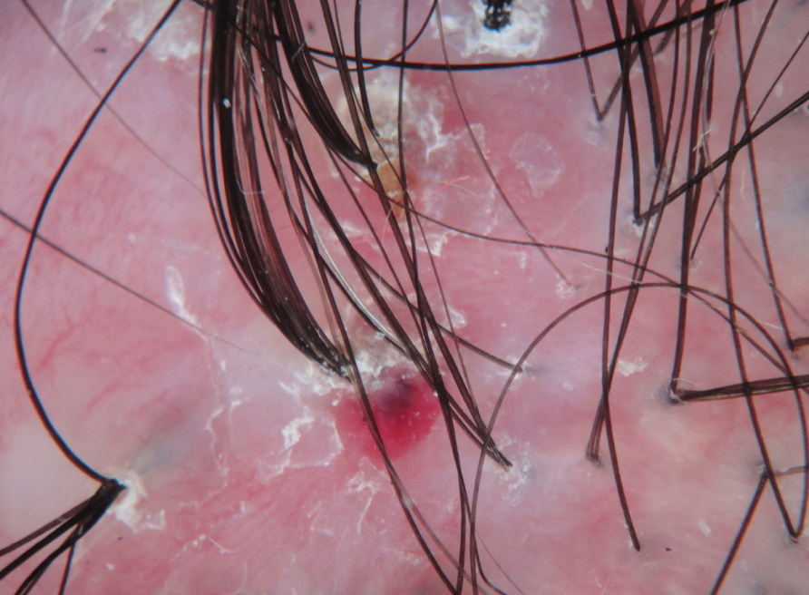
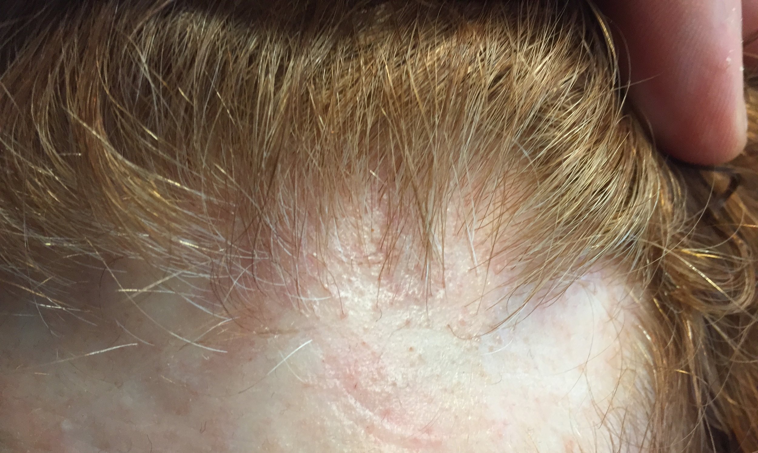
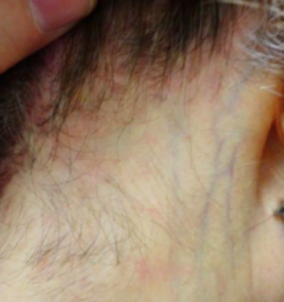
FINDING 5. Veins are seen much easier on the scalp than before and the skin itself just does not look quite normal.

FFA is a complex condition and is probably more than just a 'hair disease'.  It's likely a complex autoimmune disease that affects hair predominantly but also affects the skin as well The skin itself changes in FFA - and does so by thinning. We call this "atrophy." Some women with FFA have minimal to no atrophy but many have significant atrophy which leads to the veins becoming much more visible. Generally, the skin in the area of the hairline does not look the same as the skin of the forehead - the affected area in FFA is lighter in color and smooth.


CONCLUSION

Frontal fibrosing alopecia is a complex condition. There is no 'one way' that it appears. There are 100s of different combinations. Navigating the diagnosis oneself is frequently met with challenging. I've summarized here 5 points that patients place a great deal of emphasis on when looking at their hair - but actually don't carry all that much weight when making the diagnosis. 


This article was written by Dr. Jeff Donovan, a Canadian and US board certified dermatologist specializing exclusively in hair loss.
Share This
12 Comments

Scarring Alopecia: Loss of the follicular opening is a hallmark

Scarring Alopecias Cause Scarring

scarring

Scarring alopecias are hair loss conditions that are associated with the development of permanent hair loss. There are dozens of different types of scarring alopecia. 
Some scarring alopecias itch. Some don't. Some are associated with increased shedding. Some aren't. Some are red. Some aren't. Some bleed. Most don't. 


However what is common to all scarring alopecias is the disappearance of the follicular opening or "pore." The development of scar tissue beneath the skin leads to the destruction of the follicular pore opening.

The arrows point to an area of scarring in a subtle early scarring alopecia


This article was written by Dr. Jeff Donovan, a Canadian and US board certified dermatologist specializing exclusively in hair loss.
Share This
No Comments

Hair Loss at the Nape of the Neck: What are the main causes?

Losing Hair at the Nape: What are the main Causes?

Hair loss can either be localized (meaning one area of the scalp) or diffuse (meaning all over the scalp). There are many causes of hair loss at nape of the neck and the back of the scalp. 


1. Traction alopecia (photo 1)

Traction alopecia refers to a type of hair loss due to the tight pulling of hair. The back of the scalp is particularly susceptible to loss of hair.  Hair styling practices can frequently lead to traction including braids and weaves and pony tail.  If traction alopecia is of recent onset, hair regrowth can occur even without treatment. If traction is longer standing, the hair loss can often be permanent.  Some women with hair loss that looks like traction actually have a scarring alopecia known as cicatricial marginal alopecia. 

PHOTO 1: Traction alopecia presenting as hair loss in the nape

PHOTO 1: Traction alopecia presenting as hair loss in the nape


2. Alopecia Areata (photo 2)

Alopecia areata is an autoimmune disease that affects about 2 % of the world. Hair loss can occur anywhere. Alopecia areata can frequently cause hair loss specifically at the nape in some patients. The particular form that causes loss at a the back of the scalp is the 'ophiasis' form. The ophiasis form is frequently resistant to standard treatments although topical steroids, steroid injections and diphencyprone are typically first line. 

PHOTO 2: Alopecia areata presenting as hair loss in the nape

PHOTO 2: Alopecia areata presenting as hair loss in the nape

 3. Androgenetic Alopecia (photo 3)

Androgenetic alopecia (male and female thinning) typically causes hair loss at the top of the scalp. In men, the temples and crown are most often affected. In women, the mid-scalp region is generally affect first. The back of the scalp can also be affected although this is not typically thought of. Hair thinking along the nape is not uncommon in advancing balding in men and women. 

PHOTO 3: Androgenetic alopecia (AGA) presenting as thinning in the nape

PHOTO 3: Androgenetic alopecia (AGA) presenting as thinning in the nape


4. Frontal Fibrosing Alopecia (photo 4)

Frontal fibrosing alopecia (FFA) is a type of scarring alopecia. FFA typically affects women between 45-70. Most often hair is lost along the frontal hairline and eyebrow. However the back of the scalp (at the nape) and frequently be affected. The hair loss in the nape typically starts at the sides (left side and right side) just behind the ears. Treatments for FFA include topical steroids steroid injections, topical calcineurin inhibitors. Oral drugs include finasteride, doxycycline and hydroxychloroquine at the top of the list.


PHOTO 4: Frontal fibrosing alopecia (FFA) presenting as hair loss in the nape

PHOTO 4: Frontal fibrosing alopecia (FFA) presenting as hair loss in the nape

5.  Heat and Chemicals

Heat and chemical treatments can lead to hair shaft damage and hair loss at the nape. Frequently, heat and chemical overuse leads to an increased tendency to develop traction alopecia which si discussed above. 


6. Acne keloidalis nuchae

Acne keloidalis nuchae (AKN) is a condition that can affect both men and women. Small papule or bumps develop along the posterior scalp and are accompanied by hair loss in the region as well.  The hair loss in AKN is often permanent and can lead to thicken and thicker scars some of which are disfiguring. 


7. Hair shaft disorders

Some individuals are born with abnormalities in how the hair shaft is produced. This frequently leads to hair breakage. Monilethrix is one of the hair shaft disorders that frequently leads to hair loss along the nape. 


This article was written by Dr. Jeff Donovan, a Canadian and US board certified dermatologist specializing exclusively in hair loss.
Share This
10 Comments

Blogs by Topic


Share This
-->