h1.qusth1 { display: none !important; }

QUESTION OF THE WEEK

Dr. Donovan's Articles

QUESTION OF HAIR BLOGS


Lichen planopilaris or Pseudopelade of Brocq: Does anyone care?

Lichen Planopilaris and Pseudopelade of Brocq are Different Conditions with the Same Treatment.


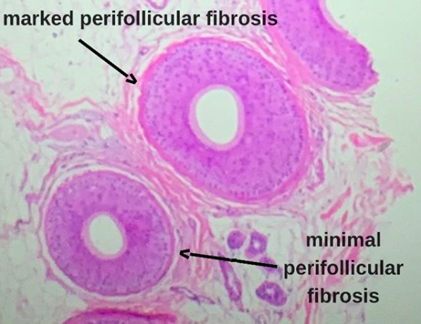
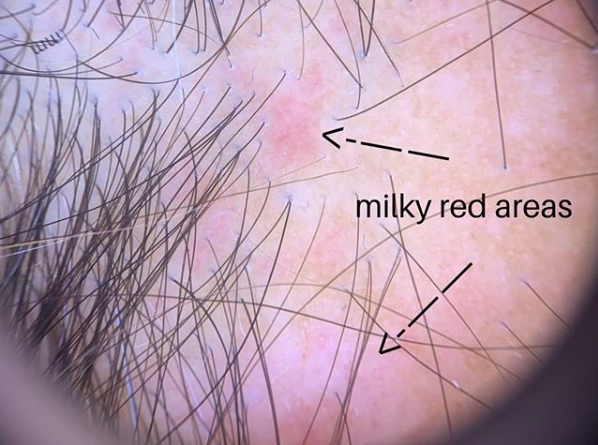
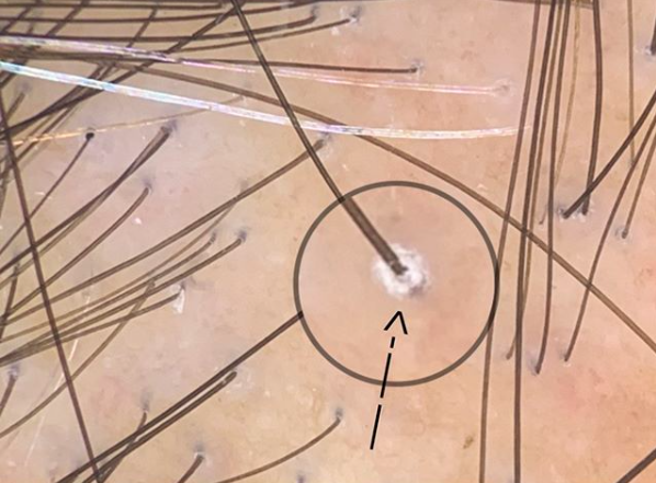
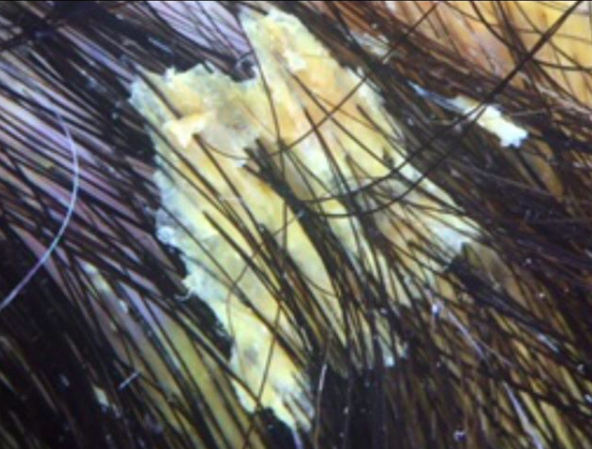
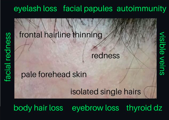
Lichen plaopilaris and pseudopelade are both types of scarring alopecia. They can look similar - but there are many unique differences as well too. Patients with lichen planopilaris often present with scalp itching and burning and the central area of the scalp is typically affected. Increased scalp shedding is common in the early stages. Other areas can be affected too. Some patients have other areas affected as well including eyebrows, and eyelashes. Some patients have oral, nail and vaginal lichen planus but this is present only in a small proportion. Examination of the scalp in a patient with LPP shows redness in the scalp and redness around hairs. There is often scaling and flaking in the scalp as well with scaling notably seen around the hairs in active disease (perifollicular scale).

Pseudopelade of Brocq: A type of primary lymphocytic scarring alopecia

Pseudopelade of Brocq: A type of primary lymphocytic scarring alopecia


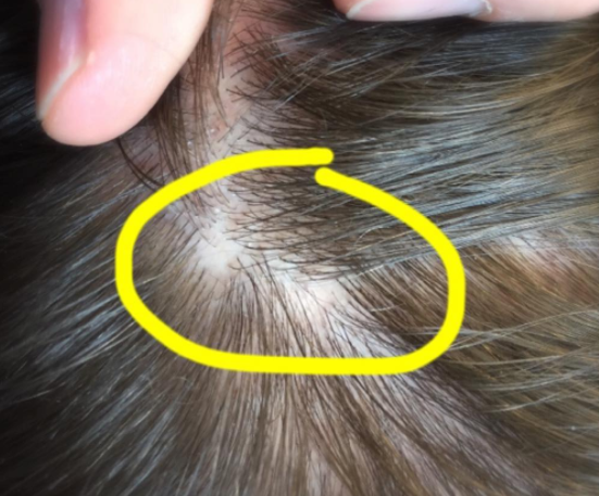
Pseudopelade is a scarring alopecia which also presents with itching and burning. The scalp is often pink in color although may be surprisingly normal in appearance despite the ongoing loss of hair. Patients with PPB often have circular or oval areas of hair loss with strikingly close resemblance to alopecia areata. In fact, pelade is the french word used for alopecia areata. Pseudopelade is different than LPP because it typically lacks the thick perifolliuclar scale seen in LPP and often has more skin atrophy. The skin dips downs with tiny depressions which are referred to as “foot prints in the snow.”

So is it LPP or PPB?

I’ve been part of countless numbers of discussions over the years with patients, physicians, and dermatology trainees as to whether the patient in question has lichen planoplaris of pseudopelade. There is often a great intensity of discussion around the subject. In formal dermatology rounds, physician can debate for extensive amounts of time as if the true answer will be found at the end of the spirited battle.

The reality is that PPB and LPP are distinct conditions. Microarray studies from 2010 showed this clearly. PPB is not just a type of LPP. Generally speaking the less scale and redness around hairs and the more atrophic the skin is - the more likely PPB will be given as a diagnosis. There are histological features too that differentiate these conditions - with PP typically showing a preserved elastic fiber network whereas LPP typically shows a destroyed elastic fiber network. Some physicians and reserachers even feel that PPB is so different from LPP that it should not even be called a scarring alopecia but rather should be called a permanent non scarring alopecia. This is subject to ongoing debate.


LPP or PPB: Leaving our Patients Confused

The active debate that physicians like to have between PPB and LPP often leaves patients confused. Discussions often go something like this;

“I went to see Dr. X and he said I don’t have Pseudopelade of Brocq and what I have is actually LPP”

“I went to see Dr. X and he said what I have is LPP”


Ending the Absurdity: Using the Term “Primary Lymphocytic Scarring Alopecia”

Sometimes, I refuse to use the terms PPB and LPP. These are usually patients with massive files and patients who have seen many physicians to date. These are the patients where 5 doctors have said LPP and 5 doctors have said PPB. These are the patients who are not getting better and what to know two things 1) What is the proper diagnosis and 2) what is the best treatment?

Many physicians fail to recogniize that there is not one LPP and there is not one PPB. It’s not a matter of selecting the right answer. There are probably 20 different presentations of LPP and 5-10 presentations of LPP. Debating over whether a patient has LPP or PPB is good for academic purposes and teaching and good for understanding potential treatments. But at the end of the day, what the patient in front of us has is a scarring alopecia that is caused in part by inflammatory cells called lymphocytes doing things they should not be doing.

In the present day, the treatments for PPB and LPP are the same!! The question that must be asked is what is the purpose of so much extensive debate when the treatments in the present day are the same. Topical steroids, steroid injections, doxycycline, retinoids, hydroxychloroquine, methotrexate are among the options. Physicians far too often spend far too long debating and one upping other colleagues with an attempt to present to the patient what they think the diagnosis is that we forget that the treatments for LPP and PPB are generally the same.

One can argue on an on as to the relevant merits of honing down the diagnosis. Knowing that a patient has LPP prompts one to ask about lichen planus of other areas. But nail, mucosal, vaginal symptoms should be asked of all our patients. Knowing that a patient has LPP might prompt one to screen for certain blood tests including thyroid abnormalities. But these same tests are still appropriate in a patient with presumed PPB.


Conclusion

As a clinician, it is helpful to practice in a manner that strives for accuracy. Accurate diagnosis are fundamental to practicing good medicine. But there are times where we can not always make accurate diagnoses because not everything is always so clear cut. In the case of LPP, and PPB there are classic presentations of these diseases and there are aytpical or non-classical presentations of these diseases. We should strive to recognize the classical cases and strive in these cases to give them the proper names (either LPP or PPB). However, there are other times were the efforts that go into pigeon holing our patients into certain disease categories only serves to remove us further from focusing on actually providing care to the patient. There are dozens of variants of LPP not one. There are many variants of PPB not one.

The term “primary lymphocytic scarring alopecia” serves us well in cases where pigeon holing and efforts to force a diagnosis just doesn’t work.


Reference

Yu M et al. Lichen planopilaris and pseudopelade of Brocq involve distinct disease associated gene expression patterns by microarray. J Dermatol Sci. 2010 Jan;57(1):27-36.


This article was written by Dr. Jeff Donovan, a Canadian and US board certified dermatologist specializing exclusively in hair loss.
Share This
No Comments

Prescribing Dutasteride in Males with Balding: Are there any criteria ? Should there be any criteria?

Prescribing Dutasteride in Males

The most effective medical treatments for male balding (at the time of this article being written) are oral finasteride and oral dutasteride. There is no debate about this particular comment. In the present day, as the world grapples with the meaning of “post finasteride syndrome”, dutasteride is increasingly a choice for many physicians where it never might have been a choice before. I see it - and I see more now than I did 2 years ago. I see dutasteride being used and 0.5 mg three times weekly. I see dutasteride used at 2.5 mg once weekly or twice weekly. I see dutasteride being used at 0.5 mg daily. I see dutasteride being used more often - and I too prescribe it more now than I did 10 years ago.

Dutasteride is not formally FDA approved for treating male balding but is still widely used. It’s used “off label” though and certainly has a large number of studies to back up its effectiveness. In some countries, dutasteride does have formal approval.

The public is also increasing asking about dutasteride - and increasingly requesting it. After all, we don’t have a great deal of data implicating dutasteride in the same set of issues that finasteride has. Furthermore, it’s more effective than finasteride. These two points lead many to turn to the drug. Some data suggests side effects like sexual dysfunction are greater with dutasteride than finasteride but certainly not all studies show this. Some in fact, show that side effects of dutasteride are similar to placebo.

We don’t yet fully understand everything behind post finasteride syndrome to even begin to dig into what might be called a post dutasteride syndrome or a general 5 alpha reductase syndrome. More studies are needed.

So, will you prescribe me dutasteride or not?

Many patients come in the clinic wanting to know if I’ll prescribe them dutasteride. Some have been on finasteride and haven’t found that it works - and they want dutasteride. Some don’t want to try finasteride at all - they want dutasteride. Some want both. Some know the dose they want.

it all comes down to understanding the medical evidence and the 20 years of science that comes before. There is not a “yes” or “no” answer to whether I will prescribe dutasteride. I don’t know when a patient walks in the door if these medications are right for them - but I do know before the patient walks out the door if these medications are right for them.

The following are the criteria I use in the clinic for determine if the patient is a candidate for dutasteride. These are my criteria and may not necessary be the guideline principles for everyone.

Top 10 Criteria for Prescribing Dutasteride (Donovan)

  1. The patient understands the treatment is life-long if he wishes to maintain active medical treatment of his androgenetic alopecia.

  2. The patient is aware of the array of possible side effects that have been reported with use of 5 alpha reductase inhibitors including mood changes, depression, anxiety, sexual dysfunction, enlargement of breast tissue (gynecomastia), penile shrinkage, loss of penile sensation, weight gain, muscle weakness and others. The potential effects of dutasteride on males wishing to father are not completely understood. The patient accepts the risk of these side effects if he chooses to use dutasteride.

  3. The patient does not currently have severe depression or currently have severe anxiety that might otherwise present a contraindication to using dutasteride. The patient has not been suicidal in the past or been hospitalized for depression and mental illness within the past 5 years.

  4. The patient is aware of reports that some patients have experienced persistent (long lasting) problems even when the drug has been stopped. These are mainly studied in the context of finasteride but should be assumed for now to be relevant to the use of dutasteride. The patient accepts the risk of these side effects if he chooses to use dutasteride.

  5. The patient is aware that class action lawsuits have been launched regarding the persistent side effects related to finasteride use.

  6. The patient is aware of alternatives for treatment including topical minoxidil, oral minoxidil, topical anti androgens (topical finasteride), low level laser, platelet rich plasma and hair transplantation.

  7. The patient has no known issues currently related to male inferility or infertility in a female partner.

  8. The patient understands the possibility of dutasteride causing a reduction in sperm count and the rare possibility that these reductions may be permanent or long lasting (even when the drug is stopped). The original dutasteride studies showed that after 6 months of stopping the drug, sperm counts had not returned to normal in all study participants and that the total sperm count in the dutasteride group remained 23% lower than baseline.

    Males who are concerned about the possibility of lower sperm count or fertility issues may consider having baseline semen analysis or baseline FSH, LH, Free T4 and testosterone measurements before starting. These lab tests may provide some guidance about baseline fertility. These issues in point 5 are relevant to males who may wish to father children in the future.

  9. The patient does not wish to donate blood and understands that blood donation is not possible for at least 6 months after stopping the drug.

  10. The patient understands that dutasteride may affect future prostate cancer screening by affecting the PSA value. These issues need to be discussed at the time of such screen and consideration might be given to baseline screening depending on the age of the patient when starting dutasteride.

Conclusion

I can’t say if a patient is a candidate for starting dutasteride when they walk in the office but after 20-30 minutes I can determine if they are likely to be a good candidate for the drug or not. The answers to the questions and issues above help guide the decision making that goes into figuring out if a patient is a candidate for dutasteride or not. It’s not something that can be ascertained in a matter of a few minutes.

References

Meeker JD, Godfrey-Bailey L, Hauser R. Relationships between serum hormone levels and semen quality among men from an infertility clinic. J Androl 2007;28:397–406.


This article was written by Dr. Jeff Donovan, a Canadian and US board certified dermatologist specializing exclusively in hair loss.
Share This
No Comments

Minoxidil, Collagen and Facial Aging: Where are we at?

Minoxidil and Facial Aging: Yes or No, …or we don’t know?

There is currently a great buzz on the internet world that topical minoxidil affects collagen synthesis and affects facial skin by promoting facial aging. To date, there is no good evidence in the medical literature that minoxidil promotes aging of the face.

As for affecting collagen synthesis - minoxidil probably DOES affect collagen synthesis in the scalp. The problem with the unsubstantiated claims on the internet is that nobody has dared to offer the potential explanation that the minoxidil-induced reduction in collagen synthesis might actually be, well... a good thing.

fibrosis

Androgenetic Alopecia and Fibrosis

The lay public is not always aware that androgenetic alopecia is associated with the body laying down scar tissue in the scalp or what we call “perifollicular fibrosis.” That’s right - male and female balding is associated with INCREASED collagen production in the form of ‘fibrosis’ around hair. And this is not a good thing as the infalmmation and scarring around hairs only serves to speed up the miniaturization process and speed up the destruction of the delicate stem cells. It seems, based on 2006 studies by Yoo and colleagues that a growth factor known as TGF beta is responsible for this increase in collagen production. A variety of studies suggest that minoxidil has the potential to REDUCE TGF beta levels and in turn REDUCE the likelihood of further fibrosis. It seems like it could be a really great thing that minoxidil reduces collagen production.

It’s certainly premature to jump to conclusions that minoxidil promotes facial aging. If it does, it’s rare - likely because not enough minoxidil reaches the facial skin. My office phone rings off the hook with people terrified that their minoxidil is causing their hair shedding or growing hair in the wrong spots. We have never received a call about facial aging concerns. But minoxidil probably does affect collagen and that’s most likely a really wonderful thing rather than a bad thing because it suppresses the formation of more and more scar tissue around hairs that ultimately destroy stem cells. Patients worried about the small unproven risk of facial aging should not of course use the product.

More research is needed to put the facial aging issue to rest or bring it to the forefront so that the FDA and various health regulatory bodies can re-examine this issue more thoroughly.


This article was written by Dr. Jeff Donovan, a Canadian and US board certified dermatologist specializing exclusively in hair loss.
Share This
8 Comments

Botox for Reducing Scalp Sweating in Frontal Fibrosing Alopecia

Botox for Frontal Fibrosing Alopecia Scalp Sweating

Frontal fibrosing alopecia (FFA) is a scarring alopecia associated with hair loss along the frontal hairline. The eyebrows, eyelashes, body hair can also be affected. A 2017 study showed that some women with FFA experience increased sweating in the areas of hair loss. Standard treatments for frontal fibrosing alopecia including topical steroids, steroid injections, and antibiotics were found to be helpful. Interestingly, botulinum toxin treatments (Botox) were also found to be helpful.

We are seeing an increasing amount of women with scalp sweating associated with their FFA. Sweating is controlled in part by the nervous system and this raises the possibility that a specific type of inflammation known as ‘neurogenic’ inflammation may be relevant in FFA. I think we will be hearing more about this area of research in the years ahead.


Treatment of scalp Sweating with Botox in FFA

For the excessive scalp sweating that some patietns with FFA experience, Botox can certainly be considered as an off label (non FDA approved indication). There is evidence that some of the inflammation in FFA is part of what is known as ‘neurogenic inflammation’ and blockade of these nerves will block signals sent to the sweat glands eccrine glands.

 My advice for FFA patients with excessive scalp sweating is to consider Botox in the hairline starting with a conservative number of units before increasing the dose. There seems to be a great (huge) variation in the amount of Botox needed to reduce scalp sweating in FFA. There is no formal protocol for FFA, but I generally recommend starting with 40 units in the affected hairline areas and waiting several months to see how well this work to reduce the sweating. One can certainly increase up to 80-100 units - and even well beyond (100-200 units).  Many patients, but not all, require 100 Units and above. 


Again, even though there is no formal protocol, one might consider diluting a 50 U vial with 1.25 mL and starting with 10 injections (of a 4U / 0.1 mL concentration) spaced 10 mm apart (i.e. 40 units total).  In 4-6 months, a decision can be made as to how well this worked and either proceeding in one of three ways


OPTION 1. Increasing to 60-80 units instead of 40 units and increasing the size of the area injected. This would involve diluting a 100 U bottle in 2.5 mL saline and proceeding with the same technique but injections covering a larger area.


OPTION 2. Diluting the Botox by one half (i.e. diluting a 50 U via with 2.5 mL saline) and injecting more sites.


OPTION 3. Doing nothing different in the case of the patient reporting success with the first trial of Botox and simply waiting for the patient to report that sweating has returned at which point the Botox can be administered again. This is typically every 6-12 months for successfully treated patients.

Reference


Harries et al. Frontal fibrosing alopecia and increased scalp sweating: Is neuorgenetic inflammation the common link. Skin Appendage Disord May 2016; 1(4):179-84


This article was written by Dr. Jeff Donovan, a Canadian and US board certified dermatologist specializing exclusively in hair loss.
Share This
No Comments

Hair Transplants for Folliculitis Decalvans: Yes or No?

Can patients with folliculitis decalvans have a hair transplant?


Folliculitis decalvans (FD) is a type of scarring alopecia. It is less common than scarring alopecias such as lichen planopilaris. Treatments, such as oral antibiotics and oral isotretinoin, are viewed as the main (so called “first line”) treatments for folliculitis decalvans. They may help slow disease progression and in some cases stop the disease altogether. However, it can takes many months to even many years to stop the disease- and permanent arrest does not happen in all cases. When the disease completely settles and stops and the patient demonstrates that they are able to come off all medications (without their disease flaring) it may be possible for a hair transplant to be considered. Until that time, a hair transplant is contraindicated.


When might a hair transplant be possible?

In general, hair transplants for scarring alopecias are sometimes associated with poor growth. However, very carefully selected patients can sometimes do well with surgery and have reasonably good results - although in my opinion far too often patients with scarring alopecias undergo surgery when they are not (yet) good candidates. Some of the patients who undergo surgery when their disease is not yet calm (ie burnt out) have less than optimal outcomes. Large studies of hair transplant outcomes for patients with folliculitis decalvans have not been published in the medical literature but certainly I see the phenomenon routinely in my clinic (Post operative Complications Clinic, POCC).

Is poor growth the worst that can happen?

In contrast to the scarring alopecia lichen planopilaris, patients with folliculitis decalvans are more likely to have thick scar tissue in the scalp which further limits growth and survival of grafts. In addition to poor growth, disease reactivation can potentially occur after surgery. Most patients, and sometimes their surgeons too, incorrectly approach hair transplantation for patients with scarring alopecia with the overriding view that “the worst that can happen is poor hair growth.” This is incorrect as sometimes the disease reactivates even worse than if the patient had not gone ahead with surgery. The worst that can happen for the patient is persistent redness, pustules, itching, scalp itching, burning, pain and marked hair loss not to mention the psychological consequences. Of course extreme examples like this are uncommon but the point has been made. Severe complications and disease reactivation/flares are unlikely if the patient and his or her disease meets the criteria outlined below. FD must be inactive before surgery is considered.

Hair transplants are not often done in folliculitis decalvans.The medical literature contains few reports of successful hair restoration surgery in FD. In 2010, Tyagi reported 40 % graft survival in a patient with folliculitis decalvans who underwent FUE (follicular unit extraction). Follow up was only 6 months in duration which is insufficient follow up time for scarring alopecia. Some scarring alopecias reactivate 1-3 years following surgery leading the patient to lose all their grafts. 6 months carries little meaning in the world of scarring alopecia. If surgery is performed, I can not emphasize enough the importance of a consultation with a dermatologist prior to the procedure to confirm that the patient truly is a good candidate (and that they meet the five criteria below). This is too often overlooked as patients are eager to move forward with surgery. Such a consultation is essential. In addition, the importance of having an experienced surgeon perform the surgery. It is essential as grafts must be prepared properly, handled properly and inserted at the correct densities.

Criteria for Hair Transplant Candidacy in Folliculitis Decalvans

Several years ago, I published a set of helpful five criteria which helps guide physicians about whether or not they are good candidates for surgery.

1.  The PATIENT should be off all hair-related medications.

2. The PATIENT must not report symptoms related to the FD in the past 24 months. 

3The PHYSICIAN must make note of no clinical evidence of active disease in the past 24 months. 

4. Both the PATIENT and PHYSICIAN must demonstrate no evidence of ongoing hair loss over the past 24 months. 

5. The patient must have sufficient donor hair for the transplant. 

interested readers can read more in the following link:

Donovan Hair Transplant Criteria - Folliculitis Decalvans

Conclusion

Hair transplants are possible for folliculitis decalvans but only for a subset of patients who have proven, beyond any doubt, that their disease is inactive. The patient and surgeon might hope for the best but be aware that outcomes and graft survival in folliculitis decalvans may be suboptimal.


Reference

Tyagi V, Singh PK: A new approach to treating scarring alopecia by hair transplantation and topical minoxidil. Indian J Dermatol Venereol Leprol 2010; 76: 215.


This article was written by Dr. Jeff Donovan, a Canadian and US board certified dermatologist specializing exclusively in hair loss.
Share This
No Comments

PADI3 mutations found in Patients with Central Centrifugal Cicatricial Alopecia

Gene Mutations in PADI3 found in about 1/3 of Women with CCCA

CCCA is a scarring alopecia that occurs in about 15 % of black women. It also occurs in some families (in an autosomal dominant manner) suggesting that there may be some underlying genes that predispose women to CCCA.

A new study by Malki et al identified mutations in the PADI3 gene in 14 of 58 patients with CCCA (24%). When the authors examined the frequency of mutations in patients with CCCA compared with a control group of 2702 women of African ancestry that did not have CCCA, it was found that mutations were slightly more common in patients with CCCA than controls (P=0.04).

Summary and Conclusion

This is an interesting study which opens up some important questions in the field of CCCA research. PADI3 plays a key role in formation of the hair shaft. The gene encodes peptidyl arginine deiminase, type III (PADI3), an enzyme that post-translationally modifies other proteins that are essential to hair-shaft formation.

Mutations in this gene appears slightly more common in CCCA than control groups. The PADI3 gene is well known to contribute to another hair shaft condition known as uncombable hair syndrome (UHS). Interested readers can review a prior article written on the UHS:

Uncombable: New Insights into a Rare Condition.

Reference

Malki et al. Variant PADI3 in Central Centrifugal Cicatricial Alopecia. New Engl J Med. 2019 Feb 28;380(9):833-841.

Donovan J. Uncombable: New Insights into a Rare Condition.


This article was written by Dr. Jeff Donovan, a Canadian and US board certified dermatologist specializing exclusively in hair loss.
Share This
No Comments

Milky Red Areas (MRA) in Lichen Planopilaris

The Milky Red Areas (MRA) in LPP


Trichoscopy (scalp dermoscopy) involves the use of a hand held dermatoscope to better assess the scalp. It has proven helpful for managing a plethora of scalp diseases. 

MRA-LPP


The field of trichoscopy is filled with bizarre terms. This photo shows the milky red areas of lichen planopilaris. These milky red areas correlate with disease activity. The more milky red areas that are seen the more activity the patient has.


Reference 


Lajevardi V, et al. Assessing the correlation between trichoscopic features in lichen planopilaris and lichen planopilaris activity index. Australas J Dermatol. 2019


This article was written by Dr. Jeff Donovan, a Canadian and US board certified dermatologist specializing exclusively in hair loss.
Share This
No Comments

Perifollicular scale in Lichen Planopilaris (LPP): Close Association with Disease Activity

The Scale in Lichen Planopilaris

Dermoscopy also called trichoscopy is extremely helpful to the proper evaluation and management of hair disorders. Hand held dermatoscopes not only help in making correct diagnoses but help in monitoring disease activity. I need my dermatoscope to properly do my job.

PFS-LPP


Perifollicular scale (scale around follicles) is one dermatoscopic feature of lichen planopilaris and frontal fibrosing alopecia. Identifying the amount of scale is important as such scaling correlates nicely with the amount of inflammation going on under the scalp as well as correlating with the so called lichen planopilaris activity index (LPPAI). The presence of perifolliclar scale in patients with LPP is not ideal as it means the patient is at significant risk to lose hair.


Reference 


Lajevardi V, et al. Assessing the correlation between trichoscopic features in lichen planopilaris and lichen planopilaris activity index. Australas J Dermatol. 2019


This article was written by Dr. Jeff Donovan, a Canadian and US board certified dermatologist specializing exclusively in hair loss.
Share This
No Comments

The Localized Telogen Effluvium

Why does everyone say I have a localized telogen effluvium?


Too often, I hear patients tell me that they have been diagnosed with some kind of localized telogen effluvium. The story typically goes something like this:


I’m losing hair in the front and my doc thinks it’s a localized telogen effluvium 

I’m losing hair in the crown and I’ve been told it’s some kind of localized telogen effluvium.


By definition, a true telogen effluvium affects all hairs on the scalp equally. The hairs at the back feel the same pressure to leave as the hairs in the front. The hairs in the crown feel the same pressures to shed as do the hairs in the sides and back. So when someone tells me they are having a localized telogen effluvium, I know to look for another diagnosis. A localized telogen effluvium does exist. So it is not that the patient or doctor is way off in how they have come to that diagnosis. It’s simply that the main diagnosis is something else.


Here are a few examples:


1. When a patient with alopecia areata sees alot of hair coming out of one area of the scalp and someone confirms they seem to be telogen hairs that are being shed- the diagnosis is still alopecia areata. It’s true there is some kind if “localized telogen effluvium” going on - but the telogen hairs are being shed due to the alopecia areata.

2. When a patient with scarring alopecia sees alot of hair coming out of one area of the scalp and someone confirms they seem to be telogen hairs that are being shed- the diagnosis is still active scarring alopecia . It’s true there is some kind if “localized telogen effluvium” going on - but the telogen hairs are being shed due to the scarring alopecia.

3. When a patient with androgenetic alopecia sees alot of hair coming out of one area of the scalp and someone confirms they seem to be miniaturized telogen hairs that are being shed- the diagnosis is still androgenetic alopecia. It’s true there is some kind if “localized telogen effluvium” going on - but the telogen hairs are being shed due to the androgenetic alopecia.


Conclusion

A localized telogen effluvium should prompt the clinician and patient to search for an underlying diagnosis.


This article was written by Dr. Jeff Donovan, a Canadian and US board certified dermatologist specializing exclusively in hair loss.
Share This
No Comments

Scalp Biopsies: Just a Piece of the Puzzle

Scalp Biopsies: Often Just a Piece of the Puzzle but Not the Whole Puzzle

Scalp biopsies are extremely helpful in some cases. When the right sized sample (4 mm) is taken from the correct spot and processed properly using the correct technique and interpreted by a dermatopathologist who understands scalp hair & scalp pathology.... a scalp biopsy can be an amazing help.

puzzle


As an analogy, take a look at the photo. I see some kind of metal object about 5 in x 2 in. It’s hard to tell what it is because it’s not representative enough of the original object. I am not so skilled in identifying electronics to know what it was when I first saw this object. I knew it was not a watch and not a toaster oven. I did not look like the insides of all the watches (or toaster ovens) that I had seen in my life.

I highly skilled electronics expert might correctly identify this as the inside of an iPhone. There are some specific design features that give clues that this is an apple iPhone and nothing else. A true iPhone expert might identify this not only as an iphone but as an iPhone5.

Biopsies of the scalp are just samples of the scalp. A good sized sample taken from the right area increases the odds that the sample will have the necessary features to make the right diagnosis. But you need an experienced pathologist too.

A good electronics technician would not just say this piece of metal is from a smartphone, they would say it’s from an iphone. A good pathologist does not just identify the patient has a scarring alopecia but helps pinpoint that the features are more consistent with lupus than lichen planopilaris. 
A biopsy is not as helpful when the principles of taking and interpreting the biopsy are not followed. A really good sample given to a less experienced pathologist can sometimes still give the right diagnosis (and usually does) and an inadequate sample given to a brilliant pathologist can also sometimes give the right diagnosis (and usually does). But really tough cases need good samples (sometimes 2 or 3), taken from carefully chosen areas and placed on the microscope stage of a pathologist who thoroughly understands hair and scalp pathology.


This article was written by Dr. Jeff Donovan, a Canadian and US board certified dermatologist specializing exclusively in hair loss.
Share This
No Comments


Pityriasis Amiantacea: A Reaction Pattern Not A Diagnosis

Pityriasis amiantacea: Many Causes

Pityriasis amiantacea, also known as tinea amiantacea, is not a diagnosis. Rather it is a reactive phenomenon that sometimes happens during the process of scalp inflammation. The scalp responds by producing thick sticky (“asbestos-like) scale that wraps around hairs like shingles in a roof. The condition was first described in 1832.

The scales can be difficult to remove and often take out hairs when the scale is removed. Many different conditions can lead to the skin responding in a manner that produces the end result of pityriasis amiantacea.

So what are some of the causes of pityriasis amiantacea? Well, the finding of pityriasis amiantacea is often seen in patients with psoriasis, seborrheic dermatitis, fungal infections, various eczemas and many scarring alopecias.

pityriasis-amiantacea


The photo here is a magnified photo of the scalp of a patient with "pityriasis amiantacea" from seborrheic dermatitis.

Treatment includes identifying the root cause and treating that scalp condition or disease. Treatment such as topical steroids, steroid injections, anti dandruff shampoos, salicylic acid, tar all play a role in treatment.  I view the treatment of pityriasis amiantacea as being somewhat urgent because this type of scale can lead to removal of hair and sometimes even chronic permanent hair loss if secondary infection and scarring are present. #pityriasisamiantacea #hair #scale#inflammation #hairclinic #trichoscopy


This article was written by Dr. Jeff Donovan, a Canadian and US board certified dermatologist specializing exclusively in hair loss.
Share This
No Comments

Air Travel and Immunosuppression: What is the effect of flying on the immune system?

Air Travel May be Slightly Immunosuppressive

Of the 2 billion people who fly on commercial airplanes every year about 1 in 5 of them report that they develop a “cold” within the first week of arriving back home. 


Is there any logic to this? Could it really be true? 


Well, the world of aviation medicine has an interest in understanding what happens to the human body in a variety of in flight situations (...let alone what happens when we all start traveling off to Mars). In 2012, a group of researchers hypothesized that hypobaric low oxygen (hypoxic) conditions associated with air travel may actually contribute to impairment of the immune system.

To test this, the researchers studied the effects of hypobaric hypoxic conditions during a simulated flight at 8000 ft cruising altitude on immune and stress markers in 52 healthy volunteers before the simulated flight and on days 1, 4, and 7 after the flight. 

flight


The findings showed that the hypobaric hypoxic conditions of a 10-h overnight simulation flight are not associated with severe immune impairment or abnormal IgA or cortisol levels. However, there were associated with short term impairment in some measurements including transient decrease in lymphocyte proliferative responses combined with an upregulation in CD69 and CD14 cells and a decrease in HLA-DR in the immediate days following the simulated flight. 


All these immune system abnormalities normalized by day 7 in most instances.

The conclusion of the study is that the 400,000 or so travellers who feel they have some kind of respiratory infection or cold after air travel could in fact be correct. A short term suppression of some immune responses could be responsible.

Reference 
Wilder-Smith A, et al. Transient immune impairment after a simulated long-haul flight. Aviat Space Environ Med. 2012.


This article was written by Dr. Jeff Donovan, a Canadian and US board certified dermatologist specializing exclusively in hair loss.
Share This
No Comments

Baby Hairs: Good or Bad?

The Three Categories of Baby Hairs

There are many type of hairs that constitute “baby hairs.” Some of these are positive signs (good signs) as they indicate that this hair has a high chance of growing longer and contributing in a few months to the overall density that a patient feels. The call these group 1 baby hairs. The upright regrowing hairs of telogen effluvium are one such example and so are the normal regrowing scalp hairs that everyone has (new anagen hair). The regrowth that is seen about 2-3 months after starting a new treatment is also an example of Group 1 baby hairs.

Some baby hairs including the vellus-like hairs of androgenetic alopecia, the sick dying vellus-like hairs of scarring alopecias and the vellus hairs of alopecia areata do not mean that baby hairs will turn into something substantial. I call these group 2 baby hairs.


baby hairs

Some baby hairs that people see can also be broken hairs. These are far less common. I call these Group 3 baby hairs. Hair breakage from excessive heat or hair breakage from trichotillomania are all examples of phenomena that can give broken hairs and in turn give the appearance of baby hairs. The exclamation mark hairs of alopecia areata are also an example. Broken hairs are important to identify but these types of hairs are often misdiagnosed. In fact, most people who think they have broken hairs are wrong. They don’t, of course, usually think they are wrong, but they are often wrong about that too. Most people with baby hairs have Group 1 of Group 2 baby hairs. Group 3 hairs are blunt at the top like a freshly cut blade of grass cut by a lawnmower. Group 1 and 2 hairs are pointy with tapered ends pointing up to the sky.

It’s always better to see one hair growing than to see none. So when a patient says “I’m seeing baby hairs but I’m not sure my hair is getting better” my concerns is that this patient may in fact have one of the hair loss conditions giving the so called Group 2 or Group 3 conditions.


This article was written by Dr. Jeff Donovan, a Canadian and US board certified dermatologist specializing exclusively in hair loss.
Share This
No Comments

Upright regrowing Hairs in Telogen Effluvium

URH Start Thinner and Thicken Up over Time

A true upright regrowing hair (URH) in the setting of a hair shedding disorder (telogen effluvium) starts out thinner than the original thickness it once. As it continues to sprout up and up and up it thickens up. As an analogy, I often explain that new trees are never massive diameter little trees. That would be strange. A new tree is quite thin and then thickens up over time. A 500 800 year old tree may reach several meters in diameter. Nobody has ever seen a fledgling tree that is several meters in diameter as it first emerges from the ground.

URH


This article was written by Dr. Jeff Donovan, a Canadian and US board certified dermatologist specializing exclusively in hair loss.
Share This
No Comments

Alopecia Areata: 3 week old Regrowing Hairs

Alopecia areata: Regrowth Can Occur Quickly

Alopecia areata is an autoimmune hair loss disease. It often shows remarkable speed - both remarkable speed for hair to fall out quickly and (sometimes) remarkable speed for hair to grow back quickly. “Patchy” alopecia areata (ie the form of alopecia areata showing just a few bald patches) has the greatest potential for rapid regrowth. Patients with one patch have a higher probability for regrowth compared to patients with two patches but overall many patients with a up to several patches of alopecia areata related hair loss do very well with treatments such as topic steroids, steroid injections and minoxidil.

aa-regrowth


This photo, taken from the scalp of a male patient with a small patch of alopecia areata, shows a few rapidly regrowing hairs just 3 weeks after the patient received steroid injections. Hairs start out thin and thicken up over time. Within another few weeks these hairs will be as thick as their neighbors and the patient will have no evidence of alopecia areata. This patient wears his hair short which allows the thickness of unaffected hairs to be easily compared to the thickness of newly regrowing hairs. #alopeciaareata #alopecia#hairloss #hairclinic


This article was written by Dr. Jeff Donovan, a Canadian and US board certified dermatologist specializing exclusively in hair loss.
Share This
No Comments

Frontal Fibrosing Alopecia: More than Simply Frontal Hair Loss

The Varied Clinical Presentation of Frontal Fibrosing Alopecia (FFA)

Frontal fibrosing alopecia (FFA) is an uncommon autoimmune condition that generally starts between 44-57 years of age and affects women much more commonly than men. The cause is still not fully understood although 4 genes were recently discovered which may have a role.

Although FFA is still uncommon, the number of women diagnosed with FFA is increasing. The age of the internet has greatly increased the number of women who worry they might have FFA. Rarely, does a day go by that a patient of mine says to me “Are you sure I don’t have that FFA condition that have read about?”

FFA- presentation

FFA is more than simply frontal hair loss. It is a scarring condition that affects the frontal hairline, back of the ears, back of the neck, eyebrows, eyelashes and body hair. It sometimes causes the face to become rough and pebbled looking and may mimic rosacea in some cases due to the redness it can create. The skin may thin are veins may be more visible on the frontal scalp. In some women, the frontal scalp hair loss is the first thing noticed; in others it’s the loss of the eyebrows. For others yet, it may be something completely different such as loss of body hair. Some women with FFA have associated other autoimmune diseases, especially autoimmune thyroid disease.

Treatment has been discussed elsewhere but includes anti-inflammatory agents (topical steroids, steroid injections, hydroxychloroquine, doxycycline, methotrexate), retinoids and hormone blocking agents (antiandrogens).


This article was written by Dr. Jeff Donovan, a Canadian and US board certified dermatologist specializing exclusively in hair loss.
Share This
No Comments

Minoxidil Toxicity in Cats

Minoxidil has Specific Toxicity to Cats

Minoxidil is a commonly used topical treatment for men and women with hair loss. It is not well known that minoxidil may have a unique toxicity to cats. Cats lack an enzyme to break down minoxidil. Given that 30% of households in the United States have cats and some 200 million or more cats are kept as pets worldwide, it’s important for cat owners to know a thing or two about minoxidil. Especially cat owners who live in a household where someone uses minoxidil. 

cats-minoxidil

In 2004, DeClementini and colleagues reported 2 cats who died after cat owners applied minoxidil to areas of hair loss on their pets. The first cat was a 3 year old cat had only one drop applied to an area of hair loss. That cat had trouble breathing, high heart rate, water in the lungs (pulmonary edema and pleural effusion) and showed increased liver enzymes. The cat died 15 hours later.

The second cat was a 7 year old cat and the owners applied an unknown amount of 5 % minoxidil solution to an area of hair loss and left the home for three days. Upon returning to the home, the owners found the cat also having difficulty breathing. Veterinarians confirmed pulmonary edema and pleural effusions. That cat died 10 hours later despite supportive care.

Several other cases of minoxidil toxicity have been reported to various animal poison control centers. The stories are similar with affected cats showing lethargy followed by fluid in the lungs and heart failure. Intensive veterinary care saves the lives of some but not all cats. 


Cat owners can use minoxidil but not without common sense and appropriate precautions. Minoxidil must not be applied directly to cats and cats should not have the opportunity to play with or lick the hair (or pillows) of owners who have applied minoxidil to their scalps as a treatment for their own hair loss. Cats must never come into contact with the actual minoxidil bottle or canister.


Reference

Suspected toxicosis after topical administration of minoxidil in 2 cats. Journal of Veterinary Emergency and Critical Care 2004; 14:287-292


This article was written by Dr. Jeff Donovan, a Canadian and US board certified dermatologist specializing exclusively in hair loss.
Share This
No Comments

Topical Minoxidil Side Effects: What can I expect?

The Side effects of topical minoxdil:

Minoxidil is FDA approved for the treatment of androgenetic alopecia in both men and women. The drug has been studied for treating hair loss for over 40 years but was formally been FDA approved in 1987.

Minoxidil is a blood pressure medication which remarkably also stimulates hair growth. Minoxidil has about 10 different mechanisms of action on hair and the precise reason in stimulates hair growth is still somewhat of a mystery. Most researchers feel that it is the effect of minoxidil on potassium channels inside hair follicles that is responsible for its mechanism of action.

Common and Uncommon Side effects of Minoxidil

Minoxidil has a number of potential side effects. The most common five side effects are headaches, dizziness, heart palpitations, hair shedding when starting and increased hair growth on the face (particularly for women)

Other side effects are less common and rarely reported but could possibly be increased when using minoxidil. Good studies of these rare side effects have not been done and were not reported in the original clinical trials. These include such issues as swelling in the ankles, ringing in the ears, among other rare side effects.

Minoxidil and Collagen: Are the Anti-fibrotic Effects of Minoxidil Actually a Good Thing?

There is currently a great buzz in the internet world that minoxidil affects collagen and promotes aging. The theory is that by reducing collagen synthesis, facial aging is sped up. To date, there is no good evidence either in the medical literature or in my practice that minoxidil promotes aging. More reserach is certainly needed but it’s not a commonly reported effect in any way whatsoever.

As for affecting collagen synthesis- minoxidil probably does affect collagen synthesis. The problem with the unsubstantiated claims on the internet is that nobody has dared to offer the potential explanation that the minoxidil-induced reduction in collagen synthesis might actually a good thing.

The lay public forgets (or was simply unaware in the first place) that androgenetic alopecia is associated with significant increases in perifollicular fibrosis. That’s right - male and female balding is associated with INCREASED collagen production in the form of ‘fibrosis’ around hair. And this is not a good thing as the infalmmation and scarring around hairs only serves to speed up the miniaturization process and speed up the destruction of the delicate stem cells. It seems, based on 2006 studies by Yoo and colleagues that a growth factor known as TGF beta is responsible for this increase in collagen production. A variety of studies suggest that minoxidil has the potential to REDUCE TGF beta levels and in turn REDUCE the likelihood of further fibrosis.

It’s premature to jump to conclusions that minoxidil promotes facial aging. If it does, it’s rare - likely because not enough minoxidil reaches the facial skin. But minoxidil probably does affect collagen and that’s most likely a really really wonderful thing rather than a bad thing because it suppresses the formation of more and more scar tissue around hairs that ultimately destroy stem cells. Patients worried about the small unproven risk of facial aging should not of course use the product.


Reference 


Yoo et al. Perifollicualr fibrosis: pathogenic role in androgenetic alopecia. Biol Pharm Bull 2006

Messenger AG et al. Minoxidil: mechanisms of action on hair growth. . Br J Dermatol 2004.


This article was written by Dr. Jeff Donovan, a Canadian and US board certified dermatologist specializing exclusively in hair loss.
Share This
No Comments

Frontal fibrosing alopecia: How long until treatment results start happening for me?

What is the timeline to see results in FFA?

I just posted a new answer to our “Question of the Week.” I was asked to explain when patients with frontal fibrosing alopecia might start seeing results if their treatment is going to help

The full answer to this week’s question can be read here:

When do treatments for FFA start to take effect?

To submit a new question for consideration of our Question of the Week, simply visit complete our online form


This article was written by Dr. Jeff Donovan, a Canadian and US board certified dermatologist specializing exclusively in hair loss.
Share This
No Comments

Blogs by Topic


Share This
-->