Discoid lupus (DLE) is an autoimmune condition affecting the scalp and skin. It can cause permanent hair loss in affected individuals. About 5% develop systemic lupus erythematosus, an autoimmune condition with the potential to affect many organs of the body. Early scalp lesions show whitish scale, follicular plugging and a perifollicular white halo. Aggressive treatment of early DLE can prompt hair growth in some individuals
This article was written by Dr. Jeff Donovan, a Canadian and US board certified dermatologist specializing exclusively in hair loss.
Telogen effluvium refers to a type of hair loss whereby the patient experiences increased daily shedding. Shedding typically occurs 2-3 months after a "trigger" such as weight loss, surgery, illness, low iron, crash diet, medication initiation or development of some internal illness.
Dermoscopy (shown here) does not have many specific findings in patients with telogen effluvium although many upright regrowing hairs (URH) may be seem along with hair follicles containing only a single hair follicle.
This article was written by Dr. Jeff Donovan, a Canadian and US board certified dermatologist specializing exclusively in hair loss.
Alopecia areata is an autoimmune disease affecting nearly 2% of the world’s population.
This condition is potentially regrowable although some patients have more challenging types of alopecia areata to regrow than others.
The photo shows typical “yellow dots” by dermoscopy. The yellow dots represent hair follicle openings that are filled with keratin. Yellow dots are very common in patients with AA. Together with black dots and short vellus hairs, yellow dots are associated with disease severity.
This article was written by Dr. Jeff Donovan, a Canadian and US board certified dermatologist specializing exclusively in hair loss.
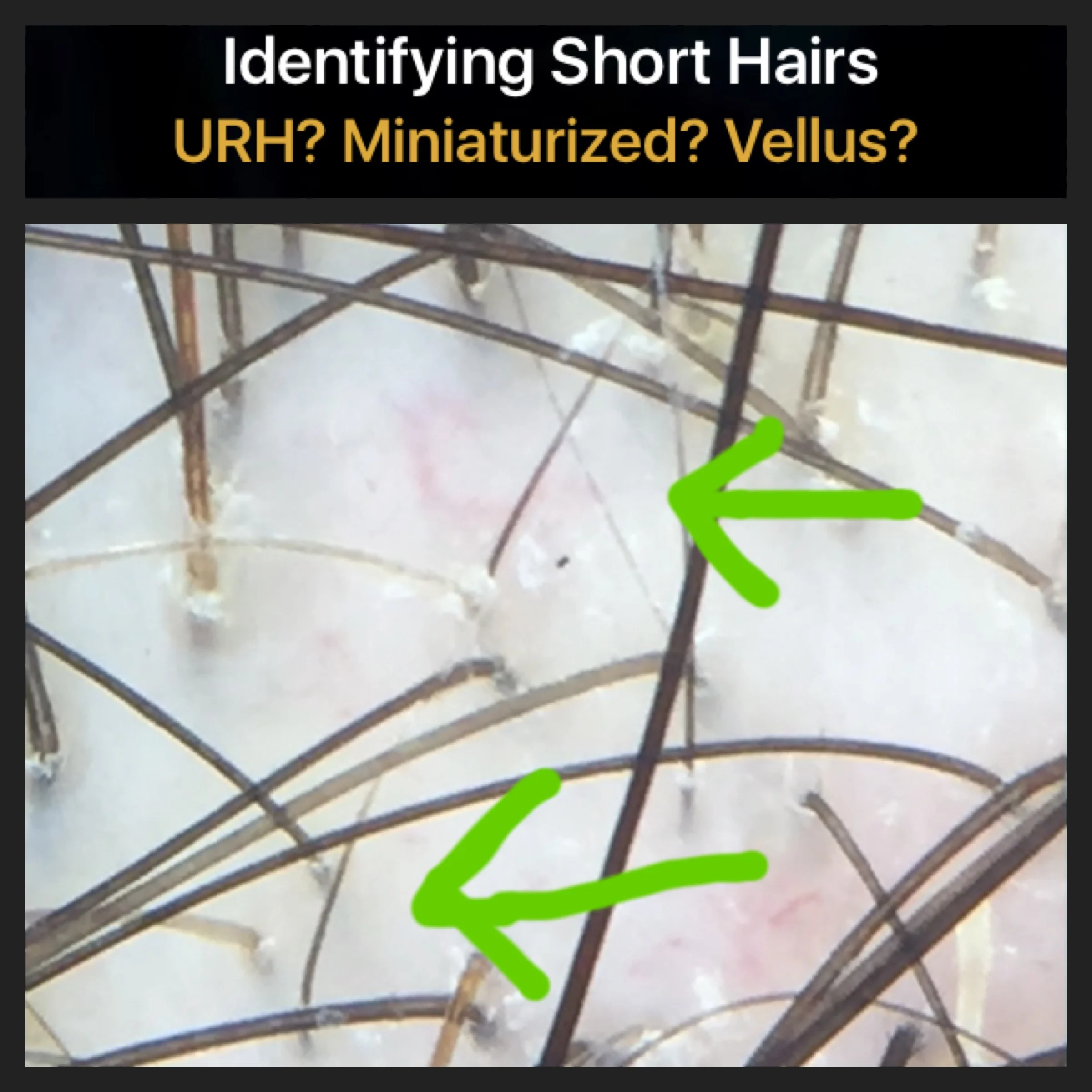
How can we tell apart the various causes of short hairs?
I'm frequently asked by patients and physicians how to determine the identify of a short 1 cm or so hair that is seen on the scalp. Looking at the scalp with dermoscopy, one often want to know "Is this a vellus hair I'm seeing or is it an upright regrowing hair as part of a telogen effluvium? ... or is it simply a normal regrowing hair ?"
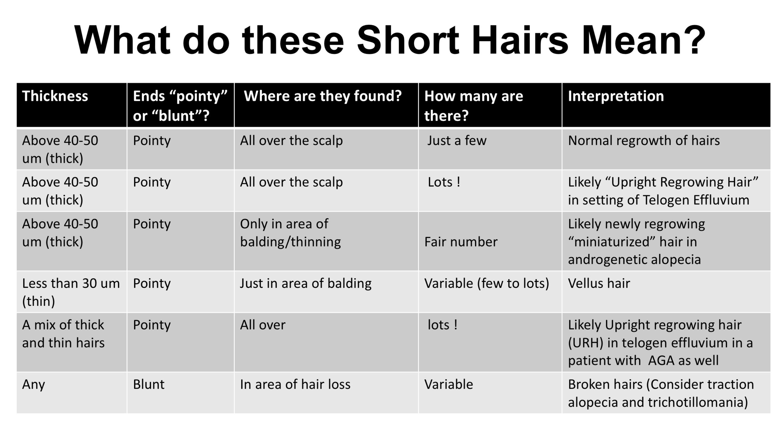
This chart below helps summarize the main things I think about when I see a short hair. The answer does not necessarily come immediately but rather it comes by asking 4 questions:
1) Is the hair reasonably thick (i.e. 40-50 um or more) or is it very thin (less than 30 um)?
2) Are the ends pointy or blunt?
3) Are these short hairs found all over the scalp or just one area?
4) Are there just a few of these short hairs or lots and lots of them?
By working through these 4 questions, I can generally determine the cause of the short hair I'm seeing on the scalp.
This article was written by Dr. Jeff Donovan, a Canadian and US board certified dermatologist specializing exclusively in hair loss.
DSC is a rare scarring alopecia. It often affects young men. A key feature is boggy tender nodules that develop in the scalp, some of which drain pus. "Sinus tracts" are another feature and this refers to the presence of small tunnels that interconnect under the scalp.
This photo shows the appearance of one such "sinus tract" after it has entered a healing phase. This area will be permanently scarred with some degree of permanent hair loss in this area.
Treatment for DSC includes isotretinoin, antibiotics, TNF inhibitors. Second line agents include zinc, dapsone, colchicine. Surgical excision and laser therapies are also considerations. Some forms are challenging to treat.
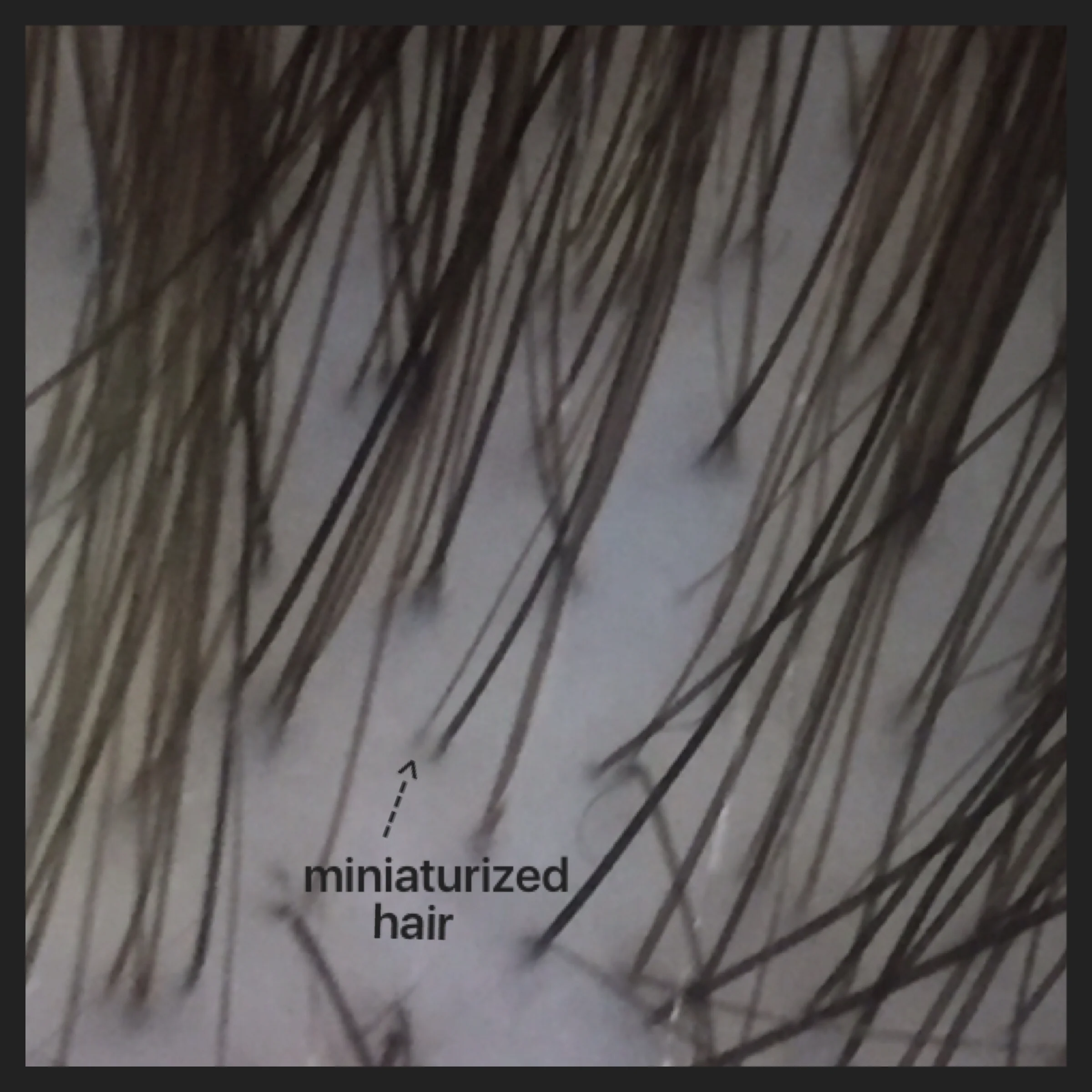
Androgenetic alopecia is common in men and women. By 50 years, about 50 % of men and 30 % of women have some evidence of androgenetic alopecia. The early features of AGA include hair shedding in some and hair loss in specific areas (temples and crown in men and central scalp in women).
When examined up close as in this photo, one can see "miniaturization" of hairs whereby some thicker hairs undergo a change to thinner hairs. Most hairs we have on our scalp as teenagers range in around 70-90 micrometers in diameter. During the process of androgenetic alopecia, the follicles become thinner and thinner and over time reduce slowly to 50 micrometers then 20 then 10 etc. Finally the fibers are so thin and short that they fail to reemerge from the scalp.
Not all hairs become thin and not all hairs thin at the same speed (rate). There is great variation in the thickness of hairs. We call this variation in hair shaft thickness "anisotrichosis." Two finding of miniaturization and anisotrichosis is a typical feature of androgenetic alopecia in both men and women.
This article was written by Dr. Jeff Donovan, a Canadian and US board certified dermatologist specializing exclusively in hair loss.
Androgenetic alopecia is often referred to as "thinning" and certainly the progessive miniaturization of hairs is a feature of AGA.
However, another important feature is the disruption of the normal architecture of how follicles are grouped. Instead of finding follicles in groups of 1, 2 or 3 hair units hairs are often seen all by themselves in more advanced stages of AGA.
The accompanying photos shows numerous single hairs in a patient with moderately advanced AGA.
This article was written by Dr. Jeff Donovan, a Canadian and US board certified dermatologist specializing exclusively in hair loss.
"Anisotrichosis" refers to a variation in the thickness of hair follicles that is commonly seen in androgenetic alopecia. In early stages of AGA, most hairs are relatively thick in caliber but some of course are thin. Over the course of AGA more and more follicles becomes thin and very thin which extends the spectrum of follicle sizes seen.
Reference
Sewell L et al Anisotrichosis: A novel term to describe pattern alopecia. J Am Acad Dermatol 2007; 56: 856.
This article was written by Dr. Jeff Donovan, a Canadian and US board certified dermatologist specializing exclusively in hair loss.
Tapered & Exclamation Hairs in AA indicate Activity
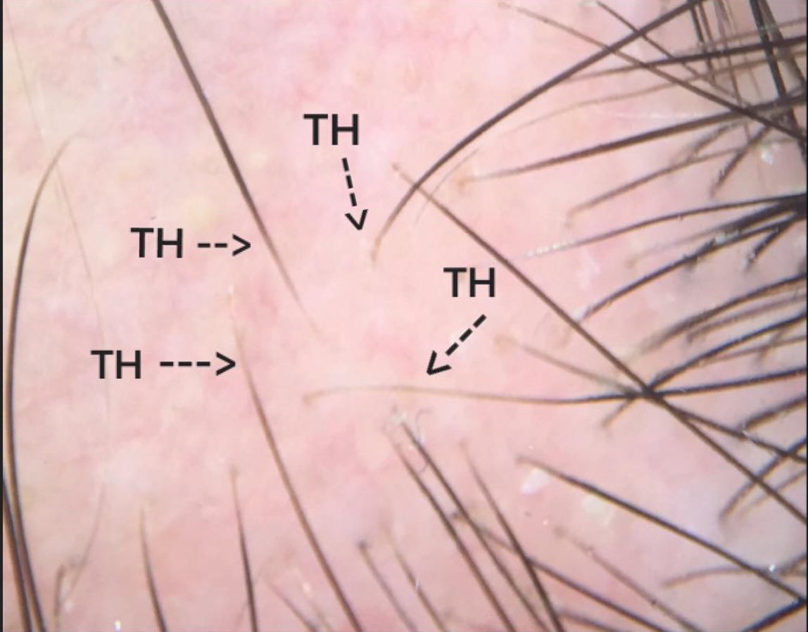
Tapered Hairs
Tapered hairs are frequently seen in patients with small circular patches of alopecia areata. In contrast to 4-5 mm exclamation mark hairs (see next post), tapered hairs are long and typically as long as neighboring hairs. As the hair enters into the skin it becomes much thinner. At the bottom of the tapered hair (deep under the skin) is inflammation.
Tapered are important findings in patients with patchy stage alopecia areata as they tell us that the condition is active and that anti-inflammatory type treatments (such as cortisone injections) are likely to help. The above photo shows several tapered hairs (TH).
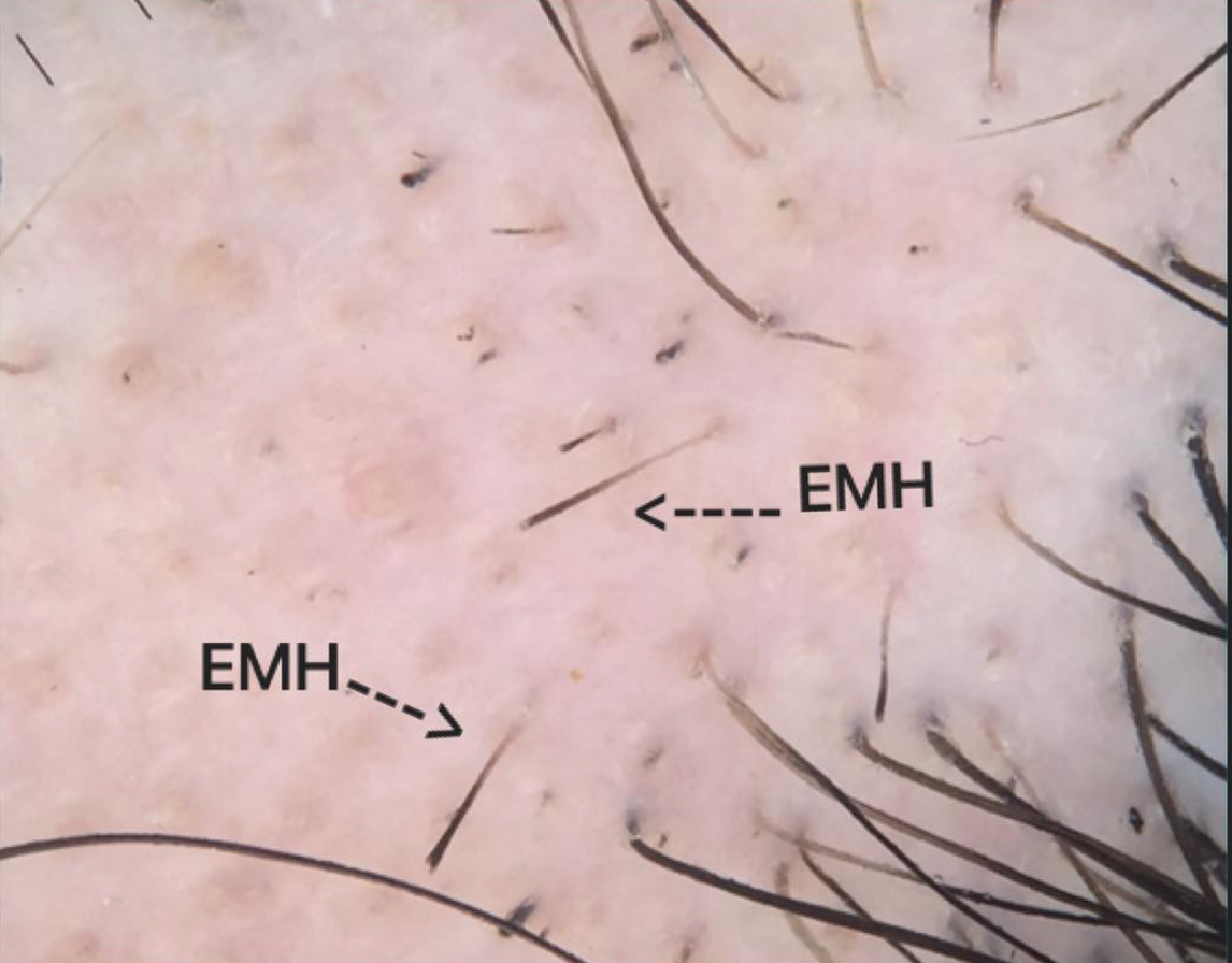
Exclamation Hairs
Exclamation mark hairs are frequently seen in patients with small circular patches of alopecia areata. These hairs a short 4-5 mm hairs and represent broken hairs. The top is thick and the end is often frayed. As the hair enters into the skin it becomes much thinner. At the bottom of the exclamation mark hair (deep under the skin) is inflammation. Exclamation mark hairs are important findings in patients with patchy stage alopecia areata as they tell us that the condition is active and that anti-inflammatory type treatments (such as cortisone injections) are likely to help. The photo shows several exclamation mark hairs (EMH).
This article was written by Dr. Jeff Donovan, a Canadian and US board certified dermatologist specializing exclusively in hair loss.
This week, we'll start a five day look at androgenetic alopecia in men (also called male pattern balding). The identification of so called "vellus" hairs is important in understanding male balding. Vellus hairs are tiny hairs less than 30 micrometers in diameter. They are present on the normal nonbalding scalp but only in low proportions. In male balding, the proportion of vellus hairs rises considerably as large "terminal" hairs are converted to tiny "vellus" hairs. In advanced balding, the vellus hairs disappear leaving a completely bald scalp in the affected areas.
This article was written by Dr. Jeff Donovan, a Canadian and US board certified dermatologist specializing exclusively in hair loss.
Vellus hairs are tiny, short non-pigmented hairs. They are fine hairs with a caliber less than 30 micrometers by definition. It is not common to find vellus hairs on the scalp in an individual without hair loss. On a normal scalp only about 1 of every 25 hairs are vellus hairs. Most hairs on the scalp are large pigmented terminal hairs. During the course of male and female androgenetic alopecia, vellus hairs become more prevalent and may even become the dominant hair type (outnumbering terminal hairs) in advanced balding cases.
Reference
Ko JH et al. Hair counts from normal scalp biopsy in Taiwan. Dermatol Surg. 2012
This article was written by Dr. Jeff Donovan, a Canadian and US board certified dermatologist specializing exclusively in hair loss.
Diagnosing hair loss in patients with darker skin types employs the same principles as for lighter skin types. However, a few features are unique including the the greater likelihood for scalp inflammation to create areas of hypopigmentation and hyperpigmemtation. In this picture of a male with androgenetic alopecia several findings are present. The redness and fine scale is consistent with seborrheic dermatitis. The patient also has folliculitis and a pustule can be seen in the upper portion of the picture. In the bottom left of the picture, areas of whitish hypopigmentation can be seen and in the bottom right areas of darker hyperpigmentation can be seen. The tiny white dots that are speckled al over the scalp represent the openings of the eccrine glands.
This article was written by Dr. Jeff Donovan, a Canadian and US board certified dermatologist specializing exclusively in hair loss.
Alopecia areata is an autoimmune condition that affects approximately 2 % of the worlds population. Many treatments are available. For patches of alopecia areata, the most effective treatment is steroid injections. When the scalp is examined a few weeks after a patient has received steroid injections a mixture of hair regrowth and hair loss is typically seen. In alopecia areata, hairs that are in the losing stage include broken hairs (arrow) and so called exclamation mark hairs (asterisk). Over time as hair growth dominates, the proportion of broken hairs and exclamation mark hairs will be reduced.
This article was written by Dr. Jeff Donovan, a Canadian and US board certified dermatologist specializing exclusively in hair loss.
Exclamation mark hairs are short hairs that are thick at the top and thinner as they enter the scalp. These hairs are known to occur in the autoimmune condition alopecia areata but also can occur in trichotillomania (shown this picture), poisoning situations (ie thallium) and have also been reported in dissecting cellulitis.
This article was written by Dr. Jeff Donovan, a Canadian and US board certified dermatologist specializing exclusively in hair loss.
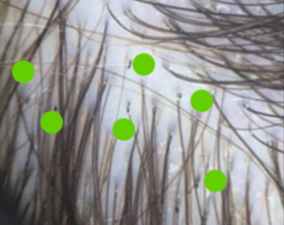
"Trichotillomania" refers to a form of hair loss where an individual pulls their own hair. It can sometimes be simply a habit - especially in very young children. In adolescents, the diagnosis of trichotillomania may signify underlying psychological illness including depression, anxiety, and eating disorders.
Trichotillomania, alopecia areata and tinea capitis are the three most common diagnoses in children followed by telogen effluvium and androgenetic alopecia. One must always at least consider this diagnosis as it is easy to miss. The presence of broken hairs, black dots, hairs of different length, and other trichoscopic features a v-sign, tulip hairs, and exclamation hairs are helpful in arriving at the diagnosis. The picture shows numerous scattered broken hairs (see green dots) in a young child with trichotillomania.
This article was written by Dr. Jeff Donovan, a Canadian and US board certified dermatologist specializing exclusively in hair loss.
Lichen planopilaris (LPP) is a type of scarring hair loss condition. Patients frequently present with scalp itching, and sometimes scalp burning and tenderness. Increased hair shedding is common in the early stages. Hair loss is generally permanent and treatment helps stop the disease or at least slow down progression.
Clinically, dermoscopy (trichoscopy) of LPP often shows perifollicular erythema and perifollicular scale (follicular keratosis).
These findings are not present in all forms of LPP. A less common presentation of LPP is shown in the photo. Patients have hair loss with scalp itching. However, by dermoscopy they have many single hair follicles growing in a base of redness. This is what I have termed the "sea of singles" (SOS) appearance to describe the numerous single hairs and absence of hair follicle units containing 2 and 3 hairs. This form of LPP is similar to Abbasi's subtype described in 2016 and fibrosing aloepcia in a pattern distribution described by Zinkernagel in 2000. The "SOS" trichoscopic appearance is important to remember and provides a clue that the patient may have a scarring alopecia.
Reference
Zinkernagel MS et al. Arch Dermatol 2000
Abassi A et al. Dermatol Surg. 2016.
This article was written by Dr. Jeff Donovan, a Canadian and US board certified dermatologist specializing exclusively in hair loss.
I frequently get asked if wearing hair extensions is okay. Often it is fine, but one needs to monitor over time if any hair damage is occurring. Individuals feeling pain, "pins and needles" should consider loosening the extensions or changing the method of application. Individuals showing clinical signs in the office of hair damage may also consider changing the method of application.
Consider the patient shown in this picture. She has been using extensions for some time now. She has a few broken hairs (labelled B) and several miniaturized hairs (labelled V for vellus) in any area that did not previously show miniaturization. These two signs are evidence of hair damage. A recommendation was made to change the extension in this case and treatment with a corticosteroid was given to reduce inflammation that is common in such cases of early traction alopecia.
This article was written by Dr. Jeff Donovan, a Canadian and US board certified dermatologist specializing exclusively in hair loss.
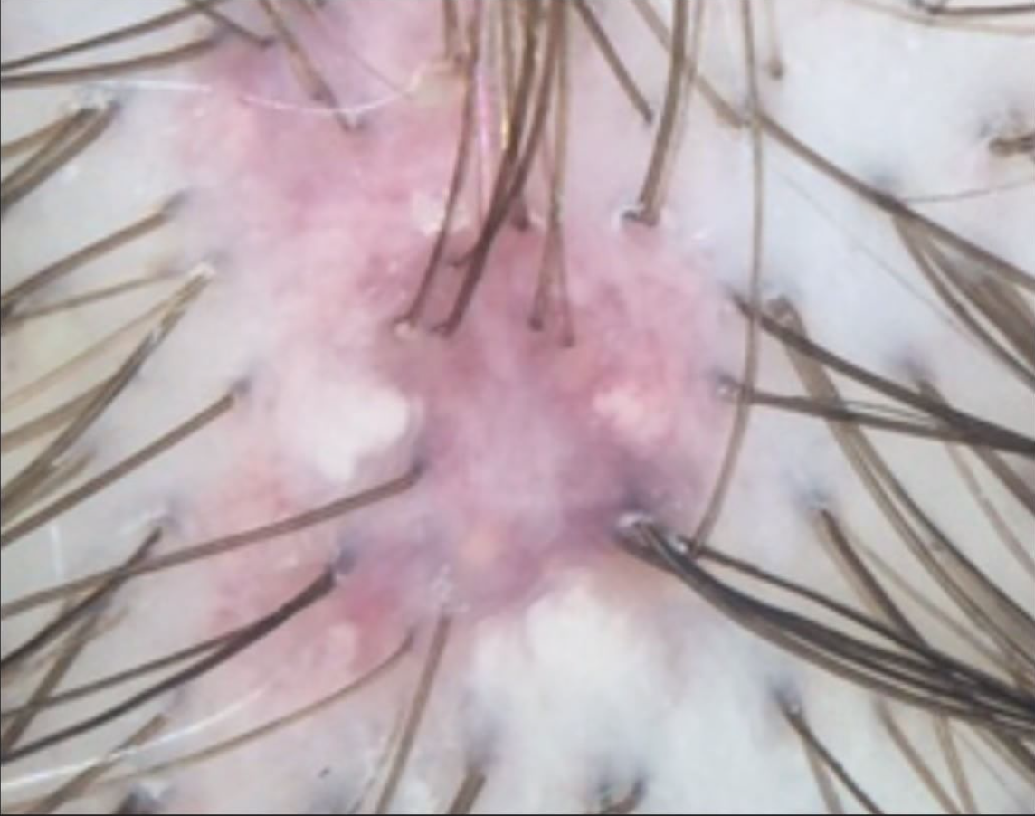
Dissecting Cellulitis (DSC), is a rare scarring hair loss condition that is characterized by deep inflammation and leads to the formation of draining sinus tracts (especially tunnels that allow pus and inflammation to escape). The diagnosis of DSC in advanced stages is easy as these openings (sinus tracts) can be seen all over the scalp. In early stages, up close exam and use of a dermatoscope can prove extremely helpful.
Early DSC is characterized on dermoscopy by large yellow dots, thin vellus hairs within the area, broken hairs and healing (covered) or open sinus tracts. This picture shows a sinus tract at an earlier stage than the picutre yesterday (panel 4 in our 5 day series). There is inflammation in the skin which gives a red color.
This article was written by Dr. Jeff Donovan, a Canadian and US board certified dermatologist specializing exclusively in hair loss.
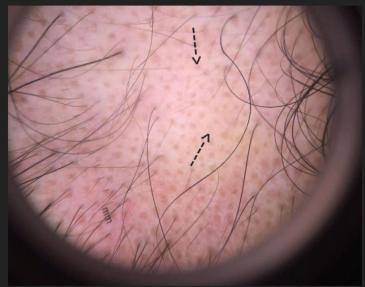
We will continue our week's theme of Dissecting cellulitis (DSC), a rare scarring hair loss condition. It is characterized by deep inflammation and leads to the formation of draining sinus tracts (especially tunnels that allow pus and inflammation to escape). The diagnosis of DSC in advanced stages is easy as these openings (sinus tracts) can be seen all over the scalp. In early stages, up close exam and use of a dermatoscope can prove to be extremely helpful.
As seen yesterday, early DSC is characterized on dermoscopy by large yellow dots, thin vellus hairs within the area, broken hairs and healing (covered) or open sinus tracts. This picture shows a healed sinus tract (arrow).
This article was written by Dr. Jeff Donovan, a Canadian and US board certified dermatologist specializing exclusively in hair loss.
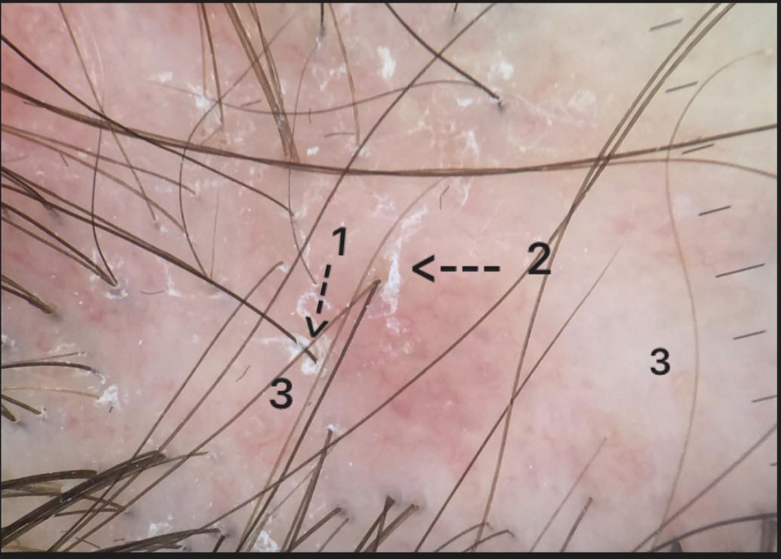
Dissecting cellulitis (DSC) is a rare scarring hair loss condition. It is characterized by deep inflammation and leads to the formation of draining sinus tracts (especially tunnels that allow pus and inflammation to escape - see number 1 and 4 in the picture). The diagnosis of DSC in advanced stages is easy as these openings (sinus tracts) can be seen all over the scalp. In the early stages an up close exam and use of a dermatoscope can prove extremely helpful.
Early DSC is characterized on dermoscopy by large yellow dots, thin vellus hairs within the area, broken hairs and healing (covered) or open sinus tracts. The early stages of the nodule can mimic alopecia areata (see top right, number 3 and 5). A swiss cheese like appearance is common as scarring progresses (number 2). Biopsies of DSC often show deep inflammation but in more advanced cases show inflammation higher up in the skin which can easily be mistaken for another scarring alopecia known as "folliculitis decalvans." Therefore, it is not uncommon for patients to be referred with a diagnosis of biopsy "proven" folliculitis decalvans only to need to explain to them after examining their scalp that what they actually have is DSC.
This article was written by Dr. Jeff Donovan, a Canadian and US board certified dermatologist specializing exclusively in hair loss.