QUESTION OF THE WEEK

Dr. Donovan's Articles

QUESTION OF HAIR BLOGS

Filtering by Category: Alopecia Areata


Is PRP an option for Alopecia Areata Treatment?

Is PRP an option for treating alopecia areata?


I’ve selected this question below for this week’s question of the week. It allows us to review some concepts in treatment for alopecia areata.


Question

I have had patches of alopecia areata for the past few months. I’d like to use something natural rather than medications and things. I am afraid to use steroid injections or topical steroids given their side effects. Is PRP an option for alopecia areata?


Answer

Thanks for your question. I would encourage you to take your time and ask lots of questions. Your views on the safety of steroid injections and topical steroids may or may not be accurate. In general, the first line treatments for ‘patchy’ alopecia areata are steroid injections, topical steroids and topical minoxidil. For small patches, the safety is quite good with these three treatments. Side effects are uncommon but of course mild temporary ones can occur.

Here are typical treatments to consider for alopecia areata in the patchy stage. You can see that PRP is a second line option rather than a first. That means that we don’t typically turn to it first as a treatment but it may be an option IF first line options don’t work or a patient does not wish to use the first line options.


Alopecia is unpredictable. I would need to know a lot more information about your story to advise what I would recommend in your specific situation. For some patients, treatment is short term and recurrences uncommon. For others treatment is lifelong. Many patients with alopecia areata regrow their hair spontaneously even without treatment within 1 year. Steroid injections is far more effective and efficient as are all the first line options. That’s why they are called first line options. Many clinics do a wonderful job scaring patients out of the first line options so that patients can start whatever the clinic offers. ( A clinic that offers laser therapy or PRP or sells other remedies may say “Oh you wouldn’t want to do injections that’s so dangerous!).

In summary, PRP is an option - yes! But it’s not usually the first step for most.

Share This
No Comments

Hair Loss: What's causing my hair loss?

What’s causing my patch of hair loss?


I’ve selected this question below for this week’s question of the week. It allows us to review some concepts regarding clinical and trichoscopic examination of acute hair loss.


Question

Hi Dr. Donovan.

I’m a 31 year old female. While giving birth I almost died, went in to septic shock and lost a massive amount of blood. 2.5 months later I lost a lot of hair, I had thinning all over but more obvious around my ears, sides of my head and on the nape of my neck. My dermatologist (and biopsy) said it was TE and gave me steroid shots and my hair is growing back normally with no thinning.

I developed seborrheic dermatitis, my head is a little itchy and I’m on ketoconazole shampoo. 5 months after birth, I had to have major surgery on my kidney, the surgery itself lasted 8 hours. On the 2nd or 3rd day at the hospital I noticed a painful bump on my parietal lobe. On the 19th day after surgery, I washed my hair and then noticed the hair loss on the same area as the swelling. Ive attached photos of the first time noticing it.

My dermatologist injected it with steroids and it isn’t growing back. It’s been 3.5 months since it fell out. There are tiny hairs in the area so my dermatologist is sure it isn’t scarred.

But there are also exclamation looking hairs so we are not sure. The area is reddish. I have no hair loss anywhere else. It hasn’t gotten bigger and I don’t have any patches elsewhere. The picture labeled February 7 is the day I noticed it.

Answer

Thanks for submitting this question. I hope that you are feeling well. At first overview, it certainly would appear that the diagnosis is alopecia areata with some overlapping findings of seborrheic dermatitis. In addition, it appears that you first had a telogen effluvium (of alopecia areata again) that settled after delivery. I still favour alopecia areata as the diagnosis in the current photos but there are a few things in your story and some of your images that cause me to pause and ask “is it possible there is anything else going on here?”

The reason I’ve chosen this question is that it allows us to review some of these features today.

There are several scalp conditions that can cause localized hair loss in this manner with possible ‘exclamation mark like hairs”. The top 4 include:

  1. alopecia areata ** most likely **

  2. dissecting cellulitis

  3. pseudocysts (alopecic and aseptic nodules of the scalp

  4. infections (syphilitic alopecia)

Other diagnoses to consider here but do not have good evidence include:

  1. tinea capitis

  2. pressure alopecia

  3. infiltrative conditions.

  4. trichotillomania

Let’s take a look first at some of the images supplied in this question and then we’ll go into these possibilities a little further and come to some conclusions.

Submitted Image 1

This image shows patchy hair loss. There are broken hairs. Inflammation is mild. The top diagnosis at this magnification would be alopecia areata. The differential diagnosis from this image might include trichotillomania, tinea capitis, and pressure induced alopecia. Alopecia areata would be the top diagnosis. Exclamation mark hairs are not clearly seen in this image but are seen in other images. There is no evidence for a scarring alopecia. Density may be reduced in the more anterior portion of the scalp (top of the photo) suggesting ongoing TE or another hair loss diagnosis happening in this area.

Submitted Image 2

This image shows a well cicumscribed area with minimal inflammation. There are vellus hairs and broken hairs. Some hairs have hair shaft changes suggestive of a pseudo-monilthrix like change (Pohl Pinkus constrictions). Exclamation mark hairs are not clearly seen in this image but are seen in other images. There is no good evidence for a scarring alopecia. Alopecia areata remains a favoured diagnosis.

Submitted Image 3

Numerous exclamation mark hairs are seen in this image. Elbow hairs are seen. Yellow dots are seen. There is an inflammatory type change with whitish scale. There is a mild pigmentation alteration which is somewhat non specific. The exclamation mark hairs make other diagnoses quite unlikely as exclamation mark hairs of this kind do not occur in pressure alopecia nor in inflammatory connective issues issues. The appearance of the scalp in this image differs quite a bit from the appearance seen in other images.

Submitted Image 4

This image shows several exclamation mark hairs with regrowing vellus hairs. There is mild yellow scale which may be in keeping with seborrheic dermatitis (of psoriasis) or an artefact of the photo itself. There is no evidence for a scarring alopecia.


Further Discussion

Thanks again for submitting this case. I favour alopecia areata but of course it’s nice to have more information and see the entire scalp eyebrows eyelashes, and nails. The most accurate way to diagnosis hair loss is to collect all the information about the patient and then examine all the scalp.

The features that support alopecia areata are the exclamation mark hairs, vellus hairs, regrowing hairs and localized nature of the hair loss.


What other conditions cause exclamation mark hairs?

As we think about this question, it’s helpful to think about all that conditions that cause exclamation mark hairs. After all, one of the key features in the submitted images are the exclamation mark hairs.

Exclamation mark hairs are seen in alopecia areata, trichotillomania, thallium poisoning, dissecting cellulitis. Syphilitic alopecia has been rarely described to have a type of tapered hair closely resembling a true exclamation mark hair. This is very rare.

Trichotillomania

There does not appear to be good evidence here for trichotillomania. The story does not fit. It’s one of the famous causes of exclamation mark like hairs. Certainly extensive broken hairs can be a feature but other findings like black dots, V hairs, coiled hairs, hook hairs, hair powder just don’t appear to be a feature of this patient’s hair loss. I don’t think we’re dealing with trichotillomania.

Thallium poisoning

Of course, thallium poisoning is rare.

Dissecting Cellulitis and Alopecic and Aseptic Nodules of the Scalp

The description of the ‘painful bump’ is a bit unusual in the submitted question. It’s not typical of alopecia areata. It may be a ‘red herring’ and unrelated to the case here or it may truly be a valuable clue. Also, it would be helpful to know more about what is meant by a painful bump and how big of a bump is the individual referring to.

As we think about painful bumps, we need to think about small bumps and things like a folliculitis. As we get into larger and larger bumps we need to consider more significant inflammatory conditions of the scalp. Dissecting cellulitis can cause a larger dome shaped bump when it occurs and is famous for mimicking alopecia. Another closely related entity is “alopecic and aspetic nodules of the scalp” (AANS). AANS can resemble alopecia areata. The condition was first called “pseudocyst” but the name AANS was proposed in 2009 by Abdennader and Reygagne when it became clear that not all of these lesions show a pseudocyst morphology under the microscope.

The back of the scalp is a common area for AANS. Often patients present with just a single painful bump. Some authors feel that AANS is closely related to dissecting cellulitis.

Exclamation mark hairs have not been described in AANS but have been described in dissecting cellulitis.

Dome shaped area of hair loss on the vertex scalp, consistent with a diagnosis of alopecic and aseptic nodules of the scalp. Image from Anna Isabel Lázaro-Simó et al. Alopecic and Aseptic Nodules of the Scalp with Trichoscopic and Ultrasonographic Findings. Indian J Dermatol. Sep-Oct 2017;62(5):515-518. Image used with creative commons license.


Trichoscopic image from alopecic and aseptic nodules of the scalp (AANS), also known as pseudocysts. There are black dots, yellow dots, vellus hairs and broken hairs. Image from Anna Isabel Lázaro-Simó et al. Alopecic and Aseptic Nodules of the Scalp with Trichoscopic and Ultrasonographic Findings. Indian J Dermatol. Sep-Oct 2017;62(5):515-518. Used with creative commons license.

Trichoscopy of alopecic and aseptic nodules of the scalp. Image from Khalil I. Al-Hamdi and Anwar Qais Saadoon. Alopecic and Aseptic Nodules of the Scalp with a Chronic Relapsing Course. Int J Trichology. 2019 Nov-Dec; 11(6): 244–246. Used with creative commons license.

There are three stages of appearance to AANS lesions as described by Al-Hamdi and colleagues. It’s important to understand this - especially in this case.

Stage 1: Firm nodule. A firm and often tender nodule is present and the nodule lasts 1-3 weeks. There may be lymphadenopathy. If the nodule is punctured, it does not usually express any fluid. But if it does, the fluid is sterile and does not grow bacteria

Stage 2: Fluctuant Nodule with Hair Loss. In this stage, the nodule becomes less tender and hair loss is clearly seen. If the lesion is punctured in this stage a yellow fluid is expressed. This stage lasts 3-7 days.

Stage 3: Patchy Hair Loss Stage. In this stage, the nodule is no longer present as it has flattened either spontaneously or by puncture. This stage may last 2-3 month at which point hair growth normally occurs. It’s common in this stage for the patchy hair loss to be given a diagnosis of alopecia areata.

Was the bump described by the patient in this case actually stage 1 or stage 2 of AANS? Clearly, more information is needed. I would say it’s still quite unlikely.

Infections (Syphilitic Alopecia)

In a case like the one presented, one must never lose sight of alopecia areata as the most likely diagnosis. Most things fit well and it could be simply that this patch is more refractory and needs further steroid injections. However, we do need to consider rare mimickers (like AANS) - and another rare mimicker of syphilitic alopecia.

I don’t think that there is much in this case that makes a diagnosis of syphilitic alopecia high on the list. However, it can be a cause of patchy hair loss - especially with tapered exclamation mark like hairs, scale and redness like we see in the photos sent in by the patient.

Atypical trichoscopy of a patient with syphilitic alopecia in a 32 year old male. Exclamation mark like hairs are seen. Tapered bended hairs, erythematous background, diffuse scaling and perifollicular hyperkeratosis were present. Testing revealed a positive Venereal Disease Research Laboratory (VDRL) at a titer of 1:256 and a reactive Treponema pallidum particle hemoagglutination assay. Image from Linda Tognetti et al. Syphilitic alopecia: uncommon trichoscopic findings. Dermatol Pract Concept. 2017 Jul; 7(3): 55–59.


Other Diagnoses to Consider


There are several other diagnoses to consider here but they do not really have good evidence. These include:

  1. tinea capitis

  2. pressure alopecia

  3. infiltrative conditions.


Tinea capitis

Tinea capitis can be a mimicker and the appearance can be altered by steroid injections. I don’t know the patient’s history well enough to know if there are predisposing factors that might make tinea capitis more likely. (In fact, I don’t have enough information in this patient’s history including information about the kidney surgery at 5 months post partum). It’s always possible that an inflammatory tinea developed and was flattened by steroid injections and persists in some manner. Of course, that’s unlikely and there do not really appear to be any trichoscopic features of tinea. There are no corkscrew hairs, comma hairs, bent hairs, i hairs, morse code hairs and no zig zag hairs. I don’t think this is tinea.


Pressure alopecia

In anyone with patchy hair loss after surgery, we need to consider pressure alopecia. It’s thought that hypoxia and altered blood flow predisposes to hair loss. Studies of patients with pressure alopecia have not suggested that exclamation mark hairs are part of the pressure alopecia diagnosis. Therefore, a diagnosis of pressure alopecia would not be likely in this case. According to Neema et al, trichoscopic findings of pressure alopecia include comedone- like black dots, black dots and area of scarring. In 2016, Francine Papaiordanou et al proposed that black dots, broken and dystrophic hairs were main features of pressure alopecia. In 2020, Tortelly et al proposed that black dots and vellus hairs were key features.

It’s not impossible that pressure from surgery facilitated the development of alopecia areata. in fact, R L Zuehlke et al in 1981 suggested that pressure may be a risk for alopecia areata too. So it’s going to be important to review if this area on the scalp shown in the photos had pressure during surgery. I don’t think it’s likely that what we’re seeing is related to pressure alopecia.

Trichoscopy of pressure alopecia showing black dots, broken and dystrophic hairs. In Image from Papaiordanou F et al. Trichoscopy of Noncicatricial Pressure-induced Alopecia Resembling Alopecia Areata. Int J Trichology. Apr-Jun 2016;8(2):89-90. Used with creative commons license.

Infiltrative conditions.

The term “infiltrative conditions” refers to a massively long list of cells that can enter into an area of the scalp (infiltrate) and cause localized hair loss. A variety of inflammatory and neoplastic cells can trigger patchy hair loss so one needs to always keep these in mind. They don’t usually cause exclamation mark hairs. Alopecia neoplastica refers to hair loss from metastatic cancer and can mimic alopecia areata in some cases.

This would not be expected in this case but this is added to the list and discussed here for completeness as we review patchy hair loss and considerations in the setting of refractory patchy hair loss. It does seem that hair is growing back in your case which makes infiltrative type causes quite unlikely. I don’t have a good sense of the time course of the photos you’ve submitted so that too would need to be carefully reviewed.

Alopecia neoplastica due to breast cancer. Image from Efthymia Skafida et al. Secondary Alopecia Neoplastica Mimicking Alopecia Areata following Breast Cancer.Case Rep Oncol. 2020 Jun 11;13(2):627-632. Image used with creative commons license.

Alopecia neoplastica due to breast cancer. Image from Efthymia Skafida et al. Secondary Alopecia Neoplastica Mimicking Alopecia Areata following Breast Cancer.Case Rep Oncol. 2020 Jun 11;13(2):627-632. Image used with creative commons license.

Conclusion and Summary

Thank you for this question. There are a few important points here in this case as we conclude. The first is that a full history and full examination are needed. This area is the area that you have photographed but one needs to always examine the entire scalp. A full history is needed of health and medical conditions over the past 31 years. The reason for the kidney surgery is completely unknown and would need to be included in the full story. I need to know everything about patients to confirm diagnoses with absolute certainty.

That said, the photos and clinical case still fit with undertreated alopecia areata as a top diagnosis. The exclamation mark hairs here and vellus hairs and regrowing hairs support this diagnosis. There are mimickers of course and these need to be considered.

For my own patients with similar stories I would first take a full history and do a full examination of the scalp, eyebrows, eyelashes and body hair. Then I might inject with 2.5 mg per mL triamcinolone acetonide (steroid) with 2 to 3 mL injected into the area. I would not be too concerned if hair does not immediately grow back as it might take 2-3 sessions one month apart. I would not do more frequent than this. There is an option to add topical minoxidil to the plan but you’d want to review side effects with your supervising doctor.

If the area was slow to regrow I might add periodic use of clobetasol and minoxidil at home while doing these steroid injections.

Your photos would suggest you are already growing back significant hair.

If the area did not respond, I might do a biopsy. I don’t see this as necessary right now. The area needs to be properly treated and then if it does not respond to proper treatment, one can move on to step 2.

I don’t see AANS as a likely diagnosis (alopecic and aseptic nodules of the scalp), or pressure alopecia as being likely. We don’t see exclamation mark hairs in most cases of pressure alopecia. It would be helpful to know just how lumpy or raised this area was when you noticed it as this might lead one to at least consider AANS. The reality is that even if it is AANS and it’s some unusual pattern of exclamation mark hairs, it should respond to steroid injections at this point. I don’t think it’s likely we’re dealing with AANS.

Finally, anyone with this story should have blood tests for CBC, TSH, ferritin, vitamin D, creatinine. An antidandruff shampoo should continue to be used. Ketoconazole is reasonable. As mentioned, a biopsy will be needed if the area is not responding to appropriate doses of steroid injections (+/- minoxidil or clobetasol).

A full scalp examination is needed to determine if there are any other issues too. The area to the front may be thinner than prior years and that needs to be evaluated. It could be part of a resolving telogen effluvium or another diagnosis. A full examination of the scalp is needed in this case (as well as full examination of eyebrows, eyelashes and body hair as mentioned)

Thanks again

REFERENCE

Anna Isabel Lázaro-Simó et al. Alopecic and Aseptic Nodules of the Scalp with Trichoscopic and Ultrasonographic Findings. Indian J Dermatol. Sep-Oct 2017;62(5):515-518.

Khalil I. Al-Hamdi and Anwar Qais Saadoon. Alopecic and Aseptic Nodules of the Scalp with a Chronic Relapsing Course. Int J Trichology. 2019 Nov-Dec; 11(6): 244–246.

Abdennader S, Reygagne P. Alopecic and aseptic nodules of the scalp. Dermatology. 2009;218:86.

Linda Tognetti et al. Syphilitic alopecia: uncommon trichoscopic findings. Dermatol Pract Concept. 2017 Jul; 7(3): 55–59.

Neema S et al. Trichoscopy of Pressure-Induced Alopecia and Alopecia Areata: A Comparative Study. Int J Trichology. Jan-Feb 2022;14(1):17-20.

Papaiordanou F et al. Trichoscopy of Noncicatricial Pressure-induced Alopecia Resembling Alopecia Areata. Int J Trichology. Apr-Jun 2016;8(2):89-90.

Tortelly et al,Pressure-Induced Alopecia: Presence of Thin Hairs as a Trichoscopic Clue for the Diagnosis Skin Appendage Disord. 2020 Jan; 6(1): 48–51.

R L Zuehlke et al. Pressure-potential alopecia areata. Am J Orthod . 1981 Apr;79(4):437-8.

Efthymia Skafida et al. Secondary Alopecia Neoplastica Mimicking Alopecia Areata following Breast Cancer.Case Rep Oncol. 2020 Jun 11;13(2):627-632.


Share This
No Comments

Can I reduce the dose of minoxidil for treating my AGA and if so, how?

Minoxidil (Rogaine) for the treatment of Androgenetic Hair loss: How soon after starting can I go down on the dose?

I’ve selected this question below for this week’s question of the week. It allows us to the review some key concepts in the treatment of androgenetic alopecia - particularly the need for ongoing lifelong treatment.


Question

I have female pattern hair loss and I’ve been on minoxidil 1/2 cap daily for about 1 year now. It seems to be helping. I was advised this week by my provider that it’s okay to go down to 3 or 4 times weekly. What protocol do you recommend for reducing the dose and how do you taper safely so as to not lose hair?


Answer

Thanks for the question. The answer is simple: If you have androgenetic alopecia and Rogaine is helping you, then this medication probably can’t be tapered to any significant degree without you losing some hair.

If Rogaine has not been helping you all these months and you’ve actually been wasting your time in the last 12 months applying it - you can stop it without any immediate negative effects. The hair won’t mind at all that Rogaine was stopped because it wasn’t helping in the first place.

However, if Rogaine was helping (which I imagine for you it probably must be), it can NOT be tapered without losing hair. I don’t know how this concept has started permeating in this world - I’ve heard it myself. But Rogaine can’t be stopped if it was helping. You can’t go from 7 days a week to 5 without some negative effect on the hair. You can’t go 7 days to 4. You can’t go twice daily to once daily. Sure you might be able to go 7 to 6 but there’s a fine line for what its acceptable.

If all a person is able to do is apply Rogaine 5 or 6 days per week, then I say try to apply it five or 6 days per week. That’s fine. It still has a chance of helping even if it’s not perfectly daily. . But to start at a higher dose and then reduce the dose at a future time just doesn’t work well for most people.

Reducing the dose of Rogaine usually leads to hair loss (if it was helping). Consider the female patient who starts using Rogaine 5% twice daily because she really wants to try to stop her hair loss, or the patient who even uses a full cap instead of the recommended 1/2 cap. Then, imagine that the patient winds up in the clinic of a specialist 12 months later and the specialist says confidently to her“Oh, Rogaine for women only needs to be used once daily at 1/2 cap - so you can reduce your dose”

Guess what happens when the patient goes from a full cap twice daily to 1/2 cap daily?

She loses hair! !!!!!!

Hair is not pleased with a four-fold reduction in the dose.


Summary and Conclusion

All the treatments for androgenetic alopecia are lifelong. If a patient is going to use Rogain to treat their AGA, then they should plan to start it with the intention to use it lifelong. If a patient is going to use anti androgens to treat their AGA, then they should plan to start it with the intention to use it lifelong. If a patient is going to use low level laser therapy to treat their AGA, then they should plan to start it with the intention to use it lifelong. If a patient is going to use PRP therapy to treat their AGA, then they should plan to start it with the intention to use it lifelong.

We would to think that it’s possible to reduce the dose of Rogaine after some time. It sure sounds like a nice plan …. and a convenient one too! It’s just that it doesn’t really work like this and reducing the dose carries a high risk of hair loss.

Now for all the people who are using Rogaine for other types of hair loss besides androgenetic alopecia - the rules are a bit different. If one uses Rogaine for alopecia areata, one can stop Rogaine once the hair grows back. Sometimes (but not always) that’s true for other types of hair loss as well. But for androgenetic alopecia the rules are pretty straight forward. If a certain dose or amount of Rogaine has helped, then that dose or amount needs to bee continued exactly the same way to maintain results. A reduction in dose will likely lead to hair loss.

The following chart is helpful to differentiate the use of Rogaine in these conditions


Rogaine in Hair Loss


Share This
No Comments

What is More Accurate for Diagnosing Early Stages of Hair Loss : A Scalp Biopsy or Clinical (Trichoscopic) Examination?

Biopsy or Up Close (Trichoscopic) Examination: What’s better for diagnosing the early stages of hair loss?

I’ve selected this question below for this week’s question of the week. It allows us to the review some key concepts in diagnosing hair loss via clinical scalp examination and through a biopsy.

trichoscopy vs bx


QUESTION


What is more accurate - a scalp biopsy or a scalp exam with a dermatoscope? My biopsy results said telogen effluvium and androgenic alopecia with the diagnosis of androgenetic alopecia being favored.

As for me, I’m a 30 year old female. My scalp is itchy, likely from seborrheic dermatitis which was diagnosed by a dermatologist. I’ve suffered from alopecia areata in the past (1 small bald patch at a time and treated with cortisone injections) . I have a lot of food and environmental allergies that I’m treating naturally. My hair started shedding excessively at the end of February 2021 after a very traumatic event in December 2020. I’m not on any prescription medications but I do take supplements (iron, vitamin D and C, coQ10, quercetin, probiotic, l-lysine, caprylic acid, and a multivitamin for hair). The shedding has been diffuse and I have lost density. My family members insist that no one would know I’m having issues with my hair. In the past few weeks I have had days with minimal shedding. I have been treating the seborrheic dermatitis with medicated shampoos. I have been treating the hair loss naturally, through dietary changes, lowering stress levels with meditation, etc; I have not used any medications.

The dermatologist that performed the biopsy said it’s “age related” (I’m a 30 year old female) and therefore not even considered an early stage AGA. The second dermatologist I saw (for a second opinion) did a scalp exam with a dermatoscope and said there was “maybe one” miniaturized follicle at the biopsy site on my crown. Throughout the rest of the top of my scalp she said about 1 in 100 follicles are miniaturized. She gave me a diagnosis of just telogen effluvium. So far all of my test results (iron, ferritin, vitamin D, vitamin B12, thyroid panel, and hormone panel) have been normal. I’m very confused and not sure if and what treatment would be best for me. Thank you!


ANSWER

Thank you for the question. In order for me to advise you on what treatment would be best for you, we need a diagnosis.

So what is your diagnosis then?

Well, in order for me to give you a diagnosis, I would need to know a bit more about your story from birth until today, and see your scalp up close myself and review your blood tests. Those are the three key steps in order to make a diagnosis for anyone!. Because I don’t have any of these pieces of information in your case, I can’t actually say what your diagnosis is.

However, there are still some very important points to be aware of and that’s why I’ve selected your question for this week’s question. It’s such a good one with so many things for us to review.

So let’s get to it.

You have what I call early hair loss. You yourself know there is a change, but your friends and family think everything is just fine. Even one of the dermatologists thinks it’s simply a telogen effluvium. This is early hair loss.

As you have correctly outlined, this can often be due to androgenetic alopecia or telogen effluvium …. or both.

As I review all your information about what your biopsy showed and what your doctors actually said, I need to know how reliable each of these three pieces of information are. If dermatologist 2 is a world expert in hair loss and doesn’t think its AGA - does this carry more “influence” as I think about your case than if dermatologist 1 thinks it’s AGA but really has only seen a handful of hair loss patients in his or her career?

Yes it most certainly does.

Your question is really all about the reliability of these three pieces of information - the 2 doctors and the 1 biopsy.

And what if the biopsy was taken from an area on the scalp that is really not so useful for making a diagnosis (like the temples) - am I to trust this result? Well, no.

So, let’s take a look at these four scenarios below in order for us to better understand when a biopsy is better than a clinician’s interpretation and when a clinician’s interpretation is to be trusted more than a biopsy report.

In general, the very early stages of hair loss can be challenging to decipher from one another. The more experience and expertise the clinician has in treating hair loss … the more reliable his or her view will be on the cause of hair loss. The less experience the clinician has, the less reliable his or her view is and the more a biopsy result is to be trusted. However, biopsies are not all the same. The only biopsy result that I really trust is one taken from the correct area of the scalp and interpreted properly by expert dermatopatholgist.


Let’s take a look at the following chart and then we’ll break it down some more.

biopsy vs clinical

SCENARIO 1. The practitioner evaluating the scalp is a VERY EXPERIENCED hair loss expert and a 4 mm punch biopsy was taken from a correct area of the scalp and interpretations were done by a VERY EXPERIENCED dermatopathologist.


In this case, both the dermatologist’s opinion and the dermatopathologist’s opinion are fairly reliable. In fact, in most cases, they are fairly equivalent. A highly experienced clinician can examine all areas of the scalp and can determine just how much variation in the caliber of hair follicles (ie “miniaturization”) is seen in the various regions including the front, middle, top and back. If the clinician appreciates that density is slightly different in one area compared to another it’s like their is some androgenetic alopecia going on - especially if the thinner area show a greater degree of miniaturization.

A clinician can also evaluate density in the frontal area and compare this to the back. If there is a subtle increase in “part width” in the frontal and mid scalp compared to the back, this gives a suggestion there could be some androgenetic alopecia going on.

aga
te


So an astute clinician can look at the scalp, look at the part width, look a the density in various regions of the scalp and look at what the trichsocopy shows and come up with a conclusion.

Clinical examinations of the early stages of hair loss are tricky to interpret. It takes expertise to appreciate subtle changes in hair follicle caliber. It’s not something that is learned overnight. It’s not a result that pops up on any sort of screen when one places a dermatoscope one the scalp. Of course, it one’s dermatoscope its connected to a computer and the caliber of follicles can actually be measured in various areas, this really increases the reliability of the interpretation for less experienced practitioners.

But if a practitioner is less experienced with hair and scalp issues, simply placing a dermatoscope on the scalp and concluding “I don’t see any miniaturization” does not give me a great amount of confidence in diagnosing early hair loss issues.

What about a biopsy? Biopsies in early hair loss can be wonderful! A biopsy taken from the area of androgenetic alopecia can also show a DECREASING terminal to vellus ratio from a normal low 7:1 or 8:1 down to 4:1 or less. In true telogen effluvium, the terminal to vellus ratio stays well above 6 or 7 to 1. An experienced dermatopathologist who interprets a biopsy from a patient with early hair loss and says ‘the T:V ratio is 3.5:1 and sebaceous glands appear enlarged and there is no real shift in catagen to telogen ratios and there is no peribular inflammation” is telling me this is likely androgenetic alopecia. I trust that report if I know the dermatopathologist is experienced.

To summarize, a very experienced practitioner can often make a diagnosis of androgenetic alopecia fairly reliably even without a scalp biopsy. However, if a scalp biopsy is done, the results should be similar trusted as the findings of a very experienced practitioner provided the biopsy is interpreted by an expert pathologist.


SCENARIO 2. The practitioner evaluating the scalp is an INEXPERIENCED practitioner and a 4 mm punch biopsy was taken from a correct area of the scalp and interpretations were done by a VERY EXPERIENCED dermatopathologist.


In this case, the biopsy report is MORE reliable than the view of the clinician. We need to remember here that early hair loss stages are really difficult to diagnose! There is no harm in saying that and I’ll be the first to point that out.

It can take anywhere from 6 months to 5 years from the time some types of hair loss first start before a patient themselves figure out that something is changing on their scalp. So, the early stages of hair loss are tricky to spot. The early stages of hair loss can sometimes look normal. The less experience the practitioner has …. the more the scalp will look normal to them ! That’s just a fact. Any practitioner who takes a quick 5-10 second glance at the scalp and says to their patient ‘your scalp looks fine to me… don't worry” is by definition an inexperienced practitioner. This is pretty much a rule. The early stages of hair loss are hard to spot sometimes and take a bit of poking and prodding in the scalp to see what all the 100,000 hairs are doing and a bit of sleuthing to gather information from the patient as to exactly what’s been happening over the past months.

If a very experienced clinician says ‘This scalp is normal” then it’s pretty unlikely there is any androgenetic alopecia. Not 100% guarantee of course….. but pretty unlikely. If an inexperienced clinician says ‘This scalp is normal” then it carries less meaning. Of course, it could be normal, but I’m a bit more skeptical. I am sent referrals every day of the year that say “ Normal scalp exam. Patient thinks they have hair loss. Please see in consultation.”

What do many of these patients end up having as a diagnosis ? Well, many have androgenetic alopecia !

Suppose I’m meeting up with a friend for dinner and I tell my friend that I have been getting some pretty bad headaches lately. If my friend tells me everything sounds fine, do I believe it? Well, if my friend is a neurologist I’m a bit more likely to trust this information than if my friend is an accountant. The quality of the information makes a difference.

So to summarize, if a clinician is less experienced with diagnosing hair loss but takes a biopsy from a correct area of the scalp (ie where the hair loss is most affected) and the biopsy lands in the hands of an expert dermatopatholgist …. then I would usually trust the dermatopathoglist report over the clinician’s interpretation of what’s causing the early hair loss.

So what’s a good biopsy in your case? Well, in your case this likely means that biopsy was taken from somewhere in the yellow area shown below. I would prefer if the biopsy was 4 mm in size. I would also like if the biopsy was processed with horizontal sections as personally that increases my confidence in these early stages of hair loss. It’s only with horizontal sections that the pathologist can give a measurement of the terminal to vellus ratio. This can’t be done with vertical sections. If your T:V ratio is less than 4:1, we might begin to think there is some androgenetic alopecia present as a diagnosis.


biopsy

SCENARIO 3. The practitioner evaluating the scalp is an EXPERIENCED hair loss expert and a suboptimal biopsy was taken from an incorrect area of the scalp and/or interpretations were done by an INEXPERIENCED pathologist.

This would be an unusual situation whereby an experienced clinician took a biopsy from a wrong spot. But this situation could be an experienced clinician is trying to decide what diagnosis a patient has and the patient brings in a biopsy report they had at another clinic showing a certain result.


In this case, I trust the result from the clinician any day over the biopsy report. Every day, I see biopsy reports that are taken form the back of the scalp or the sides of the scalp or the temples. These are not the ideal areas to be taking biopsies from if we want determine whether or not the patient has androgenetic alopecia!!!

Sometimes, the doctor does not want to cause a scar…. and so takes it from the sides of the scalp so as to hide any scar. Sometimes, a patient asks the doctor to take it from the temples because that’s where they are most worried and where they see the changes every day of their life when they look in the mirror. These are not where we should be taking biopsies to confidently assess androgenetic alopecia !

If a biopsy returns showing “no evidence of androgenetic alopecia” but was taken from the sides fo the scalp does it mean the patient does not have androgenetic alopecia? No! Not at all,. That biopsy was not helping in making the proper diagnosis.

If a biopsy returns showing “no evidence of androgenetic alopecia” but was taken from the main area of hair loss in the central scalp zone, does it mean the patient does not have androgenetic alopecia? Probably that is the correct interpretation.

SCENARIO 4. The practitioner evaluating the scalp is an INEXPERIENCED practitioner and a suboptimal biopsy was taken from an incorrect area of the scalp and interpretations were done by an INEXPERIENCED pathologist.


A particularly challenging situation is when a less experienced practitioner is not sure what the diagnosis is but proceed to take the biopsy from an area of the scalp which is less than ideal. Typically this is a well meaning practitioner who wishes to take the biopsy from an area that will best be hidden in the future should the area form a small scar. So the biopsy is taken from the sides of back of thee scalp and typically returns showing no evidence of androgenetic alopecia. The only thing that can be interpreted in this situation is that the patient does not have androgenetic alopecia down the sides of their scalp. However, we can’t conclude anything at all about what might be happening in the middle of the scalp - the area where the patient is most concerned about the hair!

bx not to take

I often use the following analogy when I explain the concept to doctors that I teach.

Suppose you have a mold of some kind in your home. The house smells like mold! Terrible, right?

And so you call a mold specialist for help. Unfortunately, all the mold specialists in town are away at a convention so you decide to call a plumber. After all, mold grows in water and damp conditions, and you figure that a plumber knows a lot about water and damp conditions in homes.

The plumber answers the call and says he or she knows how to take mold samples because they learned how to do so in a course they took.

Voila!

You are happy with the answer and invite the plumber to your home to get some help.

The plumber finds a bit of water in the basement and takes some mold samples. It all comes back negative.

You are all relieved there is no mold!

The problem is that the smell continues.

When the mold specialist in town returns from the convention, you invite him or her now into your home. Within a few minutes the source of the mold is located in the attic of the house. Their is a leak in the roof and this is causing the roof to leak and the attic to grow mold !!!! Samples are taken and the mold is finally proven.

Did it matter where the samples were taken? You bet it did!

An experienced specialist is more likely to know where to take the sample .

Conclusion

Your question is really a great one. Thanks again for submitting. It’s difficult, if not impossible for patients to know if their biopsy was taken from the correct spot or whether their clinician really has a lot of experience or not. It’s tough to navigate the medical world sometimes.

The short answer to your question , however, is that a very experienced clinician can often diagnose hair loss with a similar degree of accuracy to a biopsy interpreted by an expert dermatopathlogist. If the skills of the clinician change or the skills of the dermatopathologist now change, this no longer holds true and you’ll need to see the chart about as to which is better.

It is quite likely with your story that at least one of your diagnoses was telogen effluvium that was triggered by the stress of December 2020. With your story, I think it’s really important that someone make sure that your seborrheic dermatitis is under good control and someone keep an eye on the possibility that a diffuse alopecia areata is not part of the reason for your shedding. I think that would be unlikely given that shedding has settled now and that the biopsy did not capture this.

With this one biopsy that you do have I can’t exclude that there is not some degree of androgenetic alopecia present. There certainly is a possibility with this information you’ve given. oOf course, I would need to see the scalp or a photo of your scalp myself to know for sure one way or another.

Please keep taking photos of your scalp to show your doctors. If you feel that your hair returns to full by September 2021 and you are really pleased with the way your hair looks and feels at that time, then it’s pretty unlikely there is any AGA. However, if your hair does not return to full by September, I would encourage you to further explore ways to confirm this diagnosis with certainty one way or another so that you might get connected with the correct treatment in the event you do have androgenic alopecia.

Thank you again for your question.

Share This
No Comments

Drug Induced Alopecia Areata: Can Drugs Cause Alopecia Areata?

Can medications cause alopecia areata ?

I’ve selected this question below for this week’s question of the week. It allows us to discuss the concept of drug induced alopecia areata.

Drug induced Alopecia Areata: 14 drugs to date have been implicated in the development of alopecia areata. The association is still considered very rare even in those who take one of these medications.

Drug induced Alopecia Areata: 14 drugs to date have been implicated in the development of alopecia areata. The association is still considered very rare even in those who take one of these medications.


QUESTION

Hi Dr Donovan,

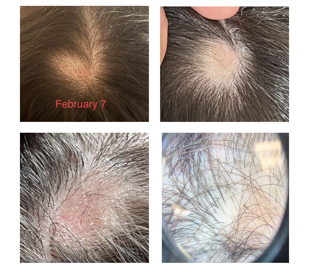
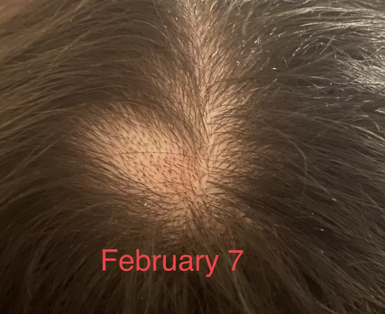
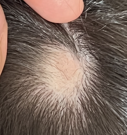
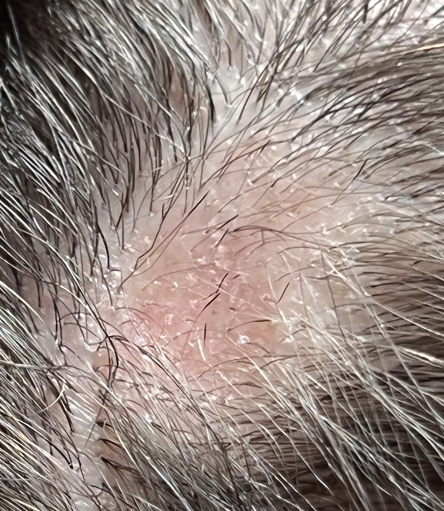
I am a 29 year old female and developed alopecia areata in late January 2021. I first developed a patch at the top of the scalp on Jan 23rd and then a second at the back of the scalp on Feb 8. They have fortunately both now grown back with steroid injections using Kenalog.

I am trying to understand this condition and what might have caused it. There is nobody in my family with this condition so I am perplexed. I am quite healthy, eat well and exercise 5 days per week. I have mild asthma and take puffers only very rarely. I developed a urinary tract infection in December and was treated with the antibiotic trimethoprim/sulfamethoxazole (Septra). I’ve been wondering if this could be the cause.

I have been researching if medications can cause alopecia areata but I’m not sure that medications are really all that commonly implicated.

Can you tell me if some medications are strongly connected with developing alopecia areata?

ANSWER

Thanks for submitting your question.

The short answer is no. Mediations are not really all that strongly connected with developing alopecia areata. That’s not to say that some medications have not been linked - it’s just the chances are very low and the vast majority of medications are not strongly associated with development of alopecia areata.

To date, there have only been about 25 patients reported in the medical journals with suspected drug induced alopecia areata. Could it be that there are more? Absolutely. But one thing is likely that drugs are probably not all that commonly connected with alopecia areata.

A nice research paper published in the journal called Clinical and Experimental Dermatology looked at these 25 patients so far who were felt to have ‘drug induced alopecia areata.’ The average age of patients with this condition was 42. Alopecia areata developed about 3-4 months after the drug was started.


Features of Drug Induced Alopecia Areata

Here are some of the key features of “drug induced alopecia areata”:

 1) There can sometimes be an itchy widespread skin rash on the body plus an itchy scalp eruption that precedes the alopecia by 2-3 weeks. Unlike many drug rashes, the patient here has no fever or joint pains. This itchy rash might not always be present.

2) The hair grows back very rapidly when the drug is stopped and further recurrences of patches of alopecia areata are unlikely to occur provided the drug is avoided.

3) Patches of alopecia areata occur again within weeks when the drug is restarted.

4) Blood tests for autoimmune tests, organ function, etc are normal.


What drugs have been implicated in drug induced alopecia areata so far? 

So far, most cases of drug induced alopecia areata that have been published in the medical journals have been with new so called monoclonal antibodies. In fact,  ¾ of cases involve a monoclonal antibody. The list of implicated drugs include 14 medications.

The monoclonal antibodies include:  Adalimumab, Denosumab, Sulfasalazine, anti PDL 1  inhibitors, Alemtuzumab, Nivolumab, Dupilumab and Secukinumab. The others include Lansoprazole, Rifampicin, Phenobarbital, Acitretin, Abacavir, and Carbocysteine.


Summary

Thanks again for your question. About 2 % of the world will develop alopecia areata during their lifetime and it seems that for 99.999% of patients with alopecia areata a drug is not the cause. But for some it could be. The list of drugs will certainly grow beyond the 14 listed above as we continue to understand this concept of drug induced alopecia areata. Perhaps it’s even a bit more common now than we even realize.

But this diagnosis is so much more complex than simply “I took a drug and then got alopecia areata.”

It’s actually closer to “I took a drug and got an itchy skin rash and then got alopecia areata and then stopped and I did not have a patch of alopecia areata at all until I chose to take the drug again.

But for most people, the chances of developing alopecia areata was present the day they were born into the world. There’s a number of genes that increase the risk of developing alopecia areata. So these are really the key factors. But there certainly are environmental factors (exposures) that cause the alopecia areata to develop in someone with the right genes. We don’t understand all these environmental factors but the factors that are being studied are include infections, viruses, stress and others. it’s possible drugs are on that list for now.

What’s striking about drug induced alopecia areata is how fast hair grows back when the drug is stopped. Now, we very rarely decide to give the drug again so it’s hard to know in many cases about criteria number 3 (hair loss occurs when drug is restarted). But if hair loss does occur rapidly when the drug is restarted, it really increases the odds that the drug is implicated.

Taken together, trimethoprim/sulfamethoxasole is not on the list of drugs currently implicated in drug induced alopecia areata. That does not mean it can’t be but there is no really convincing evidence. If a person had a rash when using this drug and developed a patch again with the use of this drug it could support a connection. But if there are patches of alopecia areata that develop when a person is completely off the drug, it really makes it less likely that a drug induced alopecia areata is the correct diagnosis.


REFERENCE

Murad A et al. Drug-induced alopecia areata? Clin Exp Dermatol. 2021 Mar;46(2):363-366.

Share This
No Comments

If I have alopecia areata, what are the chances my child will have alopecia areata?

The Inheritance of Alopecia Areata

I’ve selected this question below for this week’s question of the week. It allows us to discuss the chances of children developing alopecia areata if their parents are affected.

Here is the question….

QUESTION

Our son is 19 and he has alopecia totalis for about 16 years. Now that he has reached a marriageable age, we would appreciate professional advice on an important matter. Can alopecia pass from a parent to the kids ? If he marries a girl with alopecia are the chances for the children to develop alopecia greater?

ANSWER

Thank you for the question. I’ll explain the answer to the question in greater depth in just a moment. The short answer to your question is the following:

1) Yes, alopecia areata can pass from parents to children - but it is not common. About 6 % of your son’s children would be predicated to have alopecia areata and 94 % would not. In other words, it’ s possible for your son to have a child with alopecia areata but most likely he will not.

2) Yes, if your son marries an individual who also has alopecia areata the chances their children will develop alopecia areata is greater.

Alopecia areata is an autoimmune condition. Both genetics as well as environmental factors are important. It’s not so simple as to say that if a person has the genes they develop the condition. Not at all. Even if one identical twin has the condition, there is only a 55 % chance the other twin will develop the condition. So the inheritance is complex.

There are many genes for alopecia areata, not just one. The inheritance is polygenic not autosomal dominant like some conditions. It’s not so straight ward to say that if a parent has the condition, the child will develop the condition. In fact, if a parent has the condition, there is only a small chance a child will ever develop the condition.

Let’s look further.

The Blaumeiser et al Study of 2006

In 2006, Blaumeiser and colleagues performed an incredibly detailed study. They set out to study how alopecia areata is inherited by assessing how commonly patients with alopecia areata will have other family members affected. The study included 206 patients with alopecia areata. Permission was granted from these 206 patients to contact other members of the family to enquire about alopecia areata. A total of 1029 first-degree relatives as well as 2625 second-degree relatives were assessed.

Here are the key findings of this study as it applies to your question:

1] The estimated lifetime risks for having a child with alopecia areata if a parent has alopecia was 5.7 %.

2] It does not appear that having an earlier age of onset of alopecia increases the risk that a patient will have children with alopecia areata. However, if the patient does have a child with alopecia areata, the child is also more likely to have an earlier age of onset. Age at onset in index patients and first-degree relatives was significantly correlated in the study.

3] If a patient has alopecia totals or universalis, this does not indicate that there is a higher chance his or her children will have alopecia totalis or universalis if they do develop alopecia. In fact, in the rare event that your son has a child with alopecia areata, it is more likely that it will be a more mild form than a severe form. In other words, if a patient has alopecia totalis or universalis, there is still about a 6 % chance that his or her children will have alopecia area.

REFERENCE

Blaumeiser et al. Familial Aggregation of Alopecia Areata. Journal of the American Academy of Dermatology. 2006 Apr;54(4):627-32.

Share This
No Comments

What is causing my beard, body and scalp hair loss?

What’s causing my beard hair loss?

I’ve selected this question below for this week’s question of the week. It allows us to discuss the importance of the speed of hair loss in the diagnosis of hair loss.

Here is the question….


QUESTION

I have diffuse thinning across my entire body. I am male, under 30. The hair loss initially started as rapid thinning on the entire scalp, soon spread across facial hair and body hair. Some distinguishable features of my hair loss are that my beard and body hair only have one hair per follicle, a lot of hairs remain as very short stubble and do not grow, eyebrows experience pain during periods of shedding. I've been to 4 dermatologists and had one scalp biopsy which was inconclusive. Hair loss has been very rapid, from initial onset I lost well over 50% of my hair density within 4 months. My beard presented with patches of hair loss which have grown in.

Beard photos, before (left) and right (after) hair loss

Beard photos, before (left) and right (after) hair loss

Scalp does not itch and I do not feel any burning. However I feel tenderness and itching in my eyebrows which are constantly shedding. I've been on finasteride ( discontinued ), oral minoxidil for 9 weeks, and steroid injections in the eyebrows which have helped with regrowth. Hair loss started 3 months after I experienced a very traumatic event and has continued well over 1 year now.. After the traumatic event I broke out with very severe cystic acne across my back, scalp neck and face and hair loss soon followed. The way I look today is completely indistinguishable from what I looked like 1 year ago.

What is the likely cause ?


ANSWER

Thanks for submitting your question. There are several possible hair loss conditions that could be causing this, but the most likely cause, by far, is alopecia areata. But it’s certainly not 100%.

To help definitively figure out what’s going on, I would need to (1) ideally see photos of the scalp and eyebrows, (2) know the answers to a lot more questions I have, (3) review your biopsy and (4) review all your blood tests. I’d like to know if you’ve had patches of alopecia areata in the past, whether you have a family history of alopecia areata, how thin the eyebrows were, whether their was redness in the eyebrows too, whether the eyelashes were lost, whether you’ve had changes in your nails, weight loss, or abnormalities in your blood tests. I’d want to know if you’ve started or stopped any prescription medications and supplements in the last 12-16 months, started or stopped any anabolic steroids, and whether you’ve had any skin rashes of any sort in the last 2 years. Fevers, night sweats are important to know about as well. Of course, your entire medical history will be important.

The full list of possibilities for the hair loss includes:

  1. Alopecia areata alone

  2. Alopecia areata with a telogen effluvium

  3. Alopecia areata with seborrheic dermatitis

  4. Alopecia area with a telogen effluvium with seborrheic dermatitis

  5. Alopecia Areata with a telogen effluvium with seborrheic dermatitis with male balding of the scalp.

  6. Telogen effluvium with seborrheic dermatitis

  7. Frontal fibrosing alopecia/lichen planopilaris

  8. Rare mimickers - syphilis, cutaneous T cell lymphoma


There are many features of the story here which fit well with alopecia areata. First, the speed of hair loss is fast. The loss of 50 % density in 4 months is seen in alopecia areata and sometimes telogen effluvium but this kind of rapid hair loss is more typical of alopecia areata. it’s far too fast for androgenetic alopecia but of course this may be a part of the hair loss that is happening as well (more chronically). It’s too fast for most scarring alopecias too (and I would not expect regrowth to occur in the manner you described if this were the case). It’s not impossible for FFA, but it is an uncommon story for FFA.

I’ve written about the importance of the speed of hair loss in the past. Alopecia areata is classically quite fast and has the potential to cause more rapid hair loss than telogen effluvium if the alopecia areata is active.

speed loss

It’s possible of course, that a person has a telogen effluvium and alopecia areata too. A person can have two diagnoses or three or even four or five. The intense stress you had from the traumatic event can cause a telogen effluvium and if you are genetically predisposed, it could precipitate alopecia areata too.

The regrowth of your eyebrows with steroid injections is best in keeping with alopecia areata. I would need more information to know it it’s a little or a lot of regrowth. It would be helpful to know what the brows actually looked like before. If the regrowth has been really significant with the steroid injections, alopecia areata remains at the top of the list. That said, any inflammatory condition of the eyebrows can cause hair loss and steroid injections can help with regrowth. Seborrheic dermatitis of the eyebrow can cause a little bit of loss but it’s usually mild and steroid injections can settle down the redness and help get brows regrowing. Even frontal fibrosing alopecia can show some regrowth so the simple fact there was regrowth does not prove it is AA. Eyebrows can improve with steroid injections in quite a few conditions so this feature alone does not prove it’s alopecia areata.

The beard photos you’ve submitted are most in keeping with a diagnosis of alopecia areata. Are there mimicking conditions that can look 100% the same ? Yes, there certainly are. Rarely, a seborrheic dermatitis can cause beard loss but that’s quite unusual to be patchy in this manner. Rarely, an immune based issue can cause beard loss too (lichen planopilaris/frontal fibrosing alopecia) but regrowth is less likely in these types of situations. Frontal fibrosing alopecia really is one of the key conditions that you and your doctors need to rule out confidently. Beard hair loss or beard thinning happens in about 30-40 % of patients with FFA. Telogen effluvium affecting the beard in a patchy manner like shown in the photos is not typical so telogen effluvium would not explain the beard loss but could, of course, still be involved. Syphilis is not common cause of the hair loss pattern you are describing but this diagnosis needs to be considered by your doctors in a presentation like this. It is a great mimicker of alopecia areata. A rare condition of the blood cells (mycosis fungoides/cutaneous T cell lymphoma) needs to be considered if things don’t improve. I would not expect these latter two conditions to have spontaneous improvement you have described without treatment so they probably don’t fit well in your particular case. Alopecia areata is still at the top of the list of causes but your dermatologists can review these entities and perform a full skin examination.

The acne eruption you describe may or may not be related to the hair loss. I suspect it is related in some manner. Acne eruptions of this kind can be seen in alopecia areata. (See previous article alopecia areata and acne). In order to understand how hair could have a role in acne development, it is important to understand the function of hair. During the process of normal skin turnover, the shed skin cells from the hair follicle epithelium are carried upward in the follicular canal towards the skin surface. It is thought that the sebum that is secreted by the sebaceous glands helps in this process but helping the shed cells efficiency move out of the hair follicle canal.

Ringrose and colleagues first reported the relationship between acne and alopecia areata back in 1952. They described a male patient who developed acne, milia and cystic type eruptions only in the areas of alopecia. The authors proposed that the hair helps keep the follicular orfice open to allow sebaceous contents to be properly removed. They described the hair follicle as a “natural drain” to the removal of sebum.

These same authors performed some interesting histological studies by examining biopsies of these acne lesions. They found that acne lesions were not seen in areas that contained hair and were not seen in areas where the pilosebaceous unit was completely degenerated. The proposal here was the acne lesions of alopecia areata represented a transition period - between normal growth patterns and complete loss.

in 2007, Sergeant and colleagues proposed that the hair follicle acts as a type of ‘wick’ and acts to draw sebum up towards the skin surface. They stated that the hairs on the scalp may do this more efficiently that hairs on the face and therefore the hairs on the face may be predisposed to the formation of “micocomedones” and the typical lesions of acne. Microcomedones are a prerequisite for the ultimate acne lesion.

So in your case, there is a high likelihood a diagnosis of alopecia areata is present. It is certainly not 100 % but the likelihood is quite high. It will be really helpful to follow all hair bearing areas - as definitive signs of alopecia areata (or scarring alopecia or another condition) may show up over time. In my opinion, frontal fibrosing alopecia is the mimicker that really needs to be ruled out.

likelihood

FINAL COMMENTS

At this point, the evidence would suggest alopecia areata but I would need more information to confirm or refute that. I would recommend that you speak to your doctors about these issues as they will know your case best. I would suggest you considering asking them about blood tests for CBC, TSH, ferritin, testosterone, B12, ESR, ANA, zinc, vitamin D, RPR, creatinine, AST, ALT, urinalysis if you have not already. if any of these are missing you might get them done. If the diagnosis is not clear, a repeat scalp biopsy can be considered. It may be that with trichoscopy a dermatologist can evaluate whether alopecia areata is present although I certainly do appreciate that your story is complex and you’ve probably had many evaluations (with trichoscopy too). Biopsies of the arm hair, leg hair and eyebrows are trickier and often given less information. If a repeat biopsy is needed, it should come from the scalp. The main thing we are trying to distuish in the biopsy is alopecia areata vs scarring alopecia (ie frontal fibrosing alopecia) .

If it is alopecia areata that you have, I suspect that over time, a patch of typical alopecia areata hair loss will occur that will allow your doctor to definitively tell you if that’s what it is. There are ways to explore the diagnostic possibilities further. Certainly, the blood tests above are important. You’ll want to make sure there are no systemic issues that increase the chances for cystic acne and hair loss. We’ve spoken about the possibility of having a repeat biopsy. This should be done on the scalp and be 4 mm and be done with horizontal sections and read by an expert dermatopathologist. Alopecia areata can be tricky to diagnose in some cases. However, an increased proportion of catagen and telogen hairs and eosinophils in the tracts and peribulbar inflammation can all point to the diagnosis. A biopsy will pick up immune based issues, lymphomas, and if the percent of telogen hairs is high the biopsies will give an idea of how high it really is.

Sometimes in a situation like this, we consider a "therapeutic challenge.” A therapeutic challenge means we give certain medications to observe what happens when those medications are given. If the response to the medications is exactly what we predicted, it suggests we are probably correct with our diagnosis. I would need to know more about your story to describe exactly what might be appropriate but you and your doctor could consider therapeutic challenges like steroid injections to the entire scalp, or a 4-6 week course of oral steroids is an approach therapeutic challenge if alopecia areata is considered. If you get significant regrowth during these types of therapies, it’s a pretty good indication that there is an inflammatory issue that was blocking the growth of hair. Alopecia areata would be the most likely diagnosis in such as case.

If alopecia areata is the final diagnosis, then continued beard injections together with other systemic options would be possible including dexamethasone, methotrexate, cyclosporine, tofacitinib. A return to oral minoxidil could be reconsidered depending on exactly what your story was when you stopped it

Thanks again for submitting your case. I hope this was helpful.


Share This
No Comments

The significance of early onset hair loss in evaluating alopecia areata

Will my daughter’s alopecia Areata recover?

I’ve selected this question below for this week’s question of the week. It allows us to discuss the importance of the prognostic factors in evaluating alopecia Areata.

Here is the question….


QUESTION

What is your opinion on 'the younger a patient is diagnosed with alopecia,the less likely there is a chance of recovery/reversal?'

My daughter was officially diagnosed at 18 months with alopecia (she has complete hair loss). We were told that unfortunately the younger a patient is diagnosed with it, the less chance she will recover/have hair. I would like too know your thoughts on this and why this seems to be the case. 

She is 3 now, has had total hairloss since 18 months and prior to that had a lot of large bald patches/irregular hair growth. She currently has a few lashes and little bit of brows (these don’t seem to have any pigment/look clear- she was born with quite dark hair). Her lashes and brows dont usually stay very long when they do manage to come in. 

ANSWER

Thanks for this question. I would generally agree with the statement you have quoted, namely that the younger a child is diagnosed with AA, the lower the chances of regrowth/recovery. The way that I would word it even more accurately would be to say the following: “the younger a child is diagnosed with alopecia, the lower the chances of regrowth/recovery especially if the child is under 5 and has more advanced hair loss at the time of their visit or has other negative prognostic factors from the list of negative prognostic factors”

Over the last many 30 years, researchers have identified several prognostic factors that help hair specialists predict the likelihood of regrowth.

Over the past 30 years, several factors have been found to be associated with poorer prognosis for regrowth of hair. These factors are not absolute but do enable clinicians to be able to better predict the ilkelihood of whether patients will regrow hair. These are by no means definite but nevertheless provide a helpful guide.

The most important prognostic factors for alopecia areata are:

1] Extensive loss (especially alopecia totalis and universalis)

2] Early age of onset (especially under 5)

3] Ophiasis variant (hair loss at the back regions of the scalp)

4] Nail changes suggestive of alopecia areata

5] History of alopecia areata in a family member

6] Presence of other autoimmune diseases in the patient (eg, atopy, Hashimoto thyroiditis)

The above prognostic factors teach us that for your daughter, the diagnosis of Alopecia Areata at 18 months and especially a more severe form (complete hair loss), indicates she is less likely to regrow hair. Factors such as a family history and whether she has nail disease or other autoimmune diseases also are helpful to review but it would not alter the general opinion that chances for regrowth are lower. The chances are never zero, but regrowth is unlikely.

Good treatment options for Alopecia Areata increasing rapidly as years go by and so more and more good options are certainly on the way in years ahead as we understand more about the condition. There’s no doubt about that. You’ll want to keep close contact with the paediatric dermatologist. Organizations like the National Alopecia Arteata Foundation in the US and Canadian Alopecia Areata Foundation here in Canada can provide helpful updates on progress - and so can our website too.

Children with Alopecia Areata often have low vitamin D and other factors can be evaluated based on the full information you share with the doctor (ie. evaluation for iron issues, thyroid disease, anemias, other deficiencies). Some young children with alopecia areata have eczema (itchy patches of skin) and some have asthma and allergies so all these need to be carefully reviewed with the doctor.

Why is regrowth less likely?

We don’t understand all the factors involved in alopecia areata yet. We do know that the patient’s genetics is very relevant. In other words, the genes they are born with have a major influence over whether they will develop alopecia areata or not. Factors in the environment definitely have some role and these include things like infections, stress, and diet.

For children with alopecia areata occurring by 18 months, it’s the genetics that is guiding the development of the Alopecia Areata. There are likely strong genes for alopecia areata that are present (and were present from birth) which are providing signals for the hair to be lost from the scalp. We don’t know how to easily test these genes yet but someday it will be possible. These strong genes provide the hair follicles with messages to ‘fall’ and even with the currently available treatments we have, these messages can not be easily overcome. Someday it will be.

treatment response

Thank you again for the question. I do hope this will be helpful to you and your daughter.

Share This
No Comments

What is the cause of my alopecia?

What could have possibly caused my alopecia areata?

I’ve selected this question below for this week’s question of the week. It allows us to discuss what causes alopecia areata and the relationship between genetics and the environment.

Here is the question….


QUESTION

What could be the likely cause for my alopecia areata if the following conditions are true: 

1) I realized that I had insufficient vitamin D
2) I have been experiencing too much stress for the last 5 years resulting in poor sleep
3) I suffered from chickenpox when I was about 8 years old

How do I know if one of the above is the likely cause of alopecia areata?


ANSWER

Thanks for this question. Let’s get right to it and we’ll see that these three things you mention (stress, infections and vitamin D) might have contributed a very small amount but the chance for you to develop alopecia areata was present even on the day your were born. That’s the key message here - you were actually born with the chance to develop alopecia areata.

Alopecia areata is an autoimmune condition. There is a good amount that we still don’t understand but there is a vast amount now that we do understand in the present day and our knowledge is growing constantly. We know that fundamentally the condition is caused by inflammation that accumulates around the hairs that are trying to grow deep under the scalp. The condition is therefore an autoimmune condition meaning that the person’s immune system has become activated in such a way that the immune system generates inflammation around hairs.

In order for a person to develop alopecia areata someday, most probably need be born with the correct set of genes. Some people have the correct genes and some don’t. We know now that this is not just one gene but many genes.  As I wrote about back in 2011, there are actually eight genes that really increase a person’s chance of developing alopecia areata and then other genes that might influence it a a bit too.

READ: The Eight Genes that Markedly Affect A Person’s Risk of Alopecia Areata

We now know that most of a person’s chance to develop alopecia areata comes from their genes. Some people have these various genes and are at risk to develop alopecia later in life. Some people don’t have these genes.

In other words, it’s possible to predict to some degree if a person will develop alopecia areata from the day they are born. We may have been able to predict on the day you were born that you were at higher chance to develop it later in life. That said, we don’t test the genetics of newborn babies for their risk of alopecia areata. But we do test newborn babies for the risk of many diseases within the first hours of life. In fact, some counties test babies for their risk of dozens of health conditions by evaluating the genetics of the baby.

But someday it will be possible for parents to ask - what’s my baby’s chance of developing alopecia areata? Someday, after evaluating the baby’s genetics, we would be able to reply back to the new parents “the risk of your baby developing alopecia areata in their lifetime is very low, or we might say that the risk is medium or high or very high.” This kind of information is not routinely part of our world yet. The expense is still enormous to implement this sort of testing and we still don’t understand everything about the disease yet.

But my point is this - the risk to develop alopecia areata is in a person from the day they were born. That doesn’t necessarily mean a person with alopecia must have a family history of alopecia areata. Not at all. But there are genes that have been passed down from parents and grandparents and great grandparents that are present in the patient to give them the increased risk.

Genetics vs Environment: Genetics is important but not enough.

Now that we have reviewed the importance of a person’s genetics, we can move on to discuss what we refer to as a person’s “environment.“ By using the term environment, we are referring to all the things that happen in their life after they are born. This includes the stress the experience, the food they eat, how much sunshine they get, what types of infections they get.

Are these sorts of things relevant to alopecia areata? Yes, they are! But not nearly as much as the person’s genetics.

Both genetic and environmental factors have a role in alopecia areata - but genetic factors have the biggest and most important role

Both genetic and environmental factors have a role in alopecia areata - but genetic factors have the biggest and most important role

The sorts of things a person is exposed to can “tip” them from being a person who is unlikely to develop alopecia areata at any time in their life to a person is actually develops it. In this case, we would say that the person’s environment had a major effect on their alopecia areata. Similarly, a person’s environment can also tip them from being the sort of person that has a high chance to develop alopecia areata to a person who never every develops it. Those factors of course need to all be worked out completely but that’s how we have come to understand this disease.

This pie chart above is a helpful reminder to us all that when it comes to alopecia areata the genetic factors are much more important than the environmental ones - but everything has a role.

A Helpful Model of Alopecia Areata

So when I think of alopecia areata, I think in terms of the diagram you’ll see below. Hair follicles respond to signals that tell them to grow and signals that tell them to fall. For most people the “grow” signals are what predominate - and so that’s what hair follicles do. Even for people who are born with the chance to develop alopecia areata - most of what is heard by the hair follicle are the grow signals. Day in and day out the grow signals are what is heard. Not surprisingly, even if a person has the right genetics to develop alopecia areata - most won’t develop it any time soon because the grow signals far outweigh the fall signals. There might be few voices whispering “fall, fall fall” but there is a massive choir singing “grow, grow, grow” - and so the hair decides to grow.

Environmental factors like stress, infections, and some medications and low vitamin D like you said have the potential to tip the balance in some people and lead to the hair follicle receiving more fall signals. This probably only happens in people that have the underlying genetics to develop the condition in the first place. If the underlying genetics is not present in the person at birth, these sorts of environmental factors usually don’t cause alopecia areata.

AA pathogenesis diagram


Summary

I thank you again for your question. Your stress, low vitamin D and poor sleep and the infections you had in the past are your ‘environmental triggers.” They do have some role and in the present day and age we can’t tell you exactly how much of a role they had. But they had some role. The main message I hope that you will receive from this article is that your genetics is the main factor that influenced your alopecia. It’s a pretty complex genetics and most people don’t have strong family history of alopecia. But in your family there are probably other autoimmune diseases in parent, grandparents, great grandparents, great great grandparents or great great great grandparents that have influence the complex set of genes that you were born with. Some of these genes when they come together have influenced your risk to develop alopecia areata. All the various environmental factors come together to influence whether there are really enough “fall signals” present or not to cause thee hair to fall out.

Share This
No Comments

What is normal shedding? A Closer Look at SEVEN Key Points.

Is my shedding normal?

I’ve selected this question below for this week’s question of the week. It allows us to discuss some of the finer aspects of shedding and why 7 main considerations matter when it comes to evaluating shedding:

Here is the question….


QUESTION

I have been keeping track of my shed hairs as closely as possible for 70 days.  My 30 day moving average is a steady 40 hairs per day and my 5 day moving average ranges from 38 to 42 or so.  However, my daily shed is unstable and can range from 20 to 60 with periodic days of 70.  The 60 and especially the 70 hairs a day concern me. Is it normal for your daily shed to fluctuate this much even though your averages are stable?
I am a 37 year old female.


ANSWER

Thanks for the question. There’s a lot to discuss with your question, so let’s get to it!

Before we go further, I’d like to point out that the ideal way to diagnose hair loss is using what I termed the ”Diagnostic S.E.T.” I refer to these as the diagnostic “set” because theses 3 aspects all go together. These 3 items include:

1) the patient’s Story

2) the findings uncovered during the process of the scalp examination and

3) the results of relevant blood tests. 

The first letter of each of the three words 1) story, 2) examination and 3) tests spell out the word “S.E.T.” - again a helpful reminder of how the information obtained from reviewing each of these 3 aspects helps solidify a proper diagnosis.

I’d like to know a lot more about this story ideally but of course the magic of the “question of the week” is that I tackle questions with limited information. We’ll review 7 key questions in a moment. Other questions may also be helpful. I’d like to know what blood tests were done in your case and what the results were. I’d like to know if your menstrual cycles are now regular. I’d like to know if the person asking the question has any medical issues or takes any medications. I’d like to know about stress levels? I’d like to know her family history of hair loss. I’d like to know if the patient has lost any brows or lashes. I’d like to know if her weight has been stable? I’d like to know if the density is the same as 6 months ago or actually worse? I’d like to know if the patient has any headaches, joint pains, skin rashes, dry eyes, dry mouth, thirst, abdominal pain, fatigue, changes in libido, or ulcers the mouth. All these things matter in fully answering these questions.

With that behind us, let's return to our question of the week again!


With the information given in the question submitted, one can not get to the diagnosis. That requires a more full review of your story from A to Z …. and it requires examination of the scalp or at least photos. But let’s explore how we get to the answer.

It’s possible that the shedding here is just a variation of normal. We need to keep that in mind. Many people with your story have normal shedding. If you feel your hair density at age 37 is the same as age 25 and if you feel that your shedding rates are pretty similar now to what they were like at age 25, then it’s likely this is a variation of normal ! If not, then more work is needed for you and your doctors to get to the answer as to whether your shedding is normal or not. Hair loss conditions such as androgenetic alopecia and telogen effluvium are very much a possibility too. Conditions such as chronic telogen effluvium, alopecia areata incognito and scarring alopecia are possible with anyone with the story given in your submission, but the chances of these are pretty low overall. Statistically speaking, most likely a person with your story has either a normal variation or has androgenetic alopecia or has telogen effluvium or has BOTH androgenetic alopecia and telogen effluvium. An astute hair specialist can help you solve the mystery once they gather from you more information, examine your scalp and review some key blood tests with you.

If you really want to understand more about your shedding and what it means, you may wish to review things in detail with your dermatologist. He or she might order a 5 day modified hair wash test. This test takes time and patience to perform yourself at home, but it gives a wonderful amount of information. You can read more about it in the link above. A scalp biopsy is not advised in most cases of someone asking about shedding because the diagnosis can be determined by using the principles discussed above (the SET principles).

As well, as you think about your own shedding, you and your hair specialists can refer to the helpful table below.
Let’s take a look at this table and let’s review some key things we can learn from it.

Shedding table
  1. First - Normal shedding ranges from 20-80 hairs per day. Of course, if once shampoos every 2 days then that means the number is 40-160. If every 3 days then up to several hundreds hairs may be quite normal to be lost in the shampoo day. We lose more hair on the days we shampoo than on the days we don’t shampoo. It’s true that some lose up to 100 hairs per day but the reality is that if you average if out over a long time, it works out to under 100 hairs for most. This is the daily rate assuming one shampoos every day. If a person shampoos once per week, then they may lose 500 hairs easily that day without me even being concerned. shedding can vary across the menstrual cycle - especially after ovulation and in the days leading up to one’s period. This is normal. Other patterns are also possible.

  2. Second - shedding can occur in other hair loss conditions and that rate of shedding can range from fairly normal to quite profound. Some individuals with telogen effluvium shed a little bit more than normal. However, some with TE shed massive amounts of hair. Generally speaking the rate of daily hair shedding in androgenetic alopecia is mild - but it must never ever be forgotten that AGA is one of the most common causes of slightly increased shedding in women with hair loss. Far too often we jump to the conclusion that a person with shedding has a diagnosis of telogen effluvium - nothing could be further from the reality. AGA must be on that list for women.

  3. Third - the lengths of the hairs that are shed gives helpful information. If there are a few short hairs, one can’t conclude anything all that much. Everyone loses some short hairs and some long hairs - but mostly it’s long hairs that get shed. But if 20 %, 30 % or 40 % of the hairs that are being shed from the scalp are short less than 3 cm hairs, we need to at least start thinking about a diagnosis of androgenetic alopecia. A modified hair wash test can help quantitate this.

  4. Fourth - the types of hairs that are being lost is helpful. We’ve talked about short hairs and long hairs in the section above. But long hairs can be telogen hairs, broken hairs and anagen hairs. If anagen hairs are being lost that look pretty normal anagen hairs, then scarring alopecia needs to be considered. If the anagen hairs are a bit “strange looking” then this may be a dystrophic anagen hair that one is seeing and a diagnosis of alopecia areata or scarring alopecia need to be reviewed. Finally, long hairs can be broken hairs. If broken hairs are what’s coming out of the scalp then alopecia areata, scarring alopecia needs to be considered - as does other entities like trichotillomania and chemotherapy induced loss and over use of heat or chemical styling practices. Of course, one usually knows if chemotherapy induced loss is a possibility because the patient will tell you if they have recently received chemotherapy treatment for cancer or not.

  5. Fifth- the patient with shedding needs to figure out if they have hair loss all over or whether it’s occurring form one area more than others. If the patient feels that the back is much much less affected than the front of the scalp, the chances go up that the patient has androgenetic alopecia (AGA) as the cause of at least one of their diagnoses. Of course, they might still have TE and they might even have a scarring alopecia - but if there is a preferential reduction in density from one main area of the scalp that the person can point to with one finger - we need to consider the possibility of AGA.

  6. Six - scalp symptoms can occur in any hair loss condition, but if they are profound and disabling and interfere with life then one needs to consider a scarring alopecia as the cause of shedding. Patients with AGA can have a little bit of itching. Patients with TE can have a little bit of itching. But massive 10 out of 10 itching, burning and pain is not a feature of AGA or TE. Conditions that give marked symptoms - that prompt people to put ice bags on their scalps - include scarring alopecias, allergic contact dermatitis, and scalp burns. Others exist too but you can see that AGA and TE are not part of this list.

  7. Finally, the loss of other body hair can sometimes give clues. AGA is not associated with loss of eyebrows or eyelashes or body hair. Of course, if a patient says to me “oh, I do have eyebrow loss, come to think of it” this does not mean that they can’t have AGA. Eyebrow loss is common with age and so the simple finding of eyebrow loss does not mean that we have confirmed that the patient can’t have AGA . Not at all. It’s possible the patient has eyebrow loss as part of aging or over styling and now develops AGA too. But rapid loss of eyebrows, eyelashes and body hairs often points to an immune based reaction against hair follicles (with alopecia areata and frontal fibrosing alopecia being most common).

    SUMMARY

    I hope this helps. If you want to explore your shedding more, be sure to review with a hair specialist and pursue it methodically. You can look at the sizes of hairs being lost. You can measure the density on various areas of the scalp to determine if one area is thinner in density than another. You can review your symptoms. Together you can get a sense of whether your shedding is within the realm of normal or whether it is a reflection on an underlying scalp issue like AGA, TE or something else. If you feel that you have the same amount of hair on your head as age 27 and 17, then you are most likely dealing with the normal variations of shedding patterns. That’s really the most important question here.

Share This
No Comments

Diphencyprone (DPCP) For Patients with Breast Cancer

I’ve selected this question below for this week’s question of the week. It allows us to discuss some of the finer aspects of using a treatment for alopecia areata that some are unfamiliar with - diphencyprone .

Here is the question….

DPCP QOW


Question


I am a late 60s year old woman and I have had Alopecia Areata verging on Totalis, for 20 years. I have tried cortisone injections, minoxidil, simvastatin, etc., with varying results. Finally I used DPCP starting summer 2018 and had a good response by early Spring 2019. I even had 2 haircuts! But by mid summer 2019, some new bald patches appeared. I was diagnosed with breast cancer in the Fall of 2019, at which point DPCP was stopped. Hair shedding continued for the next 2-3 months. I had 2 surgeries to remove the cancer in the last few months of 2019, then radiation for 4 weeks in early 2020. I noticed diffuse patchy hair regrowth in January and now have approximately 70% regrowth coming in.

Question 1:
With the removal of the tumor, and associated immune stimulation, could this affect the beginning of hair regrowth?

Question 2:
I there a known link between DCPC use and my breast cancer?

Question 3:
Should I continue using DCPC in the future?


Answer

Thanks for the great question. First, I hope you are doing well after your surgery and treatments. As far as alopecia areata and breast cancer goes, we don’t have a lot of good evidence to link the removal of the breast cancer and the ability of the hair to grow back. It is certainly possible. Of course, the “how likely” this is probably depends a bit on the patient’s cancer exact histological type, size, etc. A small tumor is going to stimulate the immune system differently than a large tumor. A localized tumor is going to stimulate the immune system differently than a cancer that has spread.

We don’t have any evidence that diphenycprone enters the blood to any significant degree and we we don’t have evidence that there is no known link to DPCP and breast cancer.

See previous article : Does DPCP Get absorbed ?

DPCP is not an immunosupressing medication and has its advantages for patients with alopecia areata with a previous diagnosis of cancer. It also has advantages for patients with alopecia areata with a previous diagnosis of cancer who are going through a pandemic due to COVID 19. You’ll clearly want to speak to your dermatologist about all the facts as I don’t have all the facts in front of me with the information given in this question. But it’s quite likely that returning to DPCP is an excellent option.

Thank you again for the great question.

References

[1] Can one apply DPCP at home?

[2] DPCP for treating Alopecia areata

[3] Information for Patients on DPCP

Share This
No Comments

What is the diagnosis? Trichotillomania vs Alopecia Areata

I’ve selected this question below for this week’s question of the week. It allows us to discuss some of the finer aspects of trichotillomania and alopecia areata - both of which are common diagnoses in children and teens. It can sometimes be tough to tell them apart as alopecia areata and trichotillomania both have broken hairs and black dots. Exclamation mark hairs can be found in both although one needs to be careful, in my opinion about using the term exclamation mark hair too liberally. In my opinion a true exaltation mark hair is quite a bit thinner at the bottom than it is at the top rather than “just a bit” thinner.

Here is the question….

Question

Hi! My daughter was diagnosed with alopecia areata two years ago... her first bald spot was right behind the ear...

Photo 1: Back of the scalp behind year in 2018. This is typical of alopecia areata

Photo 1: Back of the scalp behind year in 2018. This is typical of alopecia areata

I took her to a dermatologist and she was given injections and the hair grew back.. soon after, the hair in the front of her head began to fall...

Photo 2: Front of the Scalp in 2020. As I will review below, features of trichotillomania are identified in this patch of hair loss. Alopecia areata can not be completely ruled out as a second diagnosis …. but features suggest trichotillomania witho…

Photo 2: Front of the Scalp in 2020. As I will review below, features of trichotillomania are identified in this patch of hair loss. Alopecia areata can not be completely ruled out as a second diagnosis …. but features suggest trichotillomania without any doubt.

I questioned her numerous times about wether or not she was pulling the hair, but she always denied it... fast forward to the beginning of March... I took her to a dermatologist who was well versed on alopecia and she looked at her scalp thru a magnifying glass and said that she had alopecia and it was definitely AA... then with everything happening with COVID-19 we weren’t able to see see the specialist she recommended due to being on lockdown... this past week I had a heart to heart with my daughter and she admitted that she had in fact been pulling her hair out... so now I’m questioning the validity of the exclamation hair statement... I do see hair that looks like it could be so my question is can exclamation hair be present in someone suffering from Trichotillomania? Or is it only specific to alopecia? I appreciate any words of advice u can give... we have been so desperate these past two years doing anything and trying anything we could to make this stop... and now that she has admitted to pulling, maybe there’s light at the end of the tunnel... but maybe she also has alopecia... thanks again!


Answer

Thanks for submitting your question. This is a terrific one - and a challenging one too. However, I do feel that what we are seeing here is alopecia areata in the back fo the scalp (two years ago) and trichotillomania in the frontal region at the present time. I don’t see any good evidence for alopecia areata in the frontal region but it can’t be completely excluded. I just don’t feel that’s all that likely - at least as a major feature of the loss in this area.

The story is quite typical for your daughter having alopecia areata in 2018 at the back of the scalp. First, the appearance is quite typical of alopecia areata and the location is too, Second, the robust response to steroid injections is very much in keeping with alopecia areata as trichotillomania would not grow back quite so fast.

But let’s take a closer look at this region at the back of the scalp:

Photo 3: Magnified View of the Back of the Scalp Behind the Ears. This is typical of alopecia areata.

Photo 3: Magnified View of the Back of the Scalp Behind the Ears. This is typical of alopecia areata.

This is a pretty typical photo of alopecia areata. We see many broken hairs, black dots and vellus hairs. The other finding we see is what I term the “short long” sign. This is shown in the circle - one hair is long and a neighbour right next to it is short. This is very different than the “V sign” in trichotillomania where the fingers rip our hairs in a manner such that the hairs right next to each other are the same size:

V sign of trichotillomania compared to the short long sign of alopecia areata.

V sign of trichotillomania compared to the short long sign of alopecia areata.


The Trichoscopic Features of Trichotillomania

So now that we are on the subject, what exactly are the trichoscopic features of trichotillomania. Well, they include broken hairs, v-sign, flame hairs, hair powder, tulip hairs, and coiled hairs among others. The most common are the irregularly broken hairs. The “v sign” represents two hairs that were pulled at the same time and snapped off at the surface. They are found in well over 50 % of patients with trichotillomania.

We’ve seen the V sign in the diagram above. Now let’s take a look at coiled hairs. Coiled and hook hairs represents hairs that recoil after being pulled out suddenly. They are found in well about one-third of patients with trichotillomania.

coiled

In fact, when we look up closer to the scalp, from the recent 2020 photos (from the frontal scalp), we see many features of trichotillomania including irregularly brown hairs, V hairs, hook hairs and tulip hairs and exclamation-mark like hairs.

Photo 4: Magnified view showing V sign

Photo 4: Magnified view showing V sign

Photo 5: Magnified view showing “flame hair.”

Photo 5: Magnified view showing “flame hair.”

Photo 5: Magnified view showing “tulip hair.”

Photo 5: Magnified view showing “tulip hair.”

Photo 6: A pseudo exclamation mark hair. This hair is tapered but not tapered to the degree we expect in a classic exclamation mark hair. The hair is only slightly thinner at the bottom than the top. True exclamation mark hairs, in my opinion, are v…

Photo 6: A pseudo exclamation mark hair. This hair is tapered but not tapered to the degree we expect in a classic exclamation mark hair. The hair is only slightly thinner at the bottom than the top. True exclamation mark hairs, in my opinion, are very thin as they enter into the scalp. They are less than 1/3 the thickness at the bottom compared to the hair at the top. This hair does not fit that definition so I term it a pseudo exclamation mark hair. Both true and pseudo exclamation mark hairs, can be seen in alopecia areata and trichotillomania although extremely tapered exclamation mark hairs are more common in alopecia areata.

Photo 7: Magnified view showing “hook hair.” Hook hairs are quite specific for trichotillomania.

Photo 7: Magnified view showing “hook hair.” Hook hairs are quite specific for trichotillomania.

Conclusion and Final Comments

Thanks for the submission to our Question of the Week Program. I hope this was helpful. In summary, the area at the back of the scalp back in 2018 is typical of alopecia areata and the area at the front of the scalp in 2020 is typical of trichotillomania. I can’t completely rule out “some” alopecia areata in the frontal scalp being present but it would not be the most likely scenario. A biopsy could ultimately prove whether there is any amount of alopecia areata in the front - but I don’t think this is necessary here. I know there may be a thought these are exclamation mark hairs but in my opinion they are not classic exclamation mark hairs but rather broken hairs and pseudo exclamation mark hairs. One can have exclamation mark hairs in alopecia areata and trichotillomania and this finding alone is not enough. We need to dig deeper. Here we have features of trichotillomania as the main features. It’s possible for both alopecia areata and trichotillomania to BOTH be present. It’s more likely to occur together in children younger than 5. In an older child and teen or adult, it’s more likely that trichotillomania alone or alopecia areata alone is the sole diagnosis in a case like this. But they can occur together, yes.

Your dermatologist can guide you further. One certainly needs to be humble that alopecia can always resurface in anyone who previously had the diagnosis. Ongoing surveillance is appropriate for anyone with a past diagnosis of alopecia areata. There is nothing wrong with speaking to the dermatologist about use of topical steroids or another steroid injection as there can be quite a bit of inflammation that also occurs in trichtotillomania. However, in this case, the regrowth of hair after injection does not mean the diagnosis was alopecia areata. Repeat photos fo the scalp and repeat trichoscopy can be helpful to monitor what is happening and whether in fact any amount of alopecia areata does appear again. A biopsy can be considered if there is any doubt or confusion but I’m generally against biopsies in situations like this because a) we can make the diagnosis without a biopsy and b) biopsies only add to the stress of the child or teen.

When the diagnosis of trichotillomania is made in children and teens, we need to pause and look carefully into the the child’s stressors. There can be underlying psychological and psychiatric issues present and the sooner these can be addressed the better for the child or teen. Not all children and teens with trichotillomania have underlying psychiatric issues but it’s far more likely to be present if the patient is a teenager than if they are preschoolers. A variety of issues such as depression and anxiety and eating disorders and obsessive compulsive disorders need to be explored. The dermatologist’s office may be a good place to start but the paediatric or general practitioner may be the next step and a psychologist or psychiatrist is sometimes needed if underlying psychiatric or psychological issues are present. Sometimes there are issues in the child’s life that a parent is fully aware. Sometimes there are issues that a a parent is only partly aware. And sometimes there are issues that a parent is not aware at all. I can’t emphasize enough how important this is to examine these sorts of issues more closely. If there are underlying issues, addressing them sooner can can change a child’s life forever for the better. The fact that the individual in this case has admitted to her parent that she pulls hair is a really important finding. It certainly speaks to better prognosis. A supportive rather than blame centred approach is what is needed next. It takes a lot of courage for a child or teen to admit pulling. Some individuals can stop easily and some can not. While I agree that there may be “light at the end of the tunnel” as you have said I would encourage you to be patient as this can be much more chronic than many parents first realize when the diagnosis is made. It all depends on the exact situation of course but the diagnosis is sometimes only step 1 in a long journey. Stopping the pulling may or may not relieve the underlying psychological issues. The long term measure of success here is not whether the hair grows back but whether the child or teen has been helped with whatever issues may have triggered the pulling in the first place.

Thank you again for participating in our question of the week program.

References of Interest

The “V sign” in Trichotillomania

Coiled Hairs and Hook Hairs in Trichotillomania

Trichotillomania in Children

Trichotillomania: Addressing both scalp heath and emotional health

Tulip Hairs in Trichotillomania

Share This
No Comments

Hair loss of the beard: What are the considerations?

Question

I have had beard alopecia areata/alopecia barbae since the age of 21, I am now 33. It began with one small bald patch under the chin and later in this period either simultaneously or after the bald batch grew back (which was within say the first couple of years) developed numerous very small bald patches throughout the entire beard (which on the cheeks appear slightly jagged) however are barely noticeable throughout. One bald patch larger than these very small patches developed on one cheek say 5 years ago and remains to this day. In summary, the condition has never dissipated however has remained static in its behaviour for a number of years to the present day.

My questions are as follows.

1. What is the likelihood of the condition progressing to other hair bearing areas? (I am aware of the study you quote at https://donovanmedical.com/hair-blog/beard-alopecia however this only followed up with patients after 12 months)

2. In the event that the condition did not progress to other hair bearing areas would hair transplantation surgery on the scalp (I have had male pattern hair loss since 2015/2016 and have been advised that it would be not until around age 35 when surgery could sensibly be considered - using FDA approved medications have only slowed down the condition rather than halted, or to any extent, caused its reverse) increase the risk of the condition (alopecia barbae) progressing to other hair bearing areas, especially the scalp?

Answer

The key here is …. what exactly is your diagnosis? It may or may not be alopecia areata and without actually seeing your scalp and beard up close myself, it would be a mistake to assume that it is. There are many mimickers of alopecia areata that need to be ruled out here - especially autoimmune cicatricial alopecia. If the areas are relatively unchanged for an extended period of time, the chances this is alopecia areata goes down.

Of course alopecia areata is on the list (and quite high up on the list of possibilities), but true classic alopecia areata of the beard does not stay unchanged over an extended period of time. You might consider seeing a dermatologist for expert review. A full review of your history and review of your hair loss pattern via dermoscopy is needed. A biopsy might be needed as well.

As far as chances of progression to other areas, it really depends on the precise diagnosis. If the disease has an immune basis, there is most certainly a chance of progression to the scalp. Hair transplantation of the scalp could be associated with an increased risk of the disease developing in the scalp - but it depends entirely on the precise diagnosis. For patients with isolated beard alopecia, my feeling is that there is about a 65-70 % risk over 10 years of alopecia areata being identified in the scalp. This too may be only one patch or may be more severe- but the presence of beard AA sets the stage for scalp AA to develop. Having a transplant is a small risk but only a small one. 2 % of the world will develop alopecia areata and so one generally expects 2 % of hair transplant patients to develop alopecia areata in their lifetimes. In 60-70 % of patients who do develop alopecia areata of the scalp in the years following their hair transplant regrowth happens quite readily with conventional treatments. Some do, however, have a more refractory course.

If the cause of the beard alopecia is actually a variant of immune based primary scarring alopecia (i.e. lichen planopilaris, folliculitis decalvans, lupus) or secondary scarring alopecias (sarcoid, scarring folliculitis etc), a hair transplant carries the risk of actually triggering scarring alopecia on the scalp. The concerns about proceeding with hair transplantation become magnified if the diagnosis actually turns out to be scarring alopecia.

All in all, alopecia areata is still at the top of the list here in the question that has been posed - but there are features of the story that are a bit unusual. You should be absolutely certain before moving on that this is alopecia areata and not something else. There is a risk over the next 10 years of alopecia areata developing on the scalp but in a majority of cases conventional treatments can help maintain the density. It largely comes down to understanding the risks and benefits (so called risk benefit ratio) …. and making an educated decision together with one’s dermatologist and surgeon. Making sure one has as much information as possible before moving forward with surgery is key.

Share This
1 Comment

How often can I have steroid injections?

QUESTION

ILK

QUESTION: How often can scalp steroid injections be done? I have alopecia areata and receive injections into the areas of hair loss every 4-6 weeks. Can I get these injections more often? Is my current frequency considered normal or excessive?


ANSWER

Thanks for the question. Let's first review a few things about steroid injections and then circle back to your question.

Steroid injections are used to reduce inflammation and are useful in a variety of hair loss conditions including alopecia areata, lichen planopilaris, frontal fibrosing alopecia, discoid lupus, traction alopecia, central centrifugal cicatricial alopecia and Pseudopelade of Brocq as well as a few others too.

The typical dose injected ranges from 2.5 mg of steroid per mL (ie 2.5 mg triamcinolone acetonide for every 1 mL of saline used) to a maximum of 10 mg per mL. Lower concentrations reduce the chance for “atrophy” (indentations in the scalp) and so many physicians opt for lower concentrations.

Generally speaking, steroid injections are performed as close as every 4 weeks apart. There is no maximum spacing and some patients benefit from injections every 4-6 months and others come into clinic whenever their condition flares.  Injections more often than every 4 weeks are not typical.


20 mg every 4 weeks

A limited number of studies suggest that keeping the total dose to no more than 20 mg every 4 weeks has quite a good safety profile. This means a physician can use 8 mL of steroid solution every 4 weeks if they decide to use the 2.5 mg per mL solution. If the physician decides to use a higher concentration such as 5 mg per mL, he or she only has 4 mL available to inject. If one uses 10 mg per mL (which some physicians do), one only has 2 mL.

Some studies have suggested that injecting greater volumes of steroid solution at low concentrations works better than injecting smaller volumes of steroid solution at high concentration. For many patients, I even chose 1.5 or 2 mg per mL solutions and see how it works. Use of a 1.5 mg per mL solutions allows 15 mL of fluid to inject in the scalp (that’s 5 syringes!) ... and this goes alot further than trying to figure out where to inject 2 mL of a 10 mg per mL solution. Clearly, if low concentrations aren’t working, one needs to use higher concentrations at the next visit.

In summary, steroid injections can be performed as close as 4 weeks apart but one is not obligated to continue them at this interval. Limiting the concentration, total dose and frequency of injections helps to reduce side effects. In addition to “atrophy” discussed above, side effects can rarely include fatigue, irregular menstrual cycles in women, blood sugar changes and mood changes. These are not common. Longer term side effects to monitor include osteopenia, cataracts, weight gain and changes in blood pressure. These are quite rare with proper dosing and monitoring.


Reference

Chu TW, et al. Benefit of different concentrations of intralesional triamcinolone acetonide in alopecia areata: An intrasubject pilot study. Randomized controlled trial. J Am Acad Dermatol. 2015.

Share This
No Comments

Are JAK Inhibitors Recommended for Patients with Alopecia Areata?

Question

QOW- JAKI

QUESTION: I’ve heard alot about the new JAK inhibitors for alopecia areata. Do you recommend them to patients?


ANSWER

Thanks for the question. JAK inhibitors are a groups of medications that inhibit a pathway inside cells known as the janus kinase pathway. There are actually an increasing number of JAK inhibitors being studied for human disease. The best studied by far are tofacitinib (Xeljanz) and ruxolitinib (Jakafi/Jakavi). Tofacitinib is FDA approved for the treatment of rheumatoid arthritis. Ruxolitinib is FDA approved for the treatment of myelofibrosis.

There is little doubt that the JAK inhibitors are among the more consistently effective of the 26-28 medications to date that we use for alopecia areata. However, the current high cost of the drug limits their widespead use. Furthermore they are not first line for most people meaning that they are not the first treatment to consider. The first line treatment for patient with several patches of alopecia areata remains topical steroids and/or steroid injections - not a JAK inhibitor.

Nevertheless, JAK inhibitors are finding their way into the treatment algorithms for alopecia areata. Patients not responding to topical steroids or steroid injections may consider options such as diphencyprone (DPCP), anthralin, methotrexate and prednisone. However JAK inhibitors are positioning themselves as reasonable evidence-based second or third line options.

Topical JAK inhibitors are also finding a role in the  treatment of alopecia. 2 % Tofactiinib liposomal cream and 0.6 % ruxolitinib have both shown promise. These are compounded from the pill form at a compounding pharmacy. These agents are also quite expensive and require some skill and experience from the perspective of the pharmacist in how best to make up. 

In summary, I rarely recommend a JAK inhibitor as a “first line” option to a patient who has newly diagnosed alopecia areata. However for those with refractory or progressive disease, it most certainly becomes an option to consider.

You may find these previous articles of mine helpful as well:

Tofacitinib for AA: How fast does regrowth occur?

The Topical JAK Inhibitors for AA: Update on Progress

How long do we need to use tofacitininib in AA?

Topical Tofacitininib for Alopecia Areata: How much does it help?

How does the safety of tofacitinib compare to other drugs?

Why do patients stop tofacitinib?

Tofacitinib (Xeljanz) for Children and Teens

Xeljanz in Children: How young is too young?

The JAK Inhibitors for AA: More Data

A look at Inflammatory Markers in AA Treated with Tofacitinib

What blood tests do we need to monitor for patients using tofacitinib?

Ruxolitinib for AA

Topical ruxolitinib promotes eyebrow regrow

Are responses to stress altered in users of tofacitinib?

Nail alopecia areata helped by tofacitinib


Share This
No Comments

What causes hair texture changes?

Question

texture


QUESTION: What causes hair texture changes? I used to have very soft and glossy hair. However, now after years of hairless (androgenetic, FFA & LPP) and treatments (injections, topical clobetasol & oral medications) my hair is very dry, dull, almost straw-like. Are these texture changes due to aging, the hair loss conditions or perhaps the treatments? Conditioners do not seem to help. Thank you.


Answer

Thanks for the great question. There are many causes of hair textural changes. In your case specifically, the causes are probably "multi-factorial" rather than a single cause.  Let’s take a look at some of the more common causes of textural changes and how they apply to the question you have raised. 


Consideration 1: Scarring alopecia

Many patients with scarring alopecia notice changes in their hair texture, especially a change to a drier, more brittle and slightly curlier hair texture. As the name suggests, scarring alopecia is associated with the development of scar tissue or ‘fibrosis’ under the scalp. Such fibrosis affects how hairs emerge from the scalp. Hair frequently twist and turn as they emerge from the scalp and sometimes even rotate 180 degrees. We call this twisting and turning ‘pili torti.’ Individuals with pili torti will notice a hair textural change.

Scarring alopecias are universally associated with loss of the oil glands (sebaceous glands) in the scalp. One can not have a scarring alopecia without having a reduction in the oil glands. These oil glands lubricate the hair follicle.  The destruction of sebaceous glands during the process of scarring alopecia contributes in part to the drier texture. 

Scarring alopecia also affects the quality of the hair that is produced. Commonly there is hair breakage on account of the much weaker fibers. 


Consideration 2: Hormonal changes

A variety of hormonal changes can lead to drier, coarser hair.  About 15 % of women are affected by thyroid disease and this a common cause of textural changes. The incidence fo thyroid disease is much more common in the conditions that you mention including lichen planopilaris (LPP) and frontal fibrosing alopecia (FFA) and so thyroid status should always be carefully evaluated in patients with scarring alopecia.

With approaching menopause, the declining estrogen levels and  imbalances in the ratio of androgens to estrogens also results in drier hair. Women who are predisposed to develop androgenetic alopecia may notice that the hair becomes finer and some may notice the texture changes too. About 40 % - 50% of women with frontal fibrosing alopecia (FFA) and lichen planopilaris (LPP) have androgenetic alopecia. 


Consideration 3: Heat and chemicals

A variety of products that are applied to the scalp can lead to the hair becoming drier, and more brittle. Products containing alcohol are frequently a culprit. This includes hairsprays but many other alcohol containing cosmetic products as well. Products such as minoxidil lotion, and topical steroids may contain alcohol-based ingredients which also dry out the hair.


Consideration 4: Inflammatory scalp diseases

A variety of scalp conditions that are associated with inflammation can lead to altered hair texture over time. Conditions such as seborrheic dermatitis and psoriasis can lead to drier duller hair. Many individuals with FFA and LPP have co-existent seborrheic dermatitis and if present, this should be treated. 


Consideration 5: Androgenetic alopecia (AGA)

Androgenetiic alopecia (AGA) is also a cause of hair textural changes. Although we discussed AGA in the context of hormonal changes above (see "Consideration 2"), androgenetic alopecia can also cause textural changes irrespective of any hormonal abnormalities. In fact, 85 % of women with androgenetic alopecia have normal hormone levels. In women, androgenetic alopecia is also known as female pattern hair loss and in men, male pattern balding. 

Women with AGA often notice the hair is finer and some notice the hair becomes curlier. Others notice the hair becomes flatter and less likely to hold it's original shape, curl or wave. 


Consideration 6: Aging

Hair "aging" is a poorly researched area and poorly defined in general.  Age-related changes in hair, independent of the hormonal changes that can occur with age, can also lead to textural changes in the hair. 


Conclusion

There are a variety of reasons for hair textural changes. One can usually determine the cause of the textural changes with a full review of one's story (i.e. the medical history) along with an up close examination of the scalp. Most of the time blood tests are also needed. 

Thanks again for the great question.  


Share This
8 Comments

How does an exclamation mark hair differ from a regrowing hair?

Question

I have alopecia areata and see many short hairs on my scalp and wonder if they are exclamation mark hair or regrowing hairs. How can I tell?


Answer

It's generally quite easy to tell an exclamation mark hair from a regrowing hair. A regrowing hair is thick at the bottom and 'pointy' at the top. Regrowth gets longer and longer over time. In contrast, an exclamation mark hair is wider at the top and thinner at the bottom where it enters the scalp. The exclamation hair does not get longer and longer over time. In fact, it usually falls out of the scalp.

You may wish to review these helpful articles (below) I've written in the past. Thanks again for the question. 

Exclamation mark hairs

Pointy regrowing hairs

Share This
No Comments

For alopecia areata, what treatments should I consider to boost growth?

Question:

I have alopecia areata and am currently getting steroid injections from my dermatologist. What treatments can I also be using or discussing with my doctor that could help me get back my hair faster?


Answer:

There are many treatments that are possible for alopecia areata. In fact, at last count there were 26. The so called first-line "top 3" starting points for anyone with alopecia areata are steroid injections, topical steroids and minoxidil. These should not be abandoned before they have at least been given consideration. Combining them can often help get the hair back more readily if use of one alone seems to not be giving robust regrowth. There treatments are not appropriate for everyone, but are appropriate for those with 1-8 distinct patches of alopecia that cover less than one-half the scalp. Of course, these treatments should only be considered after someone has had blood tests to check their iron (ferritin), vitamin D and thyroid levels. If these are abnormal, treatments of any kind might not work as well.

Topical steroids, steroid injections and minoxidil are helpful for many people with 'patchy' alopecia areata (which is a form of alopecia where the hair loss occurs in circles). These treatments become less helpful the more hair loss a person has. Individuals with widespread alopecia areata, alopecia totalis or alopecia universals typically require other treatments that steroid injections, topical steroids and minoxidil. Such treatments included DPCP, anthralin, methotrexate, prednisone or tofacitinib. 

You may wish to review these helpful articles (below) I've written in the past. Thanks again for the question. 


Treating alopecia areata: More than shots?

Cortisone injections: What are they and how are they used?

General articles on alopecia areata


Share This
No Comments


Share This
-->