QUESTION OF THE WEEK

Dr. Donovan's Articles

QUESTION OF HAIR BLOGS

Filtering by Category: Diagnosis


I have FPHL and have used everything imaginable. What else is there?

What are the other options for female pattern hair loss?


I’ve selected this question below for this week’s question of the week. It allows us to review some concepts in treatment of female androgenetic alopecia.

Question

I am 41 and have been diagnosed with FPHL and have used Rogaine, laser, PRP and spironolactone. Nothing works! What else is there? Have I exhausted all the options?

Answer

Thanks for the question. Let me being by saying that you’ll want to make sure that you have the right diagnosis. That’s always the first key step. My question when I see patients with a story like this is:

1) Is androgenetic alopecia the correct diagnosis ?

2) Are there other diagnoses here in addition to androgenetic alopecia ?

If there is any uncertainty, a biopsy may be needed. If you and your doctors are indeed confident it’s AGA then there are alternatives but what to use really depends on a person’s age, medical history, plans for pregnancy, emotional and psychiatric health, cardiovascular health and liver and kidney health. I’ll also assume that you have given each of these 6 months because that’s how long it takes to figure out if it’s working or not. I see patients every day who use Rogaine or spironolactone for 1-2 months and conclude it’s not working and stop. It takes a long time to evaluate effectiveness.

There are options for oral minoxidil, oral finasteride, oral dutasteride, topical finasteride, bicalutamide and hair transplantation. Be sure to give each and every treatment you try 9 months before you evaluate if it worked or not. I’ve included a list of first line, second line and third line treatments for premenopausal women. These may be a starting point for further discussions with your doctors. Some of these may not be options in young women on childbearing potential so you’ll want to discuss these in great detail. Often in a situation like you’ve described oral minoxidil or topical antiandrogens would be a next step with consideration given to a scalp biopsy to rule out any mimicking conditions.

Share This
No Comments

Hair Loss: What's causing my hair loss?

What’s causing my patch of hair loss?


I’ve selected this question below for this week’s question of the week. It allows us to review some concepts regarding clinical and trichoscopic examination of acute hair loss.


Question

Hi Dr. Donovan.

I’m a 31 year old female. While giving birth I almost died, went in to septic shock and lost a massive amount of blood. 2.5 months later I lost a lot of hair, I had thinning all over but more obvious around my ears, sides of my head and on the nape of my neck. My dermatologist (and biopsy) said it was TE and gave me steroid shots and my hair is growing back normally with no thinning.

I developed seborrheic dermatitis, my head is a little itchy and I’m on ketoconazole shampoo. 5 months after birth, I had to have major surgery on my kidney, the surgery itself lasted 8 hours. On the 2nd or 3rd day at the hospital I noticed a painful bump on my parietal lobe. On the 19th day after surgery, I washed my hair and then noticed the hair loss on the same area as the swelling. Ive attached photos of the first time noticing it.

My dermatologist injected it with steroids and it isn’t growing back. It’s been 3.5 months since it fell out. There are tiny hairs in the area so my dermatologist is sure it isn’t scarred.

But there are also exclamation looking hairs so we are not sure. The area is reddish. I have no hair loss anywhere else. It hasn’t gotten bigger and I don’t have any patches elsewhere. The picture labeled February 7 is the day I noticed it.

Answer

Thanks for submitting this question. I hope that you are feeling well. At first overview, it certainly would appear that the diagnosis is alopecia areata with some overlapping findings of seborrheic dermatitis. In addition, it appears that you first had a telogen effluvium (of alopecia areata again) that settled after delivery. I still favour alopecia areata as the diagnosis in the current photos but there are a few things in your story and some of your images that cause me to pause and ask “is it possible there is anything else going on here?”

The reason I’ve chosen this question is that it allows us to review some of these features today.

There are several scalp conditions that can cause localized hair loss in this manner with possible ‘exclamation mark like hairs”. The top 4 include:

  1. alopecia areata ** most likely **

  2. dissecting cellulitis

  3. pseudocysts (alopecic and aseptic nodules of the scalp

  4. infections (syphilitic alopecia)

Other diagnoses to consider here but do not have good evidence include:

  1. tinea capitis

  2. pressure alopecia

  3. infiltrative conditions.

  4. trichotillomania

Let’s take a look first at some of the images supplied in this question and then we’ll go into these possibilities a little further and come to some conclusions.

Submitted Image 1

This image shows patchy hair loss. There are broken hairs. Inflammation is mild. The top diagnosis at this magnification would be alopecia areata. The differential diagnosis from this image might include trichotillomania, tinea capitis, and pressure induced alopecia. Alopecia areata would be the top diagnosis. Exclamation mark hairs are not clearly seen in this image but are seen in other images. There is no evidence for a scarring alopecia. Density may be reduced in the more anterior portion of the scalp (top of the photo) suggesting ongoing TE or another hair loss diagnosis happening in this area.

Submitted Image 2

This image shows a well cicumscribed area with minimal inflammation. There are vellus hairs and broken hairs. Some hairs have hair shaft changes suggestive of a pseudo-monilthrix like change (Pohl Pinkus constrictions). Exclamation mark hairs are not clearly seen in this image but are seen in other images. There is no good evidence for a scarring alopecia. Alopecia areata remains a favoured diagnosis.

Submitted Image 3

Numerous exclamation mark hairs are seen in this image. Elbow hairs are seen. Yellow dots are seen. There is an inflammatory type change with whitish scale. There is a mild pigmentation alteration which is somewhat non specific. The exclamation mark hairs make other diagnoses quite unlikely as exclamation mark hairs of this kind do not occur in pressure alopecia nor in inflammatory connective issues issues. The appearance of the scalp in this image differs quite a bit from the appearance seen in other images.

Submitted Image 4

This image shows several exclamation mark hairs with regrowing vellus hairs. There is mild yellow scale which may be in keeping with seborrheic dermatitis (of psoriasis) or an artefact of the photo itself. There is no evidence for a scarring alopecia.


Further Discussion

Thanks again for submitting this case. I favour alopecia areata but of course it’s nice to have more information and see the entire scalp eyebrows eyelashes, and nails. The most accurate way to diagnosis hair loss is to collect all the information about the patient and then examine all the scalp.

The features that support alopecia areata are the exclamation mark hairs, vellus hairs, regrowing hairs and localized nature of the hair loss.


What other conditions cause exclamation mark hairs?

As we think about this question, it’s helpful to think about all that conditions that cause exclamation mark hairs. After all, one of the key features in the submitted images are the exclamation mark hairs.

Exclamation mark hairs are seen in alopecia areata, trichotillomania, thallium poisoning, dissecting cellulitis. Syphilitic alopecia has been rarely described to have a type of tapered hair closely resembling a true exclamation mark hair. This is very rare.

Trichotillomania

There does not appear to be good evidence here for trichotillomania. The story does not fit. It’s one of the famous causes of exclamation mark like hairs. Certainly extensive broken hairs can be a feature but other findings like black dots, V hairs, coiled hairs, hook hairs, hair powder just don’t appear to be a feature of this patient’s hair loss. I don’t think we’re dealing with trichotillomania.

Thallium poisoning

Of course, thallium poisoning is rare.

Dissecting Cellulitis and Alopecic and Aseptic Nodules of the Scalp

The description of the ‘painful bump’ is a bit unusual in the submitted question. It’s not typical of alopecia areata. It may be a ‘red herring’ and unrelated to the case here or it may truly be a valuable clue. Also, it would be helpful to know more about what is meant by a painful bump and how big of a bump is the individual referring to.

As we think about painful bumps, we need to think about small bumps and things like a folliculitis. As we get into larger and larger bumps we need to consider more significant inflammatory conditions of the scalp. Dissecting cellulitis can cause a larger dome shaped bump when it occurs and is famous for mimicking alopecia. Another closely related entity is “alopecic and aspetic nodules of the scalp” (AANS). AANS can resemble alopecia areata. The condition was first called “pseudocyst” but the name AANS was proposed in 2009 by Abdennader and Reygagne when it became clear that not all of these lesions show a pseudocyst morphology under the microscope.

The back of the scalp is a common area for AANS. Often patients present with just a single painful bump. Some authors feel that AANS is closely related to dissecting cellulitis.

Exclamation mark hairs have not been described in AANS but have been described in dissecting cellulitis.

Dome shaped area of hair loss on the vertex scalp, consistent with a diagnosis of alopecic and aseptic nodules of the scalp. Image from Anna Isabel Lázaro-Simó et al. Alopecic and Aseptic Nodules of the Scalp with Trichoscopic and Ultrasonographic Findings. Indian J Dermatol. Sep-Oct 2017;62(5):515-518. Image used with creative commons license.


Trichoscopic image from alopecic and aseptic nodules of the scalp (AANS), also known as pseudocysts. There are black dots, yellow dots, vellus hairs and broken hairs. Image from Anna Isabel Lázaro-Simó et al. Alopecic and Aseptic Nodules of the Scalp with Trichoscopic and Ultrasonographic Findings. Indian J Dermatol. Sep-Oct 2017;62(5):515-518. Used with creative commons license.

Trichoscopy of alopecic and aseptic nodules of the scalp. Image from Khalil I. Al-Hamdi and Anwar Qais Saadoon. Alopecic and Aseptic Nodules of the Scalp with a Chronic Relapsing Course. Int J Trichology. 2019 Nov-Dec; 11(6): 244–246. Used with creative commons license.

There are three stages of appearance to AANS lesions as described by Al-Hamdi and colleagues. It’s important to understand this - especially in this case.

Stage 1: Firm nodule. A firm and often tender nodule is present and the nodule lasts 1-3 weeks. There may be lymphadenopathy. If the nodule is punctured, it does not usually express any fluid. But if it does, the fluid is sterile and does not grow bacteria

Stage 2: Fluctuant Nodule with Hair Loss. In this stage, the nodule becomes less tender and hair loss is clearly seen. If the lesion is punctured in this stage a yellow fluid is expressed. This stage lasts 3-7 days.

Stage 3: Patchy Hair Loss Stage. In this stage, the nodule is no longer present as it has flattened either spontaneously or by puncture. This stage may last 2-3 month at which point hair growth normally occurs. It’s common in this stage for the patchy hair loss to be given a diagnosis of alopecia areata.

Was the bump described by the patient in this case actually stage 1 or stage 2 of AANS? Clearly, more information is needed. I would say it’s still quite unlikely.

Infections (Syphilitic Alopecia)

In a case like the one presented, one must never lose sight of alopecia areata as the most likely diagnosis. Most things fit well and it could be simply that this patch is more refractory and needs further steroid injections. However, we do need to consider rare mimickers (like AANS) - and another rare mimicker of syphilitic alopecia.

I don’t think that there is much in this case that makes a diagnosis of syphilitic alopecia high on the list. However, it can be a cause of patchy hair loss - especially with tapered exclamation mark like hairs, scale and redness like we see in the photos sent in by the patient.

Atypical trichoscopy of a patient with syphilitic alopecia in a 32 year old male. Exclamation mark like hairs are seen. Tapered bended hairs, erythematous background, diffuse scaling and perifollicular hyperkeratosis were present. Testing revealed a positive Venereal Disease Research Laboratory (VDRL) at a titer of 1:256 and a reactive Treponema pallidum particle hemoagglutination assay. Image from Linda Tognetti et al. Syphilitic alopecia: uncommon trichoscopic findings. Dermatol Pract Concept. 2017 Jul; 7(3): 55–59.


Other Diagnoses to Consider


There are several other diagnoses to consider here but they do not really have good evidence. These include:

  1. tinea capitis

  2. pressure alopecia

  3. infiltrative conditions.


Tinea capitis

Tinea capitis can be a mimicker and the appearance can be altered by steroid injections. I don’t know the patient’s history well enough to know if there are predisposing factors that might make tinea capitis more likely. (In fact, I don’t have enough information in this patient’s history including information about the kidney surgery at 5 months post partum). It’s always possible that an inflammatory tinea developed and was flattened by steroid injections and persists in some manner. Of course, that’s unlikely and there do not really appear to be any trichoscopic features of tinea. There are no corkscrew hairs, comma hairs, bent hairs, i hairs, morse code hairs and no zig zag hairs. I don’t think this is tinea.


Pressure alopecia

In anyone with patchy hair loss after surgery, we need to consider pressure alopecia. It’s thought that hypoxia and altered blood flow predisposes to hair loss. Studies of patients with pressure alopecia have not suggested that exclamation mark hairs are part of the pressure alopecia diagnosis. Therefore, a diagnosis of pressure alopecia would not be likely in this case. According to Neema et al, trichoscopic findings of pressure alopecia include comedone- like black dots, black dots and area of scarring. In 2016, Francine Papaiordanou et al proposed that black dots, broken and dystrophic hairs were main features of pressure alopecia. In 2020, Tortelly et al proposed that black dots and vellus hairs were key features.

It’s not impossible that pressure from surgery facilitated the development of alopecia areata. in fact, R L Zuehlke et al in 1981 suggested that pressure may be a risk for alopecia areata too. So it’s going to be important to review if this area on the scalp shown in the photos had pressure during surgery. I don’t think it’s likely that what we’re seeing is related to pressure alopecia.

Trichoscopy of pressure alopecia showing black dots, broken and dystrophic hairs. In Image from Papaiordanou F et al. Trichoscopy of Noncicatricial Pressure-induced Alopecia Resembling Alopecia Areata. Int J Trichology. Apr-Jun 2016;8(2):89-90. Used with creative commons license.

Infiltrative conditions.

The term “infiltrative conditions” refers to a massively long list of cells that can enter into an area of the scalp (infiltrate) and cause localized hair loss. A variety of inflammatory and neoplastic cells can trigger patchy hair loss so one needs to always keep these in mind. They don’t usually cause exclamation mark hairs. Alopecia neoplastica refers to hair loss from metastatic cancer and can mimic alopecia areata in some cases.

This would not be expected in this case but this is added to the list and discussed here for completeness as we review patchy hair loss and considerations in the setting of refractory patchy hair loss. It does seem that hair is growing back in your case which makes infiltrative type causes quite unlikely. I don’t have a good sense of the time course of the photos you’ve submitted so that too would need to be carefully reviewed.

Alopecia neoplastica due to breast cancer. Image from Efthymia Skafida et al. Secondary Alopecia Neoplastica Mimicking Alopecia Areata following Breast Cancer.Case Rep Oncol. 2020 Jun 11;13(2):627-632. Image used with creative commons license.

Alopecia neoplastica due to breast cancer. Image from Efthymia Skafida et al. Secondary Alopecia Neoplastica Mimicking Alopecia Areata following Breast Cancer.Case Rep Oncol. 2020 Jun 11;13(2):627-632. Image used with creative commons license.

Conclusion and Summary

Thank you for this question. There are a few important points here in this case as we conclude. The first is that a full history and full examination are needed. This area is the area that you have photographed but one needs to always examine the entire scalp. A full history is needed of health and medical conditions over the past 31 years. The reason for the kidney surgery is completely unknown and would need to be included in the full story. I need to know everything about patients to confirm diagnoses with absolute certainty.

That said, the photos and clinical case still fit with undertreated alopecia areata as a top diagnosis. The exclamation mark hairs here and vellus hairs and regrowing hairs support this diagnosis. There are mimickers of course and these need to be considered.

For my own patients with similar stories I would first take a full history and do a full examination of the scalp, eyebrows, eyelashes and body hair. Then I might inject with 2.5 mg per mL triamcinolone acetonide (steroid) with 2 to 3 mL injected into the area. I would not be too concerned if hair does not immediately grow back as it might take 2-3 sessions one month apart. I would not do more frequent than this. There is an option to add topical minoxidil to the plan but you’d want to review side effects with your supervising doctor.

If the area was slow to regrow I might add periodic use of clobetasol and minoxidil at home while doing these steroid injections.

Your photos would suggest you are already growing back significant hair.

If the area did not respond, I might do a biopsy. I don’t see this as necessary right now. The area needs to be properly treated and then if it does not respond to proper treatment, one can move on to step 2.

I don’t see AANS as a likely diagnosis (alopecic and aseptic nodules of the scalp), or pressure alopecia as being likely. We don’t see exclamation mark hairs in most cases of pressure alopecia. It would be helpful to know just how lumpy or raised this area was when you noticed it as this might lead one to at least consider AANS. The reality is that even if it is AANS and it’s some unusual pattern of exclamation mark hairs, it should respond to steroid injections at this point. I don’t think it’s likely we’re dealing with AANS.

Finally, anyone with this story should have blood tests for CBC, TSH, ferritin, vitamin D, creatinine. An antidandruff shampoo should continue to be used. Ketoconazole is reasonable. As mentioned, a biopsy will be needed if the area is not responding to appropriate doses of steroid injections (+/- minoxidil or clobetasol).

A full scalp examination is needed to determine if there are any other issues too. The area to the front may be thinner than prior years and that needs to be evaluated. It could be part of a resolving telogen effluvium or another diagnosis. A full examination of the scalp is needed in this case (as well as full examination of eyebrows, eyelashes and body hair as mentioned)

Thanks again

REFERENCE

Anna Isabel Lázaro-Simó et al. Alopecic and Aseptic Nodules of the Scalp with Trichoscopic and Ultrasonographic Findings. Indian J Dermatol. Sep-Oct 2017;62(5):515-518.

Khalil I. Al-Hamdi and Anwar Qais Saadoon. Alopecic and Aseptic Nodules of the Scalp with a Chronic Relapsing Course. Int J Trichology. 2019 Nov-Dec; 11(6): 244–246.

Abdennader S, Reygagne P. Alopecic and aseptic nodules of the scalp. Dermatology. 2009;218:86.

Linda Tognetti et al. Syphilitic alopecia: uncommon trichoscopic findings. Dermatol Pract Concept. 2017 Jul; 7(3): 55–59.

Neema S et al. Trichoscopy of Pressure-Induced Alopecia and Alopecia Areata: A Comparative Study. Int J Trichology. Jan-Feb 2022;14(1):17-20.

Papaiordanou F et al. Trichoscopy of Noncicatricial Pressure-induced Alopecia Resembling Alopecia Areata. Int J Trichology. Apr-Jun 2016;8(2):89-90.

Tortelly et al,Pressure-Induced Alopecia: Presence of Thin Hairs as a Trichoscopic Clue for the Diagnosis Skin Appendage Disord. 2020 Jan; 6(1): 48–51.

R L Zuehlke et al. Pressure-potential alopecia areata. Am J Orthod . 1981 Apr;79(4):437-8.

Efthymia Skafida et al. Secondary Alopecia Neoplastica Mimicking Alopecia Areata following Breast Cancer.Case Rep Oncol. 2020 Jun 11;13(2):627-632.


Share This
No Comments

What level of ferritin should I aim for ?

What level of ferritin should I aim for to keep my hair growing?


I’ve selected this question below for this week’s question of the week. It allows us to review some concepts in iron deficiency and the threshold level of ferritin to aim for.


Question

I have hair loss and my ferritin is 33. I’m a 37 year old woman and in excellent health. I am not vegetarian. I eat well and exercise every day. I’m wondering what level I should aim for? I’ve heard 40 is a good target but some say 70. What level is reasonable?


Answer

One of the biggest myths in the field of hair loss is that there is a magic number. Not so. I wish there was, but there is not.

Certainly it makes sense to aim for a ferritin level of 40-50 if one has hair loss.

However, the reality is many many people have zero iron related issues provided the ferritin is above 30. Sure, a very small proportion might. Now as ferritin levels dip down into the 20s, there will be some that have iron issues that are related to the hair. But lots and lots and lots of females have ferritin 24-32 and have zero hair loss issues at all. So it’s not a clear cut number!

The following table provides some insights into how I think about ferritin levels for most women. One can seen that as the ferritin dips down below 70, it becomes more and more likely that taking iron could help - especially if the levels are less than 26. With ferritin levels in the teens - it’s clear iron is needed!

In summary, there are hundreds of patients today with ferritin levels 32-40 who will be told “you just need to bring up your iron to 50 or 70 and you’ll be fine.” Most of the time increasing ferritin (when levels are in the 30s is simply not going to have any effect on the hair. Is it worth trying? Well that requires input from a health care provided. As ferritin gets lower and lower below 30 it becomes more and more likely that iron supplementation will help.

I would like to point out that I never just look at the ferritin. I look at B12 (which can make ferritin levels look artificially good if B12 is low) and I look at hemoglobin, MCV, MCH and RDW and transferrin saturation if available. Most importantly I look at prior labs to see how things used to be. If one wants a magic cut off number, I’d say 40 but it’s really not so simple and lots of people eat iron pills and get constipated just to reach a number that has absolutely no benefit.

If a patient has had ferritin of 34 for 20 years and now developed hair loss last year with a ferritin of 35, are we do tell the patient “Oh you lost hair because of low iron! Bring your ferritin up to 40 to 50 and you’ll be fine.” No, this is nonsense but a common scenario.

I hope this helps with understanding the complexities of iron.

Share This
No Comments

How long do I need treatment for my FFA?

How long do I need treatment for my FFA?


I’ve selected this question below for this week’s question of the week. It allows us to review some concepts in treating FFA.


Question

I was diagnosed with frontal fibrosing alopecia last month. I’ve started on hydroxychloroquine and steroid injections. How long am I going to need to do this treatment?

Answer

Thanks for your question. Some patients with FFA need treatment for 1-2 years and some patients with FFA need treatment for 10-20 years. Some never can stop treatment without losing hair. It’s difficult to say for any given person how long they will need treatment but over time you’ll come to know the answer for yourself and your specific case.

The first step in treatment is to stop the disease so that it does not keep getting worse and worse. Once your doctor stops the disease, he or she will want to continue medications a bit longer and see if it stays quiet for many years or starting acting up again and causing more hair loss. If the conditions stays quiet, it may be possible to starting slowly ‘tapering” medications or reducing the dose bit by bit. If the disease stays quiet and you don’t start losing hair again when the dose is tapered then it means that things are truly quiet.

Step 1 therefore is to stop the disease

Step 2 is to keep the disease stopped

Step 3 is to slowly taper some medications (if possible)

There is a view out there in the internet that scarring alopecia simply burn out after a year or two. This is not correct. Some patients of course do have a form that goes inactive rather quickly. But not all do. Some patients need to try many different medications before they find a combination that finally stops the disease. some get the treatment right on the first try.

Share This
No Comments

The Risk of Lichen Planopilaris in Patients with Oral Lichen Planus

What is my risk of lichen planopilaris if I have oral lichen planus?


I’ve selected this question below for this week’s question of the week. It allows us to review some concepts in oral lichen planus.


Question

I have oral lichen planus and I’m wondering what my risk of LPP is. Am I likely to develop LPP in the future? I’m feeling terrified about the whole thing actually.

Answer

Thanks for the question.

The short answer is that you probably won’t develop lichen planopilaris. The risk is not zero that you’ll develop LPP - but it’s really really low. Actually, it’s really really really low. I hope you get the point.

Oral lichen planus is a condition that develops in the 30s and 40s. Women are slightly more affected than males. F:M ratio of 1.4 to 1. Oral lichen planus affects about 1-2 % of the population. There are many different forms of oral lichan planus inlcuding variants such as the reticular, papular, plaque-like, erosive, atrophic, and bullous variants. The inner sides of the cheeks (buccal mucosa), tongue and gums are the most commonly affected.

In contrast, lichen planopilaris affects about 0.03% fo the population or roughly 1 in every 3000 to 5000 people. Most people with oral lichen planus don’t have lichen planopilaris at the time they are diagnosed with oral lichen planus and most never go on to develop it! So if you forced me to bet, I would bet you will not develop lichen planopilaris. The odds are dramatically stacked in my favour.

Most people with oral lichen planus never develop LPP. On the contrary, the risk of a person diagnosed with lichen planopilaris developing oral lichen planus is probably 1-2 %. This number is probably similar to the general population risk of 1-2 % or possibly a very slight amount higher. Good studies have not been done to really convincingly know if patients with LPP have a higher risk of oral lichen planus than the general population or not. It’s possible they do but large studies that generate good data just haven’t been done.

Share This
No Comments

How long does it take for shedding to stop once you've corrected the trigger?

How long does it take for hair shedding to stop ?


I’ve selected this question below for this week’s question of the week. It allows us to review some concepts in telogen effluvium.


Question

I think I have telogen effluvium from a drug I took. I have now stopped the drug and so it’s going out of my system. How long does it take shedding to stop once a person has found the right trigger and stopped it? I’ve heard it takes 9-12 months. Is that true?


Answer

Thanks for the question.

Shedding stops far sooner that this if truly you’ve found the right trigger. In fact, shedding should start really slowing down in 1-2 months and be quite back to normal rates of shedding by month 6 at the latest (but probably month 3-5 for most patients). There are many many patients that note that shedding seems to “shut off like a tap” when a person has really found the right trigger.

It’s important not to confuse two things:

1) The timeline for the hair shedding to slow.

2) The timeline for the hair density and thickness to come back.

These two timelines are not the same!

It takes a matter of months for the shedding to slow but it takes about 6-9 months from the time of stopping the trigger for hair density to really be growing in nicely. In other words, there will be many months where a patient will say “Ok, my shedding stopped but my hair is still so thin.” This is followed by a period where the patient notes that not only is shedding remaining low but hair thickness and volume is coming back.

I have outlined some of this timing below:

Summary

To summarize. it will take up to 6 months for shedding to return to normal once the trigger is fixed. For many patients, the shedding stops much sooner than this. It’s usually just a matter of a few months before shedding is back to the normal expected rates and for some it’s a matter of weeks rather than months. If shedding is not stopping after 6 months, one really should ask themselves?

a) do I really have the correct diagnosis?

b) do I have the correct diagnosis - but have I missed another diagnosis that is also present?

Share This
No Comments

How do we prevent progression from eyebrow FFA to scalp FFA?

How do I stop by scalp from developing FFA?


I’ve selected this question below for this week’s question of the week. It allows us to review some concepts in the management of frontal fibrosing alopecia (FFA).


Question

I am 47 years old and have eyebrow loss that my dermatologist feels is FFA. My mother has FFA and so we think my recent loss is also FFA. I have menopause at 41 which seems to fit well with the condition.

I want to know if there is anything I can do right now to stop the stop from becoming involved. My dermatologist does not see any signs whatsoever of FFA in the scalp.

My eyebrows are doing okay with Latisse and Rogaine as well as steroid injections every few months.

Answer

Thank you for this question.

The immune system has a plan for each person and we do not know what exactly that plan is for any given person and it's different for different people. If one wants to reduce the chances of scalp involvement then systemic medications may be needed.

Eyebrow loss can be treated with the options in the following chart. You are already on a solid plan with many of these. I often start with ONE OR MORE of minoxidil, bimatoproast, pimecrolimus and steroid injections and then see how the eyebrows respond. If we are successful then these are the options.

Reducing the chances of scalp involvement in the future may require one or more of the systemic agents (pills) shown in the list below. You may want to have a good discussion with your dermatologist about these various options as there are some reasons why some women can not use these medications at all. The key decisions in my mind would be for you and your dermatologist to figure out where dutasteride, finasteride, isotretinoin or hydroxychloroquine fit in. For many patients, it could be that getting on dutasteride is among the best steps and then waiting to see if any FFA develops.

Preventive Measures in FFA Have Not Been Studied

Your question is such a great one. The best way to prevent FFA of the scalp in someone with eyebrow FFA has not been well studied. For now, most physicians treat and address hair loss as it happens. This is probably not the best plan but most things we do in modern medicine are reactive rather than proactive.

Your point is a very good one and we do need to be thinking about the potential for future scalp hair loss. My feeling is that confirmed eyebrow FFA requires at least one systemic treatment and very very very close follow up. I would advise that a patient take photos every 4-6 months of the eyebrows, eyelashes, frontal hairline, crown, back of the scalp and sideburns.

If there is any evidence that FFA of the frontal hairline develops, then oral isotretinoin together with pimecrolimus cream and steroid injections can be started. Again, close follow up every 3-5 months will be needed to determine if this treatment plan is effective. If not, adding an antihistamine like cetirizine together with hydroxychloroquine would be the way I’d go in the present day (date of this post!)

Thanks again for the question

Share This
No Comments

Treatment of Beard Hair Loss in Male Frontal Fibrosing Alopecia (FFA)

Beard Hair Loss in Males with FFA

I’ve selected this question below for this week’s question of the week. It allows us to review treatments for beard hair loss in males with FFA.


Question

I was diagnosed with LPP three months ago. My beard and hair are falling. Also persistent redness is in the beard area. My dermatologist started a steroid (1 month course) and 200 mg hydroxychloroquine treatment. We’re almost in the third month, but I continue see how my beard hairs are falling. Do you think is better to go with Methotrexate instead of keep trying with Hydroxychloroquine? I do not want to lose all my beard hair.

I am male, 34 yo, with itching and redness, hair shedding. I’m on hydroxychloroquine with oral Minoxidil and isotretinoin, healthy man, I’m in the third month of treatment, the hair starts to falling in March and begins in my beard, I lost some of the beard and hair.

Answer

Thanks for this really important question. In case you have not had a chance to review it yet, be sure to review the article on male frontal fibrosing alopecia that I wrote a few weeks ago. It summarizes all the studies on FFA in males published so far. As you’ll see in that article, there is alot of information summarizing the features of male FFA, but few that actually review treatment for FFA in males and fewer yet that specifically focus on the treatment of beard FFA.

It sounds like you are on a really good starting plan. My feeling is generally that I would not abandon a ‘potentially’ good treatment too soon. There is some evidence from medical studies that hydroxychloroquine can help males with FFA. I think that you and your dermatologist may want to make sure you are on an appropriate dose of hydroxychloroquine. Depending on your weight, it might be possible to go up on the dose. The following table is a guide:

It may be possible at this stage to add some very safe treatments to ‘help out’ the hydroxychloroquine and the isotretinoin. I can’t say what is right for you specifically as I don’t know all your information but this might include a mild topical steroid once or twice weekly and the use of a non-steroid like topical pimecroliumus, topical tacrolimus or having a compounding pharmacist make up topical tofacitinib (if possible to do so in your country). I do support using the non steroid quite often because the non steroid does not cause atrophy and generally won’t cause steroid telangiectasias (redness from dilated blood vessels). The beard area in FFA can often be red so we don’t want to over do the steroid effect.

I also advise many of my own patients to start oral cetirizine as this has a pretty good safety profile overall and may provide benefit. 10-20 mg is often used.

Oral minoxidil, isotretinoin and hydroxychloroquine are part of a really solid plan. These other options (cetirizine, topical steroid and topical non steroids) may help. As far as the scalp goes, I am a big fan of getting my own patients on topical or oral finasteride or dutasteride because of how well it helps the scalp. As far as the beard goes, we don’t have any evidence finasteride or dutasteride actually helps the beard in FFA but we also don’t have any evidence that it does not. It could be that finasteride and dutasteride are not only antiandrogenic - but antifibrotic or antinflammatory in some other way.

I would encourage you to take photos of the scalp and beard and eyebrows every 4 weeks. If you have not already, I would encourage you to take a set of photographs today. This will be really helpful as this is what is going to guide you and your doctors about what to do next.

There are two choices if the plan discussed above still does not seem to be working. The first would be to stop the isotretinoin and add oral doxycycline. Doxycycline can be used with hydroxychloroquine but doxycycline can not be used with isotretinoin due to an interaction between the two drugs. This step might be considered before methotrexate but there is nothing really wrong with going to methotrexate immediately next. One of the reasons I often recommend doxycycline over methotrexate to my own patients is because it is safer overall. I often combine hydroxychloroquine with doxycycline (100 mg once to twice daily) and cetrizine (10 mg to 20 mg daily) and add on low dose naltrexone (3 mg nightly) if needed. Topical fluocinonide “gel” is used on the beard once to twice weekly and topical tofacitinib or topical tacrolimus is used 3-5 times weekly on the beard.

In refractory cases, I start methotrexate or cyclosporine or apremilast.

Thanks again for the great question. I do think you are on a really solid plan and you’ve got a good plan for second steps if this does not work. We still don’t have great research studies to guide us as to exactly what to do next but I hope these suggestions are helpful for you.

Here is a list of first, second line and third line treatment (in my opinion) for FFA. As I mentioned above, how best to treat beard FFA is not clear. But this is a general approach.

Share This
No Comments

Dry & Brittle Hair after Using Isotretinoin

Dry hair after Isotretinoin

I’ve selected this question below for this week’s question of the week. It allows us to review some of the reasons for post isotretinoin scalp dryness.

Answer

My  hair is  so  brittle  after stopping  isotretinoin. It’s also quite dry.  What  might  be  the  cause?  Was it the drug?

Answer

Thanks for the question.

It's possible, but there  are many  causes actually.  One  needs  to  ideally have a  proper scalp  examination  and have the story reviewed from start to  finish.  (By ‘start’  I  mean not just  the start of last month  up  until  today but a full history from birth). One  needs  to  consider  many things.  

Isotretinoin can cause dryness that takes a while to settle after stopping. In many patients it does but one might need to be a bit gentler on t he hair for 3-6 months than they might have otherwise. I like my patients to reduce chemical and fragrance and potential irritants that can worsen they way the scalp feels. sometimes we switch away from a sulphate containing shampoo for a few months to allow the scalp to return back to normal.

Other causes of scalp dryness and brittleness need to be ruled out. If they are ruled out, I often consider a corticosteroid oil for my own patients along with the sulphate free shampoo. For my patients, the steroid oil is used once or twice weekly for 2-3 weeks before going down to once every two weeks for 2-3 months. Periodic use of an oil like coconut oil can also help provided there is not a lot of seborrheic dermatitis that is also on the scalp. I don’t know if that’s appropriate for you because I don’t know your story but that is something you can speak to your dermatologist about if everything else is completely ruled out.

The main issue is to be incredibly gentle on the hair and scalp for 3-6 months.

But what other conditions need to be considered ?

1) Seborrheic Dermatitis

One needs  to consider  seborrheic dermatitis that has  now  flared after stopping Accutane.   For  some people,  Accutane  treats some coexisting seborrheic dermatitis without  the  patient even knowing  and  then the  condition  flares when stopping.  

2) Hair styling Issues

One  needs  to consider  the  possibility that  overprocessing, coloring and/or heating of hair  is leading to  the  increased dryness. In  other  words, how  has  one’s  hair styling  practices  changed recently?  

3) Shampoo and Conditioner Issues

 One  also needs to consider  irritation from shampoos and conditioners and other topical products.   Has  a shampoo  or  conditioner or styling product changed?  

4) Autoimmune and inflammatory Issues

Of  course, with dryness one  needs  to  consider  inflammatory  scalp  issues  including autoimmune  issues,  psoriasis  and scarring alopecias  that have activated  or even  flared  post  stopping  Accutane. Fortunately,  they are rare  in  a situation like this.  But a dermatologist  can properly evaluate.  

5) Hormonal and Metabolic Issues

Finally, once needs to  consider  a variety  of metabolic and/or hormonal  issues  including those related to thyroid hormones,  estrogen  hormones or  androgen  hormones.  These  can  affect dryness  in  a  dramatic way.   Other  inflammatory scalp conditions  are  possible and  can be ruled  out  following proper examination.   

Share This
No Comments

How does my treatment plan for LPP sound to you?

Treatment of LPP

I’ve selected this question below for this week’s question of the week. It allows us to the review some key concepts in the treatment of lichen planopilaris and the importance of photos in the long term evaluation of LPP disease activity.


Question

I have been diagnosed by biopsy 18 months ago with lichen planopilaris. I am a 74 year old woman and I noticed about three years before that I was thinning at the front of my head. My dermatologist has tried to help me with an antibiotic which I took for 6 months, to help the inflammation, or lower it. It wasn't effective and I don't think one should remain on this antibiotic for a longer time. The hair loss is also behind my ears and a little on my crown.

Since the beginning of April, I have been taking Hydroxychloroquine 200 mg every day in an attempt to stem the hair loss. I have not had any of the possible side effects thankfully. In fact, in the two months since I started this medication, I notice that I really don't have an itchy scalp any longer and much less frequently, an itchy back, My scalp isn't tender and not red close to the hairline as it was before this medication. Thankfully, I do not have any health problems and the only medication I take is for an under active thyroid.

Can you please advise if this would be a plan of action that you would advise?

Answer

Thanks for the great question. I’m so glad things seem moving in the right direction for you.

I can’t tell you if you are using the perfect plan because not enough time has elapsed. What I can say is there are good things in your story above that indicate you are indeed on track.

However, if you take a photo of your scalp in 2 years from now and it looks 100 % the same (or better) than the that photo that I’m going to enoucrage you to please take tonight ….. I can tell you that you are indeed on the right track!

Let’s look at some important points.

When we successfully treat scarring alopecia, we want several things to happen:

a) We want the rate of hair shedding to return back to ”normal”

b) We want scalp symptoms to disappear such that there is no itching, burning or tenderness

c) We want any scalp redness to disappear

d) We want hair loss to stop such that it’s not getting worse and worse over time.

e) Finally, if we catch LPP “early enough” in its disease evolution, we even want a bit of improvement in hair density to occur.

So are you successfully treating your scarring alopecia ?

It certainly sounds like you are treating your LPP quite well so far in your treatment journey. However, I can’t know for sure if the disease activity has “stopped”. Sometimes there will be clues when your dermatologist examines your scalp up close with trichoscopy that the disease activity has been reduced. For example, if you once had perifollicular scale (scale around the hairs) or had perifolliicular redness (redness around the hairs) when the scalp was examined by trichsocpy and now you do not have these findings, it means that your LPP disease activity has been reduced. If it was once possible to easy pull a great number of hairs from the scalp (i.e. a positive pull test) and now it is not so easy to extract hairs, then it may mean your disease activity has reduced.

The mistake that many patients and physicians make is assuming that zero redness, zero shedding and zero scaling means zero scarring alopecia disease activity. That’s not quite accurate. However, it sure sounds good. But it’s certainly a really important “first step.”

You see, it could be that for some patients that the redness goes away, and shedding reduces and flaking goes away and still the hair loss continues a little bit. That’s why photos are so important! I would encourage everyone with scarring alopecia to take photos or have someone take them for you.

I often get referrals from doctors who ask me to help the figure out if their patient’s disease activity is zero or not. If I don’t see much activity on the scalp when I perform a scalp examination, I reply to the patient and referring doctor by saying “things seem quiet today but I’ll give you my final answer in two years.”

I’ll take a photo now and then again 6 months, 12 months, 18 monks and 2 years. Sometimes we will take even more photos. If the photos look the same over a 2 year period of careful observation and there has been no further loss at all … then the scarring alopecia disease is deemed inactive.

I will point out at this juncture that the word inactive is not the same as burnt out. Inactive means the disease is quiet but it could be that the use of medications brought it to that state. Burnt out means the disease is completely over. One can take away all medications from true burnt out scarring alopecia and the disease still stays quiet.

Is your disease active?

You are only a few months into hydroxychloroquine treatment. It could be that the medication has slowed your LPP disease activity down or it could be that the medication has completely stopped the disease. We won’t know for sure a while. For some patients, it takes just a matter of months to see that the hair loss has in fact still continued. For others, it will take a full 1 or even 2 years to really get an appreciate that the hair loss is still continuing. It just depends one the speed of the hair loss.

If one’s LPP is progressing along fast, I can tell in 1- 2 months if the disease is still active

If one’s LPP is progressing along moderately fast, I can tell in 4-6 months if the disease is still active

If one’s LPP is progressing along moderately slowly, I can tell in 9-15 months if the disease is still active

If one’s LPP is progressing along really slowly, I may need 18-24 months to tell if the disease is still active

So I would encourage you to take lots and lots of photos every 3-6 months. When it comes to treating scarring alopecia, photos are so so important.

Of course, you’ll want to check in sooner with your doctors than 2 years in order to figure out how well you are doing on your treatment. In fact, you’ll probably want to be checking in with your doctors every 4-6 months. I’m glad you have no side effects with hydroxychloroquine but do keep in mind that some side effects with hydroxychloroquine happen after 1-2 months (ie changes in blood counts or liver enzymes) and some side effects don’t really become all that common until 5-10 years have gone by (ie retinopathy) So, if you do stay on this medication, be sure to have periodic follow up to monitor side effects.

If you find that hair loss seems to be continuing when when see your doctors at the 4-6 months follow up or the 1 year follow up, then that means you may want to bring on board other treatments to help the hydroxychloroquine. There are many treatments for LPP.

Sometimes, a patient keeps losing hair despite the fact that the every doctor thinks the scarring alopecia is quiet. In some cases, the scarring alopecia is not quiet and the impression of the doctors is wrong. In other cases, the scarring alopecia is, in fact, really quiet but other types of hair loss are present in the patient too that nobody has picked up on.

For example, if a patient’s scarring alopecia has been brought under good control but they have a telogen effluvium present from some other medication they recently started then guess what? Hair shedding is going to continue and hair density is not going to get better. Every hair loss condition needs to be addressed!

If a patient has both a scarring alopecia and a telogen effluvium, then both the scarring alopecia and the telogen effluvium need to be properly treated. If a patient has scarring alopecia and androgenetic alopecia, then both the scarring alopecia and the androgenetic alopecia need to be properly treated in order to maximize regrowth. If a patient has scarring alopecia and a telogen effluvium and androgenetic alopecia (all three), then a treatment plan needs to be created that addresses all three of these conditions.

Conclusion

Thanks again for the great question. I am glad things have improved. I would encourage you to take photos as this issue going to be the best way to get a really good sense of how well you are doing in the long run. I am always encouraged when patients report they are improving but what matters most to me is really how well a person is doing over a very extended period of time.

Doctors and patients can sometimes debate about whether they are better or not after a few months. However, there is absolutely no debate after 1 or 2 years. A photo captures the essence of the disease activity completely.

Share This
No Comments

What is More Accurate for Diagnosing Early Stages of Hair Loss : A Scalp Biopsy or Clinical (Trichoscopic) Examination?

Biopsy or Up Close (Trichoscopic) Examination: What’s better for diagnosing the early stages of hair loss?

I’ve selected this question below for this week’s question of the week. It allows us to the review some key concepts in diagnosing hair loss via clinical scalp examination and through a biopsy.

trichoscopy vs bx


QUESTION


What is more accurate - a scalp biopsy or a scalp exam with a dermatoscope? My biopsy results said telogen effluvium and androgenic alopecia with the diagnosis of androgenetic alopecia being favored.

As for me, I’m a 30 year old female. My scalp is itchy, likely from seborrheic dermatitis which was diagnosed by a dermatologist. I’ve suffered from alopecia areata in the past (1 small bald patch at a time and treated with cortisone injections) . I have a lot of food and environmental allergies that I’m treating naturally. My hair started shedding excessively at the end of February 2021 after a very traumatic event in December 2020. I’m not on any prescription medications but I do take supplements (iron, vitamin D and C, coQ10, quercetin, probiotic, l-lysine, caprylic acid, and a multivitamin for hair). The shedding has been diffuse and I have lost density. My family members insist that no one would know I’m having issues with my hair. In the past few weeks I have had days with minimal shedding. I have been treating the seborrheic dermatitis with medicated shampoos. I have been treating the hair loss naturally, through dietary changes, lowering stress levels with meditation, etc; I have not used any medications.

The dermatologist that performed the biopsy said it’s “age related” (I’m a 30 year old female) and therefore not even considered an early stage AGA. The second dermatologist I saw (for a second opinion) did a scalp exam with a dermatoscope and said there was “maybe one” miniaturized follicle at the biopsy site on my crown. Throughout the rest of the top of my scalp she said about 1 in 100 follicles are miniaturized. She gave me a diagnosis of just telogen effluvium. So far all of my test results (iron, ferritin, vitamin D, vitamin B12, thyroid panel, and hormone panel) have been normal. I’m very confused and not sure if and what treatment would be best for me. Thank you!


ANSWER

Thank you for the question. In order for me to advise you on what treatment would be best for you, we need a diagnosis.

So what is your diagnosis then?

Well, in order for me to give you a diagnosis, I would need to know a bit more about your story from birth until today, and see your scalp up close myself and review your blood tests. Those are the three key steps in order to make a diagnosis for anyone!. Because I don’t have any of these pieces of information in your case, I can’t actually say what your diagnosis is.

However, there are still some very important points to be aware of and that’s why I’ve selected your question for this week’s question. It’s such a good one with so many things for us to review.

So let’s get to it.

You have what I call early hair loss. You yourself know there is a change, but your friends and family think everything is just fine. Even one of the dermatologists thinks it’s simply a telogen effluvium. This is early hair loss.

As you have correctly outlined, this can often be due to androgenetic alopecia or telogen effluvium …. or both.

As I review all your information about what your biopsy showed and what your doctors actually said, I need to know how reliable each of these three pieces of information are. If dermatologist 2 is a world expert in hair loss and doesn’t think its AGA - does this carry more “influence” as I think about your case than if dermatologist 1 thinks it’s AGA but really has only seen a handful of hair loss patients in his or her career?

Yes it most certainly does.

Your question is really all about the reliability of these three pieces of information - the 2 doctors and the 1 biopsy.

And what if the biopsy was taken from an area on the scalp that is really not so useful for making a diagnosis (like the temples) - am I to trust this result? Well, no.

So, let’s take a look at these four scenarios below in order for us to better understand when a biopsy is better than a clinician’s interpretation and when a clinician’s interpretation is to be trusted more than a biopsy report.

In general, the very early stages of hair loss can be challenging to decipher from one another. The more experience and expertise the clinician has in treating hair loss … the more reliable his or her view will be on the cause of hair loss. The less experience the clinician has, the less reliable his or her view is and the more a biopsy result is to be trusted. However, biopsies are not all the same. The only biopsy result that I really trust is one taken from the correct area of the scalp and interpreted properly by expert dermatopatholgist.


Let’s take a look at the following chart and then we’ll break it down some more.

biopsy vs clinical

SCENARIO 1. The practitioner evaluating the scalp is a VERY EXPERIENCED hair loss expert and a 4 mm punch biopsy was taken from a correct area of the scalp and interpretations were done by a VERY EXPERIENCED dermatopathologist.


In this case, both the dermatologist’s opinion and the dermatopathologist’s opinion are fairly reliable. In fact, in most cases, they are fairly equivalent. A highly experienced clinician can examine all areas of the scalp and can determine just how much variation in the caliber of hair follicles (ie “miniaturization”) is seen in the various regions including the front, middle, top and back. If the clinician appreciates that density is slightly different in one area compared to another it’s like their is some androgenetic alopecia going on - especially if the thinner area show a greater degree of miniaturization.

A clinician can also evaluate density in the frontal area and compare this to the back. If there is a subtle increase in “part width” in the frontal and mid scalp compared to the back, this gives a suggestion there could be some androgenetic alopecia going on.

aga
te


So an astute clinician can look at the scalp, look at the part width, look a the density in various regions of the scalp and look at what the trichsocopy shows and come up with a conclusion.

Clinical examinations of the early stages of hair loss are tricky to interpret. It takes expertise to appreciate subtle changes in hair follicle caliber. It’s not something that is learned overnight. It’s not a result that pops up on any sort of screen when one places a dermatoscope one the scalp. Of course, it one’s dermatoscope its connected to a computer and the caliber of follicles can actually be measured in various areas, this really increases the reliability of the interpretation for less experienced practitioners.

But if a practitioner is less experienced with hair and scalp issues, simply placing a dermatoscope on the scalp and concluding “I don’t see any miniaturization” does not give me a great amount of confidence in diagnosing early hair loss issues.

What about a biopsy? Biopsies in early hair loss can be wonderful! A biopsy taken from the area of androgenetic alopecia can also show a DECREASING terminal to vellus ratio from a normal low 7:1 or 8:1 down to 4:1 or less. In true telogen effluvium, the terminal to vellus ratio stays well above 6 or 7 to 1. An experienced dermatopathologist who interprets a biopsy from a patient with early hair loss and says ‘the T:V ratio is 3.5:1 and sebaceous glands appear enlarged and there is no real shift in catagen to telogen ratios and there is no peribular inflammation” is telling me this is likely androgenetic alopecia. I trust that report if I know the dermatopathologist is experienced.

To summarize, a very experienced practitioner can often make a diagnosis of androgenetic alopecia fairly reliably even without a scalp biopsy. However, if a scalp biopsy is done, the results should be similar trusted as the findings of a very experienced practitioner provided the biopsy is interpreted by an expert pathologist.


SCENARIO 2. The practitioner evaluating the scalp is an INEXPERIENCED practitioner and a 4 mm punch biopsy was taken from a correct area of the scalp and interpretations were done by a VERY EXPERIENCED dermatopathologist.


In this case, the biopsy report is MORE reliable than the view of the clinician. We need to remember here that early hair loss stages are really difficult to diagnose! There is no harm in saying that and I’ll be the first to point that out.

It can take anywhere from 6 months to 5 years from the time some types of hair loss first start before a patient themselves figure out that something is changing on their scalp. So, the early stages of hair loss are tricky to spot. The early stages of hair loss can sometimes look normal. The less experience the practitioner has …. the more the scalp will look normal to them ! That’s just a fact. Any practitioner who takes a quick 5-10 second glance at the scalp and says to their patient ‘your scalp looks fine to me… don't worry” is by definition an inexperienced practitioner. This is pretty much a rule. The early stages of hair loss are hard to spot sometimes and take a bit of poking and prodding in the scalp to see what all the 100,000 hairs are doing and a bit of sleuthing to gather information from the patient as to exactly what’s been happening over the past months.

If a very experienced clinician says ‘This scalp is normal” then it’s pretty unlikely there is any androgenetic alopecia. Not 100% guarantee of course….. but pretty unlikely. If an inexperienced clinician says ‘This scalp is normal” then it carries less meaning. Of course, it could be normal, but I’m a bit more skeptical. I am sent referrals every day of the year that say “ Normal scalp exam. Patient thinks they have hair loss. Please see in consultation.”

What do many of these patients end up having as a diagnosis ? Well, many have androgenetic alopecia !

Suppose I’m meeting up with a friend for dinner and I tell my friend that I have been getting some pretty bad headaches lately. If my friend tells me everything sounds fine, do I believe it? Well, if my friend is a neurologist I’m a bit more likely to trust this information than if my friend is an accountant. The quality of the information makes a difference.

So to summarize, if a clinician is less experienced with diagnosing hair loss but takes a biopsy from a correct area of the scalp (ie where the hair loss is most affected) and the biopsy lands in the hands of an expert dermatopatholgist …. then I would usually trust the dermatopathoglist report over the clinician’s interpretation of what’s causing the early hair loss.

So what’s a good biopsy in your case? Well, in your case this likely means that biopsy was taken from somewhere in the yellow area shown below. I would prefer if the biopsy was 4 mm in size. I would also like if the biopsy was processed with horizontal sections as personally that increases my confidence in these early stages of hair loss. It’s only with horizontal sections that the pathologist can give a measurement of the terminal to vellus ratio. This can’t be done with vertical sections. If your T:V ratio is less than 4:1, we might begin to think there is some androgenetic alopecia present as a diagnosis.


biopsy

SCENARIO 3. The practitioner evaluating the scalp is an EXPERIENCED hair loss expert and a suboptimal biopsy was taken from an incorrect area of the scalp and/or interpretations were done by an INEXPERIENCED pathologist.

This would be an unusual situation whereby an experienced clinician took a biopsy from a wrong spot. But this situation could be an experienced clinician is trying to decide what diagnosis a patient has and the patient brings in a biopsy report they had at another clinic showing a certain result.


In this case, I trust the result from the clinician any day over the biopsy report. Every day, I see biopsy reports that are taken form the back of the scalp or the sides of the scalp or the temples. These are not the ideal areas to be taking biopsies from if we want determine whether or not the patient has androgenetic alopecia!!!

Sometimes, the doctor does not want to cause a scar…. and so takes it from the sides of the scalp so as to hide any scar. Sometimes, a patient asks the doctor to take it from the temples because that’s where they are most worried and where they see the changes every day of their life when they look in the mirror. These are not where we should be taking biopsies to confidently assess androgenetic alopecia !

If a biopsy returns showing “no evidence of androgenetic alopecia” but was taken from the sides fo the scalp does it mean the patient does not have androgenetic alopecia? No! Not at all,. That biopsy was not helping in making the proper diagnosis.

If a biopsy returns showing “no evidence of androgenetic alopecia” but was taken from the main area of hair loss in the central scalp zone, does it mean the patient does not have androgenetic alopecia? Probably that is the correct interpretation.

SCENARIO 4. The practitioner evaluating the scalp is an INEXPERIENCED practitioner and a suboptimal biopsy was taken from an incorrect area of the scalp and interpretations were done by an INEXPERIENCED pathologist.


A particularly challenging situation is when a less experienced practitioner is not sure what the diagnosis is but proceed to take the biopsy from an area of the scalp which is less than ideal. Typically this is a well meaning practitioner who wishes to take the biopsy from an area that will best be hidden in the future should the area form a small scar. So the biopsy is taken from the sides of back of thee scalp and typically returns showing no evidence of androgenetic alopecia. The only thing that can be interpreted in this situation is that the patient does not have androgenetic alopecia down the sides of their scalp. However, we can’t conclude anything at all about what might be happening in the middle of the scalp - the area where the patient is most concerned about the hair!

bx not to take

I often use the following analogy when I explain the concept to doctors that I teach.

Suppose you have a mold of some kind in your home. The house smells like mold! Terrible, right?

And so you call a mold specialist for help. Unfortunately, all the mold specialists in town are away at a convention so you decide to call a plumber. After all, mold grows in water and damp conditions, and you figure that a plumber knows a lot about water and damp conditions in homes.

The plumber answers the call and says he or she knows how to take mold samples because they learned how to do so in a course they took.

Voila!

You are happy with the answer and invite the plumber to your home to get some help.

The plumber finds a bit of water in the basement and takes some mold samples. It all comes back negative.

You are all relieved there is no mold!

The problem is that the smell continues.

When the mold specialist in town returns from the convention, you invite him or her now into your home. Within a few minutes the source of the mold is located in the attic of the house. Their is a leak in the roof and this is causing the roof to leak and the attic to grow mold !!!! Samples are taken and the mold is finally proven.

Did it matter where the samples were taken? You bet it did!

An experienced specialist is more likely to know where to take the sample .

Conclusion

Your question is really a great one. Thanks again for submitting. It’s difficult, if not impossible for patients to know if their biopsy was taken from the correct spot or whether their clinician really has a lot of experience or not. It’s tough to navigate the medical world sometimes.

The short answer to your question , however, is that a very experienced clinician can often diagnose hair loss with a similar degree of accuracy to a biopsy interpreted by an expert dermatopathlogist. If the skills of the clinician change or the skills of the dermatopathologist now change, this no longer holds true and you’ll need to see the chart about as to which is better.

It is quite likely with your story that at least one of your diagnoses was telogen effluvium that was triggered by the stress of December 2020. With your story, I think it’s really important that someone make sure that your seborrheic dermatitis is under good control and someone keep an eye on the possibility that a diffuse alopecia areata is not part of the reason for your shedding. I think that would be unlikely given that shedding has settled now and that the biopsy did not capture this.

With this one biopsy that you do have I can’t exclude that there is not some degree of androgenetic alopecia present. There certainly is a possibility with this information you’ve given. oOf course, I would need to see the scalp or a photo of your scalp myself to know for sure one way or another.

Please keep taking photos of your scalp to show your doctors. If you feel that your hair returns to full by September 2021 and you are really pleased with the way your hair looks and feels at that time, then it’s pretty unlikely there is any AGA. However, if your hair does not return to full by September, I would encourage you to further explore ways to confirm this diagnosis with certainty one way or another so that you might get connected with the correct treatment in the event you do have androgenic alopecia.

Thank you again for your question.

Share This
No Comments

Do I just need patience or is my hair density not going to fully return?

Is my hair density going to return?

I’ve selected this question below for this week’s question of the week. It allows us to the review some key concepts in the diagnosis of hair loss in the early stages.


QUESTION


I am a 40 year old female. I have always had a lot of hair, and coarse hair. I have always been a shedder, but it never made a difference on how dense my hair was. Until now. I had my "normal to me" hair up until August/September 2020. But, in September/October, I started to see a lot more hair coming out in the shower/brushing afterwards/when blowdrying.

Handfuls would come out in the shower when in the shower. It was definitely the worst/at it's peak in November 2020.

I remember after one shower the entire wall was covered with the hair I collected. By this time I started to freak out a bit. It was definitely making a difference on my head now as far as density. I went to my family doc, and he did blood work. My ferritin came back at 21, but my hemoglobin was ok. I started taking iron supplements at the end of November. At the end of Jan 2021 I went to see a dermatologist. She only had a physical look at my scalp, did not do a biopsy and did not look at my scalp with any sort of magnifying tool or anything. She said based on my story she thought it was either Androgenetic Alopecia or Telogen Effluvium. She had my vitamin D tested. It was a bit low, so I started taking 2000IU of vitamin D daily. The hair shedding continued like this until end of February-ish/beginning of March. (The lost hair was mostly long hairs, some medium length, barely any short hairs)


In March/April 2021 the hair fall slowed down a lot, and now I would say it is back to a normal amount with each wash.

But, I can definitely see a difference on my head. It is most noticeable on the top/sides of my head, and down the back of my head (I have weird parts all along the back of my head). It also sort of looks like I lost hair at the nape of my neck. My part has not gotten wider at all, but sparser. I do have a lot of new growth at the top and back of my head, but it doesn't seem like enough to make a difference in terms of overall thickness, even when it grows longer.

My scalp hurts often, as if it has been in a super tight ponytail, even though it has not. Sort of hurts to move it around. My scalp can be quite dry/itchy at time (always has been like this, even before hair loss)

I am still taking iron supplements, as when I was re-tested in February my ferritin had only gone up to 30. I am also still taking the vitamin D daily. I should mention I take 2.5 or Ramipril daily.

My question is … would Androgenetic Alopecia happen that quickly and then taper off that quickly? And, if it is Telogen Effluvium would I expect to have more re-growth by now? Or, is there any chance I could have some sort of diffuse Alopecia Areata, based on what is happening at the nape of my neck and the weird parts down the back of my head? I have attached some photos. I am trying to be patient, as I know hair takes a long time to grow.

Thank you for your input!!

Image 1. Hair density in the central part.

Image 1. Hair density in the central part.

Image 2: Hair density in the crown.

Image 2: Hair density in the crown.


Image 3: Hair regrowth.

Image 3: Hair regrowth.

Image 4: Hair regrowth.

Image 4: Hair regrowth.

ANSWER

Thanks for the great question. The short answer is that many diagnoses are possible for you. I’ll get into these in just a moment.

I would need to see your scalp and know more about your full story to tell you which diagnosis (or diagnoses) you actually have…. but the 6 possibilities are outlined below. Each of these possibilities has different probabilities for being your actual diagnosis. If I was to see your scalp, these ‘estimated’ probabilities would change. However, with the information provided, we have six scenarios. The most likely is scenario 1 and 2 followed by scenario 3.


Six Possible Scenarios for Your Hair Loss


There are six possible scenarios for your hair loss. The most likely is scenario 1 and 2 followed by scenario 3.

Scenario 1) You have a telogen effluvium due to low iron or low vitamin D. This has now been fixed and you need to give it until October/November in order for your density is going to come back.

Scenario 2) You have a telogen effluvium for some other reason (other than simply low iron and vitamin D) and it has now somewhat resolved and you need to give it until Oct/November in order for your density is going to come back. Causes of telogen effluvium that could be relevant for you would include stress last summer 2020, low iron (which you might have), thyroid problems, medications started last summer, weight loss last summer, COVID infection last summer. Other causes are possible too.

Scenario 3) You have actually had a hint of subtle “subclinical” androgenetic alopecia for a while and this recent telogen effluvium has “unmasked” the subtle androgenetic alopecia. Your density is going to improve by the Fall 2021 now that your telogen effluvium is resolving but you might or might not get back all your density - but you may come pretty close.

Scenario 4) You have an inflammatory scalp condition that has been present for a while and is now acting up to give periods of hair shedding. The iron and vitamin D are unrelated in this particular scenario and are simply a true red herrings. Your inflammatory scalp condition has now settled again but you need to give it until November/December to see if things will fully settle. Such inflammatory condition could include seborrheic dermatitis, psoriasis, scarring alopecia or contact allergy (ie to some ingredient in a shampoo, conditioner, hairstyling product or dye). This scenario number 4 carries a risk of flare again.

Scenario 5) You have an inflammatory scalp condition that has been present for a while but it’s not enough to give hair loss. A new telogen effluvium has come along that will resolve and time will tell whether the inflammatory scalp condition also settles fully. If the inflammatory scalp condition is a low grade scarring alopecia, density won’t come back fully but still will improve to some degree when the current telogen effluvium resolves.


Scenario 6) You have an inflammatory scalp condition that has been present for a while but it’s not enough to give hair loss. You also have a subtle amount of androgenetic alopecia that has now been unmasked by the new telogen effluvium. If the inflammatory scalp condition or androgenetic alopecia is active enough it may prevent density from coming back to your full normal by Fall 2021. 


Detailed Review of the INITIAL Situation (August 2020 to Dec 2020)


Let’s go further into the situation that you describe in your question. Before we do, let me point out that there are three stages of hair loss for most people. At least that’s a helpful way that I view hair loss. These stages are important to appreciate because it impacts how I approach your question.

In “stage 1” of hair loss, the patient has hair loss but doesn’t really know it. For all practical purposes, the patient feels the hair looks the same as it always did and feels the same as it always did. Perhaps when they look at a photo from years gone by they might say something like “Wow, I can’t believe how much hair I had back then!” Otherwise stage 1 of hair loss is unrecognizable by anyone - patient, doctor, specialist or hairstylist.

In “stage 2” of hair loss, patients themselves realize they have hair loss - but others around them don’t believe it or don’t realize it. The patient feels the pony tail is smaller or the scalp is more see through or something is just not the same. A spouse, sister, parent, daughter, son, barber, hairstylist or friend usually say the same thing - “You’re exaggerating ! Everything looks fine to me! Sometimes that sentence is delivered by the doctor or other hair expert that has been asked to help.

Stage 2 is sometimes frustrating and lonely and anxiety provoking. Patients feel something is wrong but the world around them says repeatedly that everything is just fine.

Now, some patients in stage 2 resolve their hair loss and go back into stage 1 and so they do end up feeling they were exaggerating because everything resolves itself. Some patients stay in stage 2 and eventually find an answer to their hair loss issues. If specialist A does not believe them, they move on to specialist B. If specialist B does not believe them, they move on to specialist C.

Some patients in stage 2 do progress on to stage 3 of hair loss where hair loss becomes more noticeable to others. With hairstyling and camouflage a patient in stage 3 might still be able to hide their hair loss. With treatment of course, a patient may be able to return to stage 2 or even stage 1.

3 stages of hair loss


With these stages in mind, let’s delve a little further into this situation you have mentioned in your question.

There are two main scenarios that may have been present before you noticed hair loss in August. The first is that your hair density was completely normal and the same as it was when you were 20. You then lost hair in the August - December period and the density went down. In other words, you went from no hair loss to stage 2. This is shown below.

scenario  1


The second scenario is that you felt that your hair density was completely normal but it was not, in fact, completely the same as it was 20 years ago. You then lost hair in the August - December period and the density went down. In other words, you went from stage 1 of hair loss into stage 2. This is shown below

scenario 2


Both of these situations above would appear identical to you. In the first situation, you had normal hair to start and then you lost density. In the second situation, you had (what you thought was) normal hair to start and then you lost hair. The only difference is that in the second sitatution you actually didn’t have quite normal hair - it just seemed that way to you (and everyone else).


Detailed Review of the RECOVERY (April 2021 to Dec 2021).

Your hair loss is now in a recovery phase. Your shedding has stopped. You are sprouting hair everywhere!

Let’s spend some time looking at the recovery of your hair loss and how the hair might respond over the next few months. The most likely are the following 2 scenarios.

If you don’t have any underlying issues that are affecting how hair grows, then it’s likely that this telogen effluvium will continue to settle and a you’ll get a return to full growth by the end of the year. In other words, you’ll go from stage 2, into stage 1 and back to full hair. The chapter on hair loss will be closed

scenario three


Even if you do have some kind of “subclinical” hair loss situation happening in your scalp, there is still a good chance that you’ll recover your density by the end of the year and you’ll return feeling that your hair feels ‘full’ to you. In other words, you’ll move from stage 2 into stage 1. Stage 1 of hair loss looks just as good of having no hair loss at all so for all practical purposes it does not matter.

scenario four


What happens if my density does not recover by the end of the year?

The final scenario is a bit trickier to explain. If you did in fact have some sort of subclinical hair loss situation going on in the scalp before August 2020 and this condition got a little bit worse from August 2020 through summer 2021, then you might not find that you have a full recovery by the time the Fall 2021 comes around. This could be due to several situations including

a) you had some subclinical androgenetic alopecia prior to August 2020 and the androgenetic alopecia got a bit worse from August 2020 to August 2021.

b) you had some subclinical scarring alopecia prior to August 2020 and the scarring alopecia got a bit worse from August 2020 to August 2021.

c) you had some subclinical psoriasis or contact dermatitis prior to August 2020 and the inflammatory issues got a bit worse from August 2020 to August 2021.

In these situations, it’s possible you stay in stage 2.

scenario five

This final scenario is the least likely but a proper scalp examination and full review of your story is going to help me decide just how likely it is in your specific situation. For now, I estimate it as unlikely (but not zero).

What you can see here in these examples above is that you really need some definite diagnoses. If you allow time to help you with a solid diagnosis then that’s one good strategy. For example, if your density comes back perfectly to normal by the end of the year, then there’s probably no real hair issues at all that need treating or need any kind of workup. In other words, if your density returns back to full by December 2020 (and you enter stage 1 or no hair loss), it’s pretty unlikely there’s any other hair loss issue going on.

But if density does not return, I strongly believe that you need to have some formal diagnoses put on paper for BOTH the hair loss and the scalp symptoms. The reason we need different diagnoses is because every hair loss condition is treated differently. Unless we have a diagnosis, we can’t formulate the right treatment plan.


My Final Comments

Thanks again for the great question.

I’m really glad you are seeing all this hair growth sprouting everywhere as it’s a really good sign. The hairs are about 5-6 cm so it seems that the telogen effluvium you had in Aug/Sept 2020 is settling down. It could be that the iron and/or vitamin D is helping or that could just be a coincidence. It’s difficult to prove.

I do feel your scalp symptoms (dry, itchy, hurts to move) needs a formal diagnosis. Your scalp symptoms need a name of some kind. Now, keep in mind that the diagnosis of that situation might not be anything concerning given how long you have had it, but it still needs a formal diagnosis. If nobody is sure of what to call your itching and soreness, then you need a scalp biopsy. That is pretty clear in my mind. There is flaking present in some of your photos so there is some kind of inflammatory issue present. I would need to see the scalp up close to give a diagnosis. Please be sure to follow up on that.

I am glad you are taking photos as that will be key over the next 6 months. If you feel by November/December 2021 that you are really happy with your hair and how the density has returned, then this chapter of your hair story is likely done: you had a telogen effluvium and it resolved. It went away. In this situation, it’s unlikely there is some subclinical androgenetic alopecia present but it does not really matter much. I wouldn’t treat hair loss if you go back to feeling good about your hair. I’d simply repeat photos in 1 year. Put the shedding episode behind you for now.

If your density does not return to normal by the end of the year, then there is a good chance that there is some androgenetic alopecia that has entered the picture. No, it’s not 100% but that becomes increasingly likely. There’s very small chance that another diagnosis besides androgenetic alopecia is responsible but that’s pretty rare. Of course, the itching and tender issues on your scalp need to be diagnosed properly. That may or may not have any role here. But someone needs to give it a name.

But if you are pleased with your density in November/December 2021 and your shedding is completely back to normal (and stays normal), and your scalp symptoms are not worrisome to you and your doctors …I would put it all to rest and simply take a photos again in 1 year. It’s helpful to have your doctors follow you closely but nobody really knows their hair better than you. If you feel your hair has not returned back to normal, then you have remained in stage 2 and need a solid convincing diagnosis.

I hope this helps.

Many thanks for the question.

Share This
No Comments

Hair Breakage and Hair Growth in Central Centrifugal Cicatricial Alopecia (CCCA)

CCCA: How do I overcome my hair breakage?

I’ve selected this question below for this week’s question of the week. It allows us to the review some key concepts in the management of central centrifugal cicatricial alopecia (CCCA) and the importance of hair breakage in patients.


QUESTION

I recently was officially diagnosed with CCCA. I am a 35 year old black woman. I started having hair breakage on the vertex of my scalp since around June of last year. I stumbled across your papers, watched your seminars on youtube etc etc. and I had a strong suspicion that I had a diagnosis of central centrifugal cicatricial alopecia (CCCA).

I went to see the physician but by that time, my hair was growing back in that area (I had to wait several months to see a specialist) so the specialist did not think it was CCCA and diagnosed me with another hair condition called Trichorrhexis Nodosa as there were nodules on my hair shaft and I had a positive pull test.

Long story short, my hair seemed to be doing relatively well for awhile but around the end of March of this year, what seemed like overnight, I had hair breakage in my vertex again with the hair shaft super short. Basically I had a peach fuzz in that area. So I immediately called my dermatologist again and pushed for a biopsy. Three weeks later I was diagnosed with an inflammatory scarring alopecia, with the differential being CCCA.

This is what my biopsy said:

Microscopic Examination: There is concentric perifollicular fibrosis and the level of the isthmus, with associated patchy perifollicular lymphocytic inflammation. There are decreased sebaceous glands and focal interface dermatitis involving follicular epithelium. The features are of a lymphocytic inflammatory scarring alopecia, with the differential including lichen planopilaris and CCCA. Clinical correlation is recommended.
- Final Diagnosis SKIN BIOPSY SCALP: INFLAMMATORY SCARRING ALOPECIA"


So, now to my question.

I do not have any external scarring....as in I am not bald on any area of my scalp. I just have a small island maybe the size of a golf ball of short hair while the surrounding hair is significantly longer. At the end of March when I noticed the breakage, it was a peach fuzz. Since getting the biopsy and waiting on the results it has grown a little. It is now measured at a little less than 1.5 inches. So the hair in that area seems to be growing. However, will it grow continuously OR will it grow to a certain point and break off? I have internal follicular scarring at the level of the isthmus and my dermatologist told me that that is irreversible (though I want to explore the 10% metformin in Lidoderm that is hypothesized to reverse scarring).

So, assuming I had CCCA since last year, I am afraid that I will go through a cycle of hair growth to a couple inches and then breakage even if I am able to get the inflammation under control with the oral doxycycline and topical clobetasol that I was prescribed. I assume that the follicular fibrosis at the level of the isthmus has probably affected the formation or structure of the hair shaft and that's why some patients experience hair breakage early on in the disease? So, in your experience, have you seen continuous hair growth when CCCA is caught at the hair breakage stage or did it go through a cycle of grow and break?

Also, should I be taking clobetasol breaks? My dermatologist prescribed it once a day, every day indefinitely. Well, I suppose we will reassess when I see him again in 4 months but I remember reading on your site that you suggest steroid holidays every couple weeks. I use very little in that area daily as I am aware of, and afraid of any side effects.


ANSWER

This is a great question.

Hair breakage is very common in CCCA. In fact, hair breakage can be one of the very first clues that a patient has to the disease process. Hair breakage can happen prior to hair loss and prior to the appearance of ‘patches.’ You may wish to review a previous article I wrote:

Hair Breakage in Black Women: Not to Be OverLooked!

It’s true that “trichorrhexis nodosa” is associated with the hair breakage as you write - but the reason the hair is breaking in these situations is partly due to the underlying scarring alopecia. Heat and chemicals and hair styling might still have a role in some so that needs to be investigated in your case too. You and your dermatologists can review that.

The pattern that you describe of these short hairs that don’t seem to grow very long is very typical of CCCA. Usually these areas on the scalp are about the size of a tennis ball, although sometimes the area is bigger and sometimes smaller.

Hair breakage is common in CCCA and may appear as small patches about 2- 6 cm in diameter that simply fail to grow longer. Proper treatment of the CCCA reduces  the  inflammation under the  scalp and sometimes helps the hairs to elongate and areas to  fill in more normally.

Hair breakage is common in CCCA and may appear as small patches about 2- 6 cm in diameter that simply fail to grow longer. Proper treatment of the CCCA reduces the inflammation under the scalp and sometimes helps the hairs to elongate and areas to fill in more normally.

This hair breakage issue is so important to recognize because it can be a sign of very early CCCA (or what I term subclinical CCCA).


Screen Shot 2021-05-31 at 9.17.52 AM.png


Will my hair grow longer?

These areas of short hair might grow longer - yes! It’s possible that’s for sure. However, the overall chances this area will regrow is higher if you choose to aggressively treat your scarring alopecia than if you choose to less aggressively treat your scarring alopecia. By “aggressive treatment”, I mean the use of a plan that really works to chase away inflammation. This plan might include periodic topical steroids with steroid injections and oral doxycycline. You and your doctors can decide what is right for you and if you have any specific contraindications to these medications (ie reasons why you should not use). Topical 10 % metformin is an option too but no it’s not a “first line” treatment. It is a newer treatment and it’s not clear how many it helps. Maybe someday it will be first line treatment, but it’s not right now. Topical metformin does not reverse scarring, that would not be correct. Nothing reverses scarring - that’s permanent. However, topical metformin most certainly can help with stopping inflammation and getting hairs to sprout back up. There is a big difference between helping with hair growth and reversing scarring. Many treatments for early staged scarring alopecia can get hairs growing and help the patient achieve improved density. But that’s not because it reverses scarring.

To review the treatments for CCCA, you might wish to review the prior article but I’ll go into it a bit more here to review.

CCCA: Diagnosis and Treatment

It could be that topical steroids like clobetasol alone (as the sole treatment) might be sufficient to treat CCCA but for many women with CCCA, clobetasol alone is not enough to stop the disease. An aggressive approach might involve use of topical clobetasol with periodic steroid injections and a 3-6 month course of oral doxycycline. I am also a big fan of topical tacrolimus ointment as well in treating CCCA and it can be used with the clobetasol. How exactly these are used depends on the patient and her story and how much inflammation seems to be present. Treatment might initially involve daily clobetasol in the morning and tacroliumus in the evening or clobetasol three times weekly (Monday, Wednesday and Friday) and then Tacrolimus the other days. This is always re-evaluated every 3-5 months. No treatment is forever, except perhaps minoxidil if the patient also has a diagnosis of androgenetic alopecia.

I’m all very much a big fan of reducing heat and chemical as much as possible for at least 6 months. I know it’s not always possible but it does seem to help a great deal during this period where the hair is really weak. Your hair will be better able to tolerate heat and chemicals in the near future if we can get rid of the inflammation.

The ‘gold medal’ or first line treatments for CCCA are shown below together with second line (silver medalist) and third line treatments (bronze medalist):

IMG_3619.jpg

You can see from the list above that 10 % metformin is certainly an option, but jumping on board with this as the first step would not be considered “first line”. There are only a small number of patient around the world treated with topical metformin so if that’s the route you want to take that’s fine. But know ahead of time that you are going at this without the backup of thousands upon thousands who have used the other first line treatments. It still could be a good option.


It may take 3-5 months to see changes but the patient often sees less breakage, less itching, less tenderness and this is followed by some degree of improvement. Early stages of CCCA are more likely to have some degree of improvement in terms of a bit of hair regrowth. Late stages usually do not.

I do feel that periodic breaks in using steroids are needed. I like my own patients to initially treat as aggressively as they are comfortable with. This means topical steroids daily for 4-6 weeks and then a 2-3 week holiday whereby no topical steroid are applied. This ‘holiday period’ is a great time for applying tacrolimus ointment as tacrolimus does not thin the skin and has very little in the way of absorption. After the two week clobetasol drug holiday is over, I might advise the patient to do another 4-6 weeks. However, if things are going well, we’ll often drop down to using clobetasol 3-4 days per week (and possibly use the Tacrolimus on some of the other days). So, to answer your question, use of clobetasol is not lifelong for patients with CCCA. Clobetasol is used until the point in time that we feel we don’t need daily treatment any more because of how good the scalp looks or feels or how well things are growing. But for those who need it, I have no issues with using clobetasol longer term provided we have some “steroid holiday” periods mixed in there.

It’s okay to be afraid of side effects, but periodic topical clobetasol, steroid injections, tacrolimus, and topical metformin are very well tolerated for the vast majority. Even doxycycline is well tolerated for most, although you’ll want to be comfortable with all the side effects of doxycycline if you go down that route.

All patients with CCCA need blood tests for CBC, TSH (thyroid studies), ferritin (iron), and 25 hyroxyvitamin D, glucose and hemoglobin A1c. If any of these are abnormal, they need to be addressed.

I hope this helps. If you treat the inflammation and limit injury to hairs for the next 6 months, you can break this hair breakage cycle.

Thank you again for the question.


Reference

Callender V et al Hair breakage as a presenting sign of early or occult central centrifugal cicatricial alopecia: clinicopathologic findings in 9 patients. Arch Dermatol. 2012 Sep;148(9):1047-52.

Share This
No Comments

Increased Shedding: Should I start treatment for Male Pattern Balding?

Does increased shedding indicate that a male should start finasteride?

I’ve selected this question below for this week’s question of the week. It allows us to the review the diagnosis and work up of increased hair shedding in males.

QUESTION

I am a male, 30 years old who has been monitoring his hair for quite some time as I don't want to lose my hair to androgenetic alopecia (AGA). I have been counting the hairs that I shed in the shower since around 2016. Since I have usually midlength to long curly hair I stretch out the days I wash my hair, otherwise it gets dry. I have always shed around 210/220 hairs when showering every third day. This has increased to 280 since a few weeks now.

Since I said every third day this means the average on a day was: 210/3=70 . Now it has become 280/3=93 a day, for the last month or so. I know that 100 hairs a day is normal, but considering that the number I was shedding was consistently around 70 for years I am a little worried. The majority of the shed hairs look healthy and thick and long. My hairline, crown and density are still the same. Could this be temporary and could the shedding go down within the next few months?

When should I start finasteride? As soon as I see recession / lessening in density / thinning?

In short, is a little increase in shedding a reason to go on treatment?


ANSWER

Thanks for submitting this question.

I’d like to discuss several important things in the question you ask and the information you have submitted.

Before we do go further, I’d like to point out that the ideal way to diagnose hair loss is using what I termed the ”Diagnostic S.E.T.” I refer to these as the diagnostic “set” because theses 3 aspects all go together. These 3 items include:

1) the patient’s story

2) the findings uncovered during the process of the scalp examination including trichoscopy

3) the results of relevant blood tests. 

The first letter of each of the three words 1) story, 2) examination and 3) tests spell out the word “S.E.T.” - again a helpful reminder of how the information obtained from reviewing each of these 3 aspects helps solidify a proper diagnosis. I don’t have a full story in your case and I don’t have photos (or a chance to examine the scalp) and I don’t have the opportunity to review any tests …. so I am limited to some degree in my helpfulness. Nevertheless, I do think the discussion here will be helpful.


The main phenomenon you are describing is increase shedding. You have gone from 70 hairs per day of shedding to 93 hairs per day . Although both are considered within the realm of normal, I would agree that in your case this likely represents a true increase in shedding. This does not necessarily mean that this will be long term shedding or that it will actually translate into a loss of hair density or thickness. There are some unknowns here.

There are 3 potential considerations for why you are shedding more hair in the last month:

a) a telogen effluvium has occurred

b) the initiation of androgenetic hair loss has taken place

c) an autoimmune mimicker of telogen effluvium is present rather than telogen effluvium

d) combination of the above


Let’s take a look at these in more detail.

a) Is this a true telogen effluvium and if so what is the cause?

It’s quite possible that you are having a minor telogen effluvium. Hair shedding does not need to be over 100 hairs per day to have a telogen effluvium - it just needs to be more than one’s original shedding rates. If a person shed 38 hairs per day before and now sheds 68 - it’s abnormal. In your case, your shedding from 70 to 93 is abnormal. Of course, there is a big difference between something being abnormal and something being serious. Your hair shedding may not be serious and might only be temporary. We’ll get to that in moment. But this shedding suggests a potential diagnosis of telogen effluvium. As we’ve reviewed in the list above, a diagnosis of telogen effluvium is not the only cause of shedding. So it would be a mistake to assume otherwise.

A variety of ‘triggers’ are responsible for telogen effluvium. These triggers include stress, low ferritin levels, thyroid problems, medications, weight loss, seborrheic dermatitis, scalp psoriasis, immunizations, infections and internal illness inside the body.

You and your doctors really need to go through each of these potential triggers step by step very carefully to see if anything fits with your story. If your shedding started at the beginning of April, then you’ll want to go back to January or February (2-3 months prior) and ask yourself if something changed in your life. If this is a telogen effluvium, that’s probably when the body felt some sort of a trigger.

So, you and your doctors will want to evaluate that answers to the following

1) Did you experience higher stress levels in January or February?

Stress in January can trigger shedding 8-12 weeks later.


2) Did levels of any of your key blood test parameters change in January or February?

Clearly, blood tests are going to be needed if you really want to get to the bottom of why you are shedding more. There are over 75 blood test abnormalities that can trigger shedding. Fortunately, most of those 75 are rare and we don’t go about even ordering them. The ones we mainly check are iron (ferritin levels) and thyroid (TSH test) along with basic hemoglobin level and 25 hydroxy vitamin D level. However, your doctor will need to review your story from start to finish and examine your scalp and ideally perform aa physical examination too. If there’s anything that crops up as a concern, more blood tests besides just ferritin and TSH might be needed. For example, patient who has lost 25 pounds in 2 months and has no idea why they are losing so much weight is likely going to need an extensive work up.

3) Did you start any new medication or vitamin or supplement in January or February?

Starting medications can sometimes be a trigger - and the same is true for some supplements, herbal medications, teas, and vitamins. This all needed to be reviewed.


4) Did you stop any sort of medication or vitamin in January or February?

Stopping medications can sometimes be a trigger - and the same is true for some supplements, herbs, teas, vitamins. This all needed to be reviewed. In addition to stopping medications. sometimes even changing brands can be an issue. For some people, a change in drug A from brand name to generic is enough to trigger a shed. You can see that we’re going to need aa pretty detailed understanding of what’s been happening in your life to determine why you area shedding.


5) Did you receive any sort of immunization in January or February?

Immunizations are not common causes of shedding but that does not mean they are not on the list. Immunizations of all kinds have a slight chance to trigger some temporary shedding. COVID vaccination rarely does too. I don’t know if you received a vaccine or not, but your doctors will need to review this carefully. If you were vaccinated in January or February, it most certainly could give a shed now. Fortunately, it’s pretty rare - but we’ve seen it.


6) Did you get any kind of infection in January or February?

Infections of any kind can trigger shedding. Granted, severe infections associated with someone being quite ill are more likely overall to trigger shedding than less severe infections. But even minor infections can trigger shedding. Tooth infections, flus, COVID 19, all can trigger shedding. Many patients who have been infected with COVID 19 do not even know they had COVID 19 but some still get shedding. In fact, about 10 % of patents with hair shedding due to COVID 19 didn’t really have any COVID 19 symptoms. You and your doctors are going to want to evaluate that carefully and whether there is any possibility of an asymptomatic COVID 19 infection happened in January or February. Antibody tests can help address this question in some patients.

Other infections may also be relevant to ask about. Again, over 300 infections can cause shedding but most of the time, none are all that relevant for the patent in front of the doctor. But a variety of bacterial, viral, protozoal infections can cause shedding. In some of my patients from outside of North America, Dengue fever (spread by dense virus) is a common cause of shedding. There are estimated to be 390 million Dengue viral infections every year in the world. Sometimes we test for infections like HIV and syphilis in patients with risk factor who have chronic shedding. So depending on your story, more broad testing could be needed. For most people, we don’t need much in the way to these sorts of tests.


7) Did you develop any kind of illness inside my body in January or February?

Any illnesses inside the body can trigger shedding. Most often these are illnesses that a significant impact on the body. For example, an illness that causes diarrhea or a bad cough or an intensively sore joint that affects the motion in the joint all have the potential to cause shedding.


8) Did you have any kind of surgery or procedure in January or February?

Any proper evaluation for shedding examines whether or not the patient had any type of surgery or hospitalization in the months leading up to the shedding. Medications used in surgery, blood loss during surgery are well known triggers of telogen effluvium.


In summary, you can see that if you want to get to the bottom of what is happening with your hair, you are going to need a proper history and examination and you are going to need blood tests. Whether you get blood tests now or simply wait a month or two if things don’t improve is a clinical decision that is left to you and your doctors. But the only way right now to be sure that an abnormality in some blood test is not the reason why your shedding went from 70 to 93 is to order these blood tests.

Three Important Patterns of Telogen Effluvium

Before we leave the concept of telogen effluvium, it’s important to mention a few more points regarding the patterns of shedding that you might see. There are a few scenarios that might happen. If the diagnosis of your hair shedding is telogen effluvium and there is some well defined trigger that happened to you in January or February and then went away you will have some shedding in April through July but then the months of August September and October will be associated with less shedding and things should return to normal by November or December (assuming you don’t have genetic hair loss). If you do have genetic hair loss, that’s a bit of a different story as patients who are en route to slowly developing genetic hair loss may not find there shedding goes back completely to normal once the trigger of the telogen effluvium is fixed. We say in this case that the TE precipitated or unmasked an androgenetic hair loss.

TE1


Let’s take a look at scenario 2 now. If the diagnosis of your hair shedding is telogen effluvium and there is some well defined trigger that happened to you in January or February but is not going to end up being fixed until July then you will have a bit more prolonged shedding. This could be a scenario whereby an individual went on a diet from January to July or had intense stress from January to July or developed a thyroid problem in January and could not get into the doctor until July to get it fixed. If scenario 2 applies in your case, you will not slow your shedding quite as quickly. You will have some shedding in April through July but then the months of August September and October will be associated with less shedding and things should return to normal by November or December (assuming you don’t have genetic hair loss). If you do have genetic hair loss, that’s a bit of a different story as patients who are en route to slowly developing genetic hair loss may not find there shedding goes back completely to normal once the trigger of the telogen effluvium is fixed.

TE2


Let’s take a look at scenario 3 now. If the diagnosis of your hair shedding is telogen effluvium and there is some well defined trigger that happened to you in January or February but it simply goes unfixed then you will have a prolonged shedding and it’s not going to stop. This could be a scenario whereby an individual developed a chronic illness like inflammatory bowel disease in January and does not get connected with the right treatment because either the diagnosis has not yet been even uncovered or the medications are not working. Another example would be a patient who experience high stress starting January because they are caring for a terminal ill member of the family and the stress just continued to run high. Another example is patient who develops an unknown chronic illness in January but just can’t get the right diagnosis and shedding just goes on and on because the issue is not addressed.

If scenario 3 applies in your case, you will not slow your shedding at all until the problem is fixed. You will have some increased shedding starting in April but then it will persist. Only when the trigger actually gets fixed will you shedding resolve.

TE3

b) Is this the start of androgenetic hair loss?

You are very wise to consider the possibility that your increased shedding is actually a reflection of androgenetic alopecia arriving on the scene. Very wise. More hair specialists and patients need to get into the frame of mind that hair shedding means potentially so much more than simply a diagnosis of telogen effluvium. What’s the most likely cause of chronically increased shedding in a healthy 30 year old male with normal blood tests? Androgenetic alopecia is the answer.

Now, are you a 30 year old male with chronically increased hair shedding and normal blood tests? No, you are not. You are a 30 year old male with acutely increased shedding (just a few weeks so far) and I have no idea what your blood tests show.

So you could have the early stages of endogenetic alopecia and this diagnosis becomes more and more likely if your shedding does not reduce as time goes on and more and more likely if all your blood tests come back normal.

Is now the right time to start finasteride?

You have asked a great question - is now the right time to start finasteride? In my opinion, a male should only every consider finasteride if five conditions are met. Interested readers can read more about these in a previous article that I wrote last year.

Criteria 1: The individual has a proven (confirmed) diagnosis androgenetic alopecia

Criteria 2: The individual does not have any contraindications to finasteride

Criteria 3: The individual accepts the potential side effect profile of this drug

Criteria 4: The individual has considered the other treatment options but feels they are not better than finasteride as a starting point (topical minoxidil, oral minoxidil, laser, PRP)

Criteria 5: The individual plans to use the medication lifelong.


A little bit of shedding that is due to AGA is certainly a reason to consider starting treatment but it does not necessarily mean it has to be with oral finasteride. It could be topical minoxidil, topical finasteride, laser or PRP . There’s nothing wrong with being conservative in the early stages and seeing if the AGA can be halted with minimal coaxing.

Before leaving the topic, let me reiterate how important criteria one actually is. If you had some major stress in January or February and telogen effluvium is actually the cause of your shedding rather than a diagnosis of androgenetic alopecia, starting finasteride would not be the right step, If you are shedding 93 hairs because your diet is poor and you have a micronutrient deficiency, starting finasteride is not the right step, However, fixing the diet or starting a vitamin in the meantime would be .


c) Is this a mimicker of telogen effluvium ?

There are many conditions that look like telogen effluvium, sound like telogen effluvium but aren’t telogen effluvium. Unfortunately, they can be tough to spot for even clinicians. Fortunately, they are not common. Alopecia areata incognito is not common but presents with shedding. Trichoscopy is needed for the diagnosis (or a biopsy). It’s usually not so perfectly symmetrical as seen with AGA but it can be of course. It responds to topical steroids and Rogaine. Lichen planopilaris and folliculitis decalvans are scarring alopecias that can mimic AGA in some cases. They generally have associated itching, burning or scalp tenderness. Some patients have all three symptoms and some have only two and some have no symptoms at all .

The other common mimicker of classic TE is seborrheic dermatitis and scalp psoriasis. These are common in the population so every hair specialist needs to know everything there is to know about scalp seborrheic dermatitis and scalp psoriasis because these are common concerns. . About 5-10 % of people have seborrheic dermatitis and 2-3 % of all people have psoriasis. In mild cases, seborrheic dermatitis and psoriasis don’t give much in the way of shedding. In moderate to severe cases, they most certainly do. These can mimic a telogen effluvium.


Conclusion

Thanks again for your great question. I hope this helped.

In summary, it’s likely your shedding is abnormal but it could be temporary. A full exam together with review of your story and completion of some key blood tests would be part of an ideal plan. Taking photos every 3 months is probably the most helpful thing you can do for yourself. If your shedding settles but density seems to be dropping off it’s likely there is some AGA present and starting one of the treatments is a good idea if your goal is to halt hair loss. If your shedding stabilizes in the next few months and density stays high, watchful waiting is probably the way to go. This later situation probably means you had some form of telogen effluvium.


Share This
No Comments

Daily Shedding... with high DHEAS.... What should I be doing?

Why am I still shedding ?

I’ve selected this question below for this week’s question of the week. It allows us to discuss shedding issues in young women with high DHEAS.


QUESTION.

Dr. Donovan, I'm hoping you may be able to shed some light onto my hair shedding situation. I started experiencing increased hair shedding 6 months ago. Prior to the shedding, I'd started a birth control pill (low androgen) 2.5 months before, and Spironolactone 5.5 months before (for hirsutism). I experienced mild stress but it was nothing out of the ordinary. A month into the shedding, I began to experience tingling, crawling, and pain in my scalp. 1.5 months after the hair loss started, I went off of Spironolactone, and 2.5 months into the shedding, I went off birth control. My scalp became itchy as well.

I went to 3 dermatologists, all of whom diagnosed me with telogen effluvium. I recently had a biopsy done that stated telogen effluvium as well. I was also diagnosed with seborrheic dermatitis (which I've never had before in my life). I tried ketoconazole shampoo. I've had blood work done that stated my thyroid was normal, ferritin within normal range, (was 95 in October due to brief supplementation and then 26 in January), and I was deficient in vitamin D. My vitamin B12 level was too high, I'm not sure what that means.

I have symptoms of androgen excess (acne and excess hair growth on my face / body), dating back to my teen years (17), I'm 21 now, soon to be 22. The facial hair growth appeared in November of 2019 at age 20. I have elevated DHEAs (512), and had an ultrasound ruling out PCOS (no irregular periods, no polycystic ovaries).

Photo of the patient’s scalp.

Photo of the patient’s scalp.


Since the hair shedding began, I experienced massive emotional stress as a result. I was diagnosed with anxiety and depression - which I'm sure I've had for years but got extremely bad once my hair started to fall out. I am thin (weighing only 104lbs) but lost 10 pounds presumably due to stress of the hair loss. I've had two episodes of what was probably telogen effluvium in the past - one was related to low iron (ferritin of 7) back when I was 15, and one episode when I was 18 most likely due to a bad case of the flu. Those episodes only lasted 3-6 months and I grew all of my hair back.

This time, I've been consistently shedding for 6 months with no sign of improvement. My scalp is very tender, sensitive, flaky and itchy still. On an average day, I used to shed maybe 5-10 hairs, now I am shedding roughly 50-150 hairs - which is not normal for me. I'm at a loss for what could be causing this and what I can do about it. Any advice would be greatly appreciated


ANSWER

Thanks for the question.

I’d like to discuss several important things in the question you ask and the information you have submitted.

Before we do go further, I’d like to point out that the ideal way to diagnose hair loss is using what I termed the ”Diagnostic S.E.T.” I refer to these as the diagnostic “set” because theses 3 aspects all go together. These 3 items include:

1) the patient’s story

2) the findings uncovered during the process of the scalp examination including trichoscopy

3) the results of relevant blood tests. 

The first letter of each of the three words 1) story, 2) examination and 3) tests spell out the word “S.E.T.” - again a helpful reminder of how the information obtained from reviewing each of these 3 aspects helps solidify a proper diagnosis.

There is lots more to your story that I need. I would want to know about other medications you have started and stopped. I would want to know about other symptoms like joint pains, headaches, fatigue, weight loss, eyebrow changes, eyelashes changes, body hair changes, nail changes, and rashes.

The 2 key questions here in your case are:

a) Is the diagnosis ONLY telogen effluvium … and …. if so what is the trigger?

b) Is this a telogen effluvium with the starting stages of androgenetic alopecia?


Let’s go further into your story.

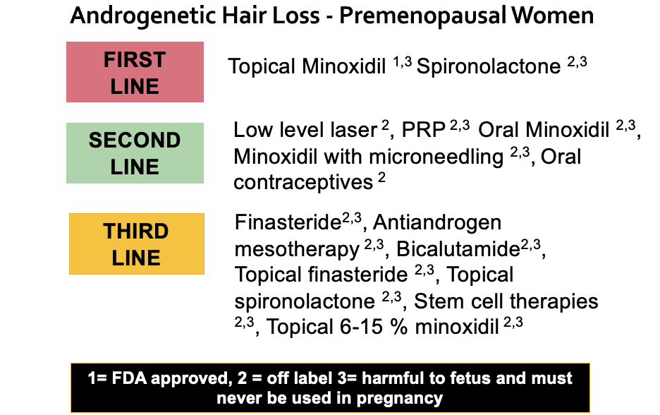
POINT 1. Many people who take birth control pills shed for the first few months.

First, I think there’s little doubt that at least one of your diagnoses is telogen effluvium. We don’t actually need to debate that. The debate we will get into in a moment is whether anything else is going on.

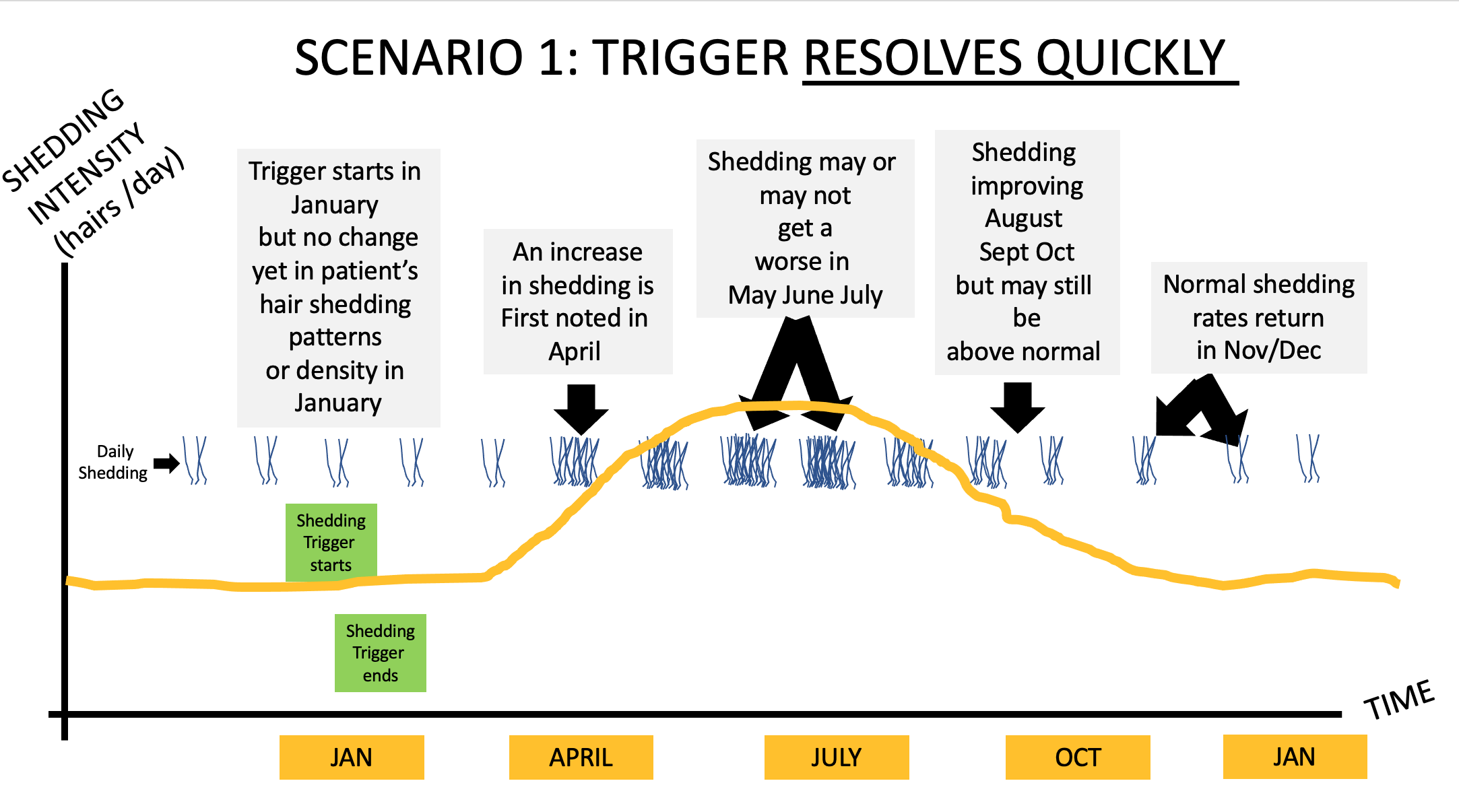
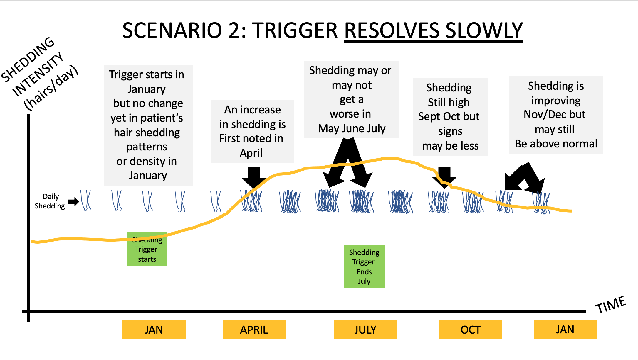
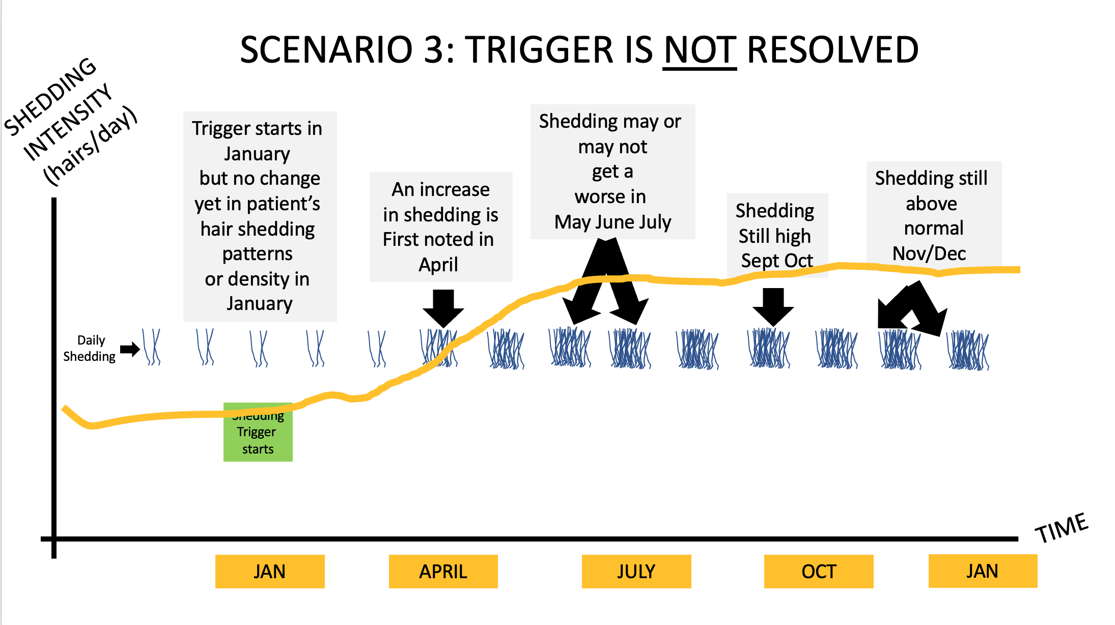
You have several reasons why you could have a telogen effluvium, including starting the birth control pill and starting spironolactone. A large proportion of women shed when starting these treatments, especially birth control pills. The shedding starts 2-3 months after taking the first pill and the shedding lasts 3-6 months provided the pill is continued every day. A lot of women shed when stopping these pills too, especially the birth control pill. The shedding starts 2-3 months after stopping the pill and the shedding lasts 3-6 months provided the pill is not restarted.

So what would I expect to hear from a 21 year old woman who starts spironolactone and then starts a birth control pill? Shedding.

And what would I expect to hear from a 21 year old woman who starts spironolactone and then starts a birth control pill and then stops these pills? Shedding.

What is your story? Shedding.

So in some ways, it’s possible this is entirely consistent with your story.


POINT 2: All patients with hair loss, acne and hirsutism and androgen excess need a proper work up. A work up should be done off birth control.


You have DHEAS 512 (which is 13.9 umol/L in SI units). Any female age 21 with DHEAS 512 and acne and hirsutism and hair loss needs a thorough endocrine work up in my opinion. We need to rule out PCOS and late onset CAH.

With the limited information you have provided here, it would be false to say that you “don’t” have PCOS.

The correct way to say it is more likely “you have a low likelihood of having PCOS.”

Women with PCOS who are thin with low BMI often have regular periods and often have no cysts visualized on ultrasound examinations … but still have elevated androgens. This is a bit more advanced type of knowledge, but I think it’s important especially since you have hyperandrogenism. Anyone who claims there is zero chance you have PCOS is wrong. Anyone who thinks there is a very low chance you have PCOS is correct. I have seen many women with your story exactly who go from being thin to being heavier in their 20s and 30s and ‘develop’ PCOS. I’m not saying that is your case, but sometimes weight gain brings about insulin resistance that then promotes a fuller PCOS clinical presentation.

So what work up do you need? Well, I would advise a proper work up on day 3-5 of your cycle for my own patients that come to see me with a story like yours. The fact that you are off birth control again is a good time to do this. We can’t do these tests when women are on birth control.

The tests that I order on day 3-5 of the cycle for my patients with similar stories are: LH, FSH, estradiol, testosterone, free testosterone, SHBG, glucose, insulin, hemoglobin 1A1c, AM cortisol, prolactin, androstenedione and 17 hydroxyprogesterone, AST, ALT, and cholesterol. These should be done fasting and day 3-5 of the cycle. You have already had your DHEAS measured so there is not a lot of good reason to do this again unless someone suspected levels could be climbing. I would probably include it again for completeness.

What am I looking for in these tests?

a) a high 17 OHP level on day 3-5 that would lead us to a diagnosis of late onset congenital adrenal hyperplasia

b) a high testosterone, high fasting insulin, high LH that would point is towards a PCOS like state

c) A normal prolactin and AM cortisol that reassures us that no other issues are present

In your case, I would want more blood tests if I was your doctor. I would want to know if there is any evidence of insulin resistance that would push me towards PCOS. I would want to know if the other hormones were normal. I would want to know your free testosterone and SHBG. I would want to know your 17 OHP levels to rule out late onset congenital adrenal hyperplasia before moving on.

POINT 3: When it comes to ultrasound examinations, there is a lot that patients don’t realize.

When I hear that a patient had an ultrasound that showed no cysts, my first response is usually that I’m glad to hear that news. But there are a few points to keep in mind. First, it depends on whether the ultrasound examination was a transabdominal ultrasound or transvaginal. There is a big difference in the quality of the studies and what it all means. Transvaginal studies with modern ultrasound techniques are the most helpful. Many people don’t have these studies done. Transvaginal studies can pick up a lot of cysts that the transabdominal can not. Of course, it’s very unlikely this is even an issue with your story but it’s something that we need to keep in mind.

With your ultrasound, I would want to know where it was done (what center? what clinic?) and whether it was transabdominal or transvaginal? What was the volume of the ovaries noted in the report ? Were any ovary measurements more than 10 mL ?

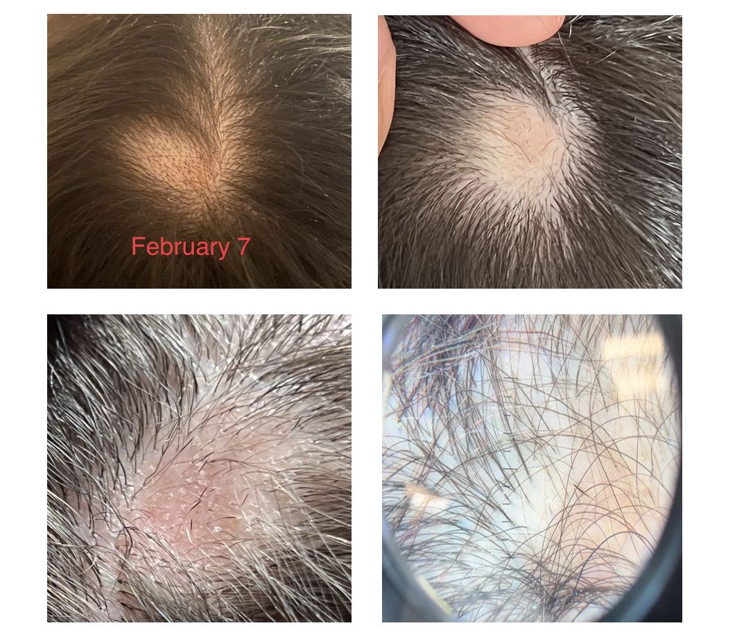
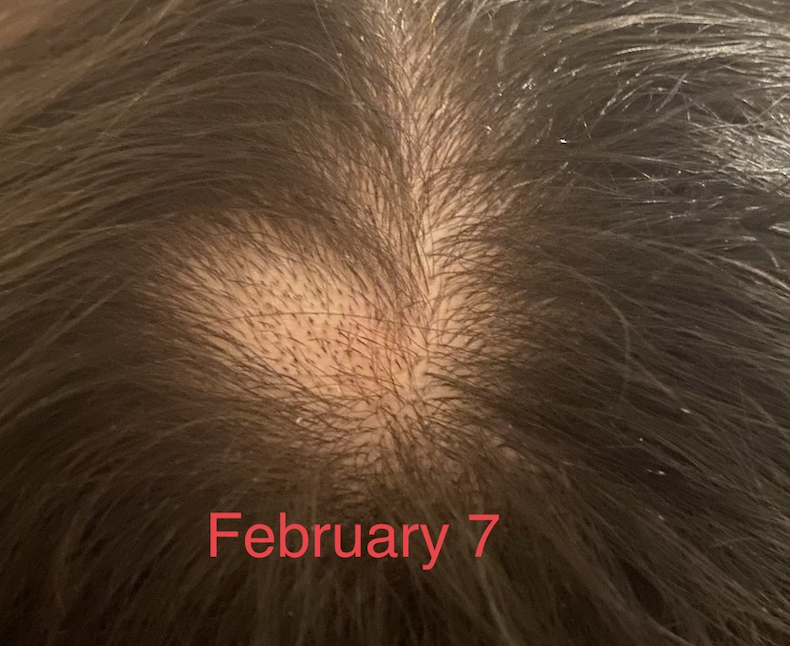
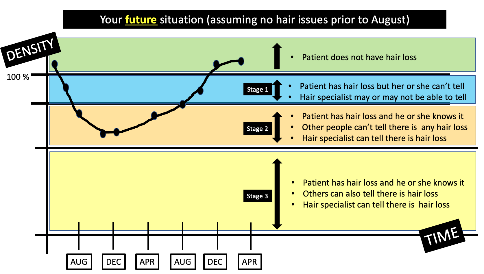
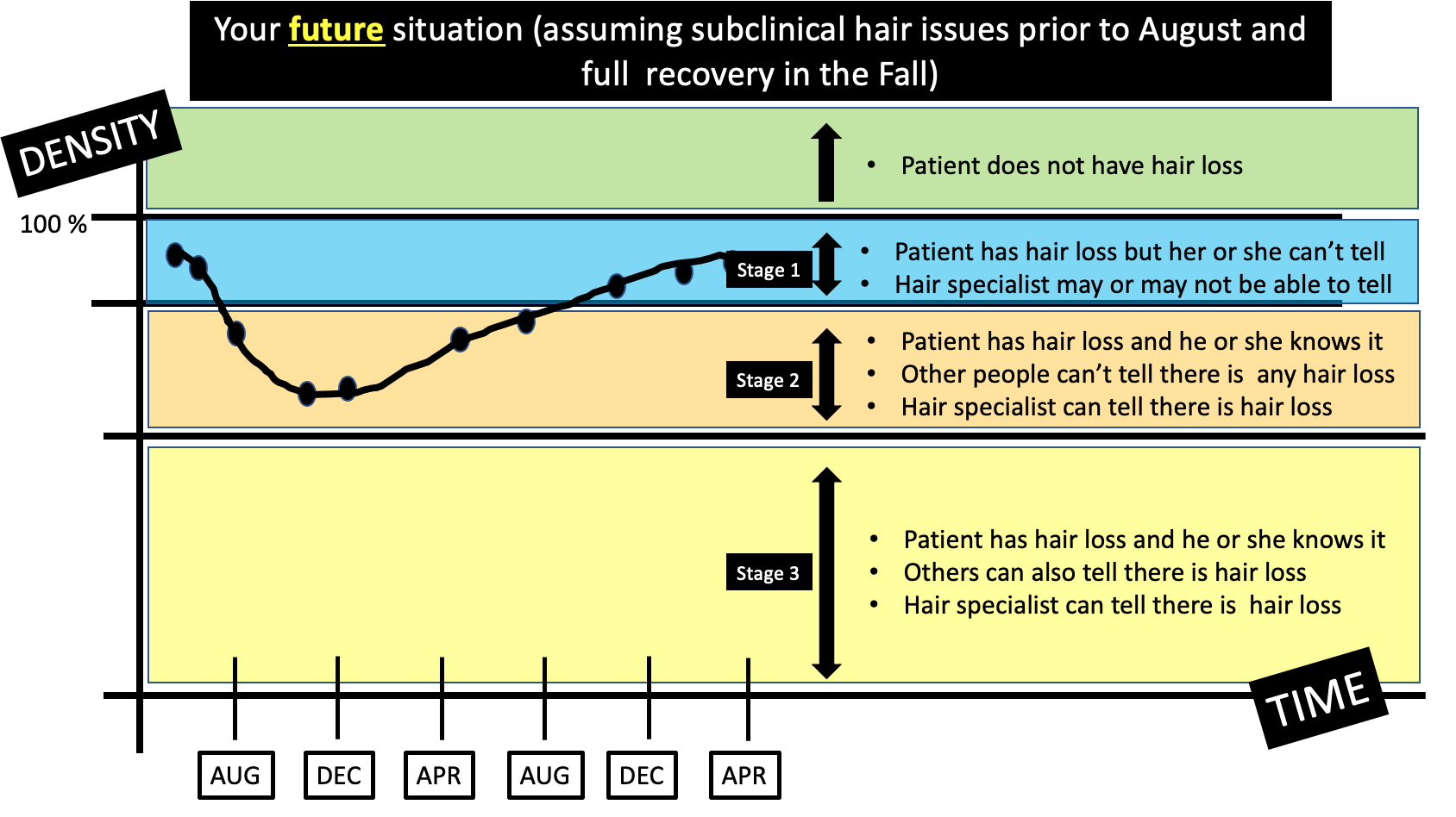
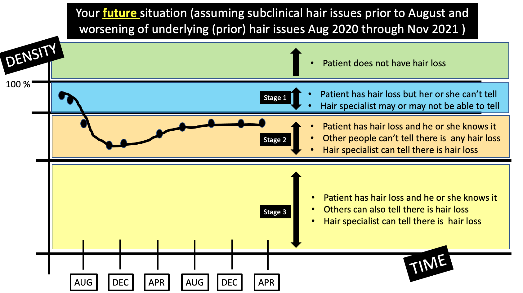
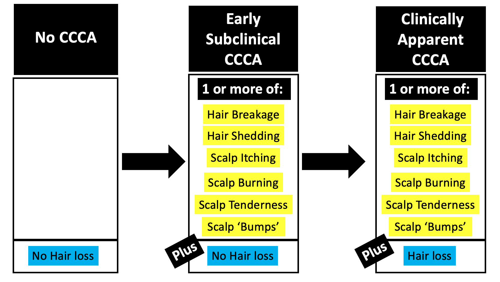
POINT 4: In the early stages of hair loss, TE and AGA can look the same and have the same story. Rarely, they can have a similar biopsy too.

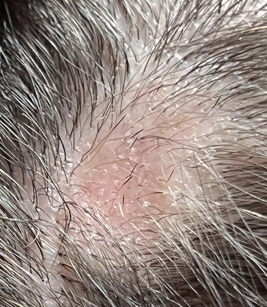
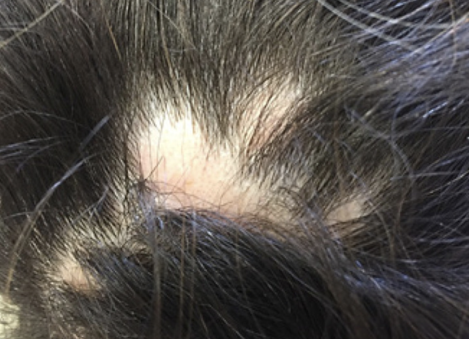
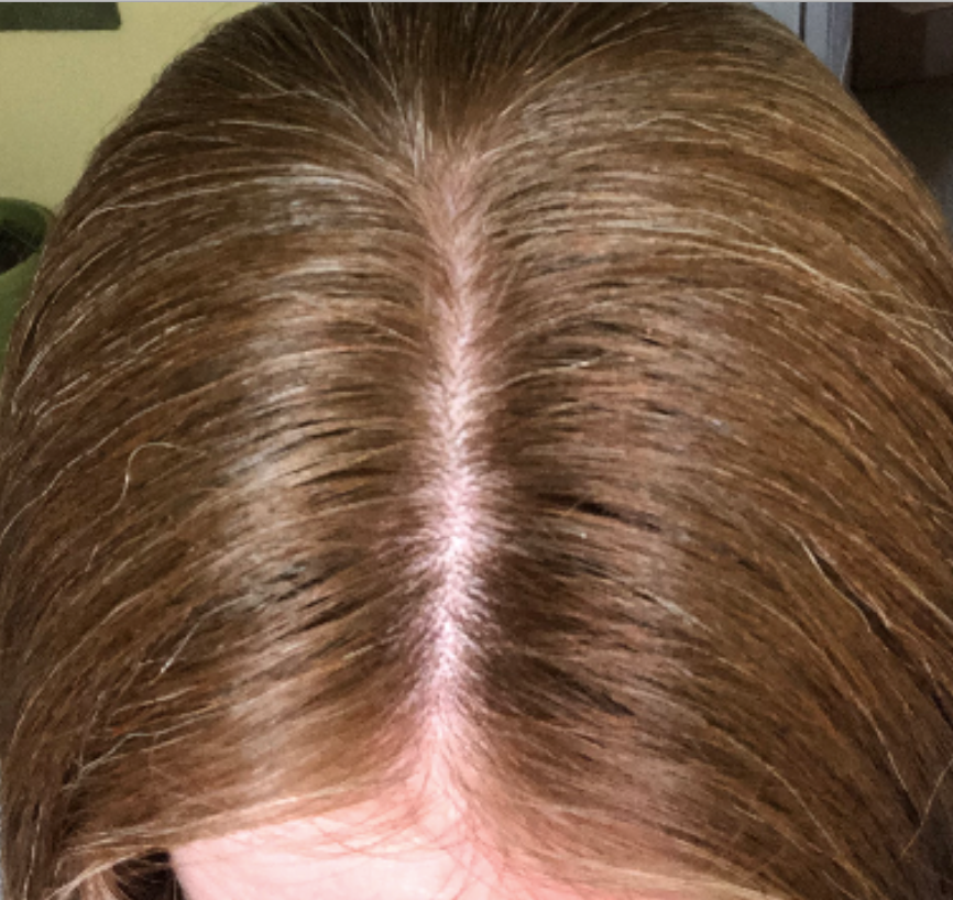
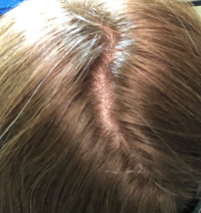
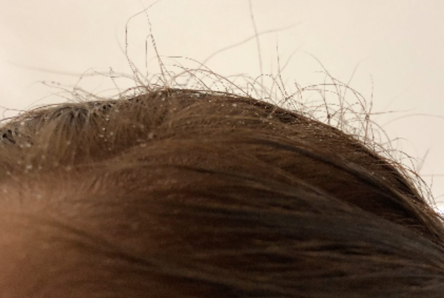
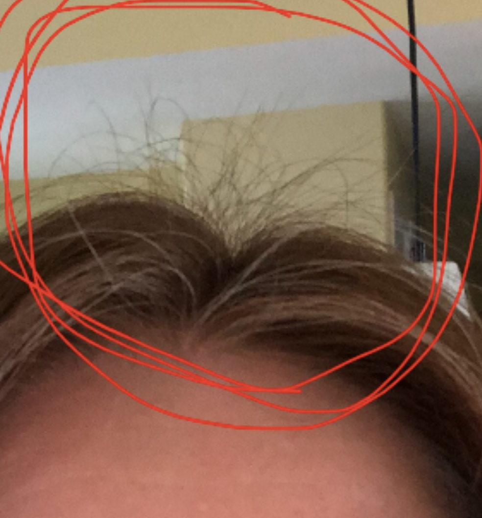
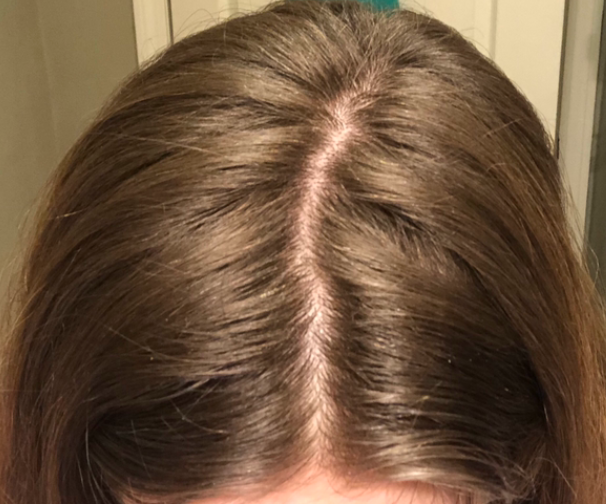
When I look at your photos, I immediately say to myself :

Could this person have AGA?

Could this person have TE?

Could this person have both conditions?

(Also …. whenever we use the word TE, we need to immediate shout out hey could this be diffuse alopecia areata …. but I don’t think that’s what this is. But I include this for completeness of this write up).

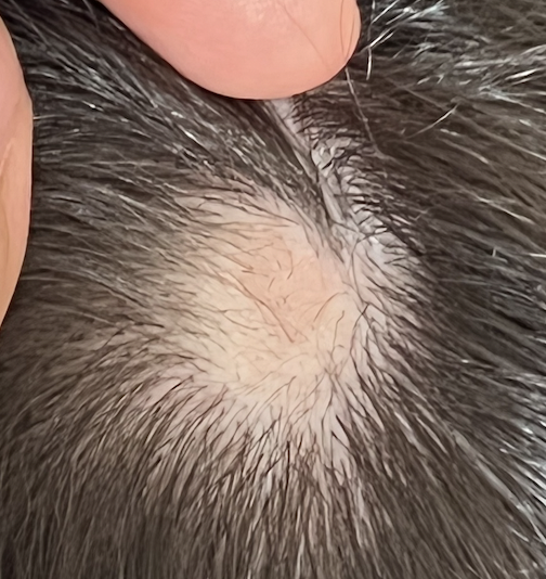
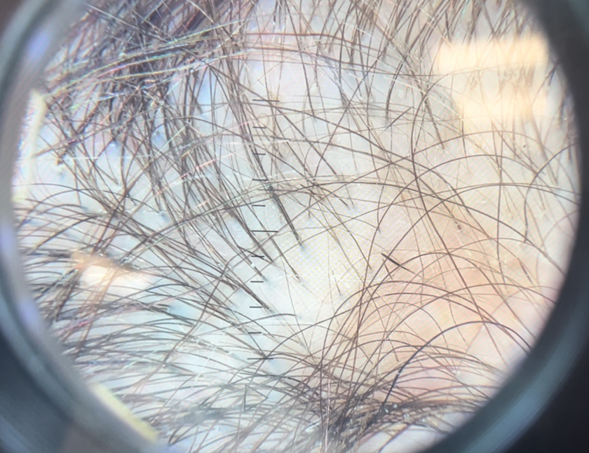
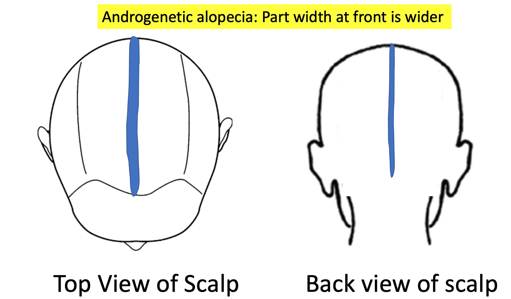
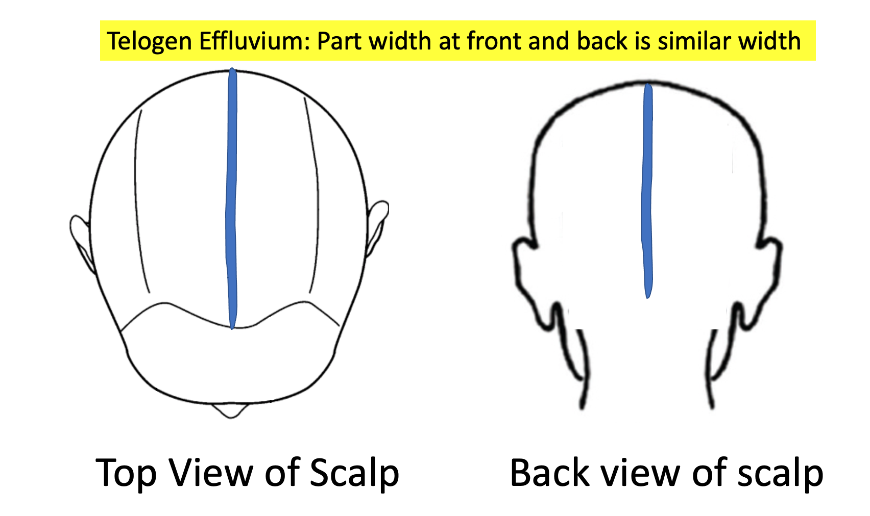
In the early stages TE and AGA look the same. Of course, an up close examination with use of trichoscopy is going to help in your situation. In fact, it’s critical this be done! If the back of the scalp is convincingly thicker than the front of the scalp, I am pushed more towards thinking this could be AGA (with your TE). If the back and front areas are similar density, we are more likely thinking about a sole diagnosis of isolate TE. If there is no evidence of “follicular miniaturization” or variation in the caliber of your hairs when your scalp is examined with trichoscopy, we are likely dealing with an isolated TE. If there is a convincing variation in the caliber of hairs, it could be an early AGA.

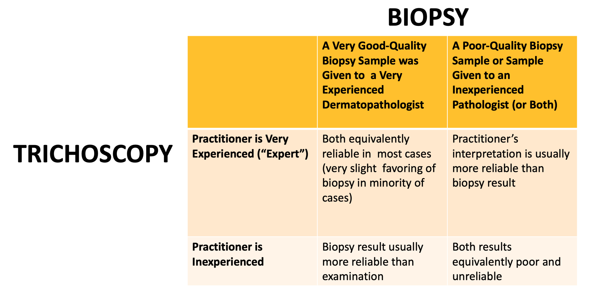
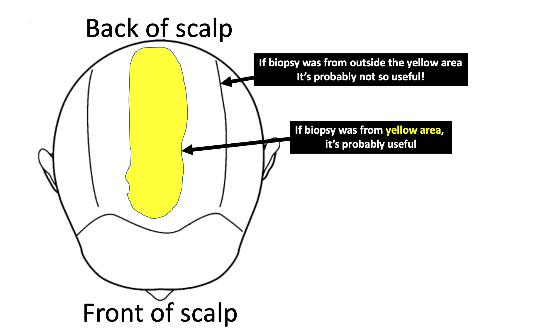
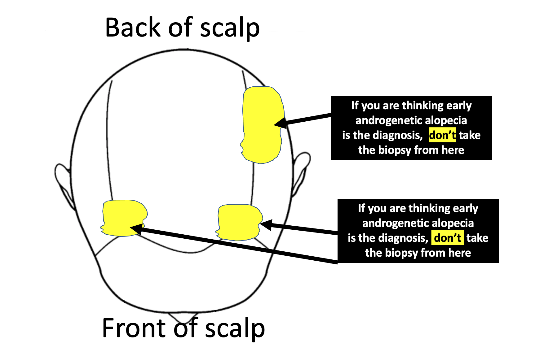
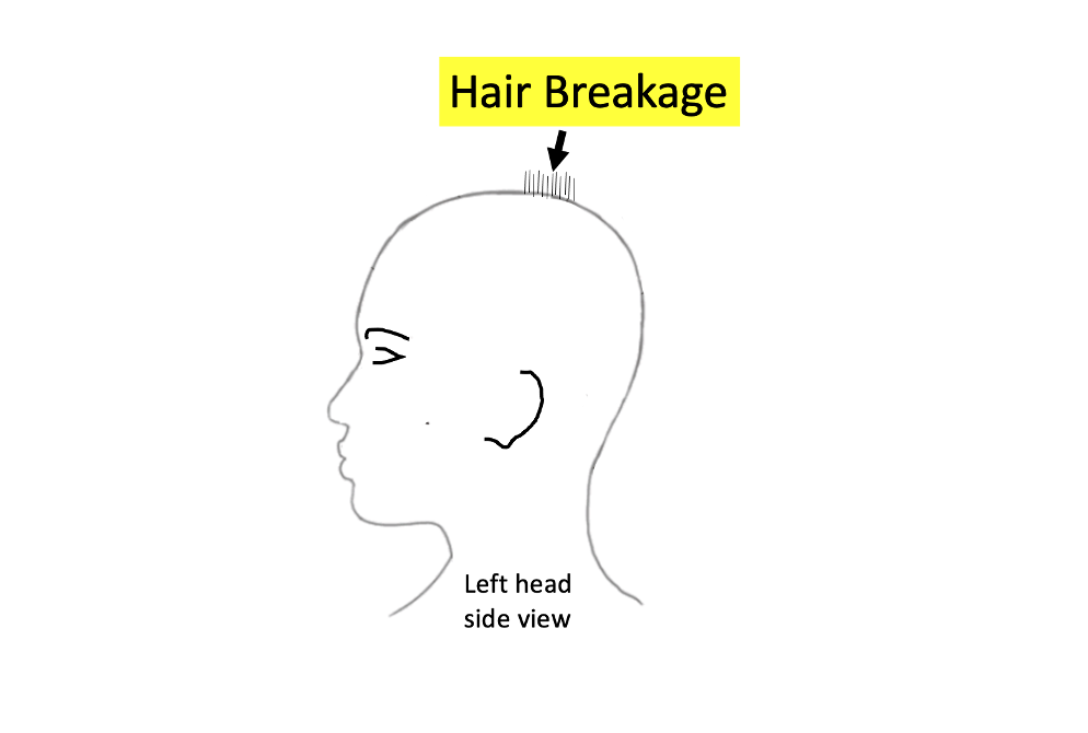
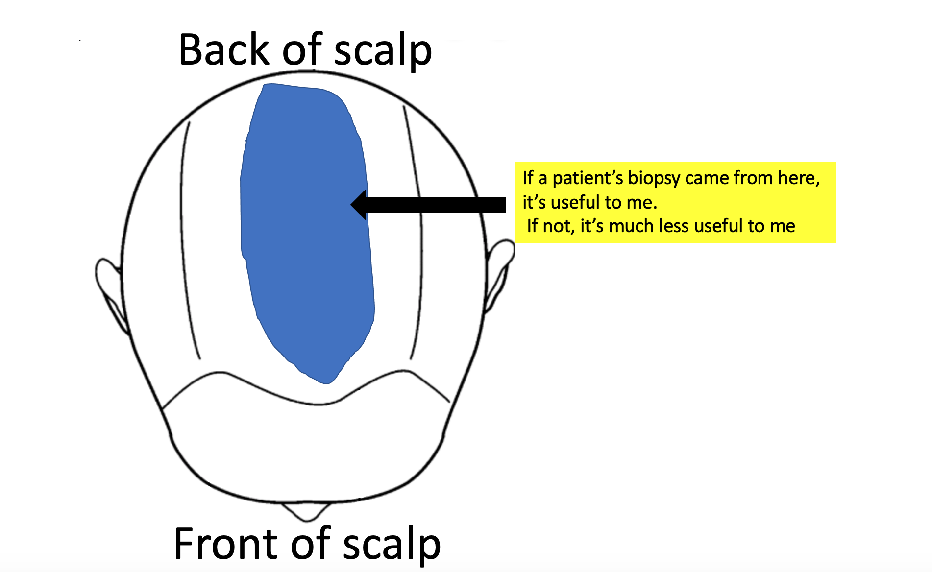
Biopsies can be tricky. There is so much more to doing a biopsy than just doing it and so much more to interpreting a biopsy than just reading the information that comes printed on report. My ability to accurately interpret a biopsy depends where on the scalp it was taken from!. It depends how it was processed (horizontal vs vertical section). It depends who read the biopsy (dermpath vs general path). If a biopsy was taken from the sides or the back or somewhere just to prevent the patient from having a visible scar, then the biopsy is often useless. Biopsies in your case need to come from the top.

Here is where I would need your biopsy to have been taken from for me to feel better about the situation:


sites of biopsy

Also, if someone is going to tell me all you have in your biopsy is a telogen effluvium, I’m going to hope that horizontal sections were used. It’s a huge stretch to diagnose a telogen effluvium confidently from vertical sections. In horizontal sections, the pathologist gets to see 20-40 hairs in order to give their best guess about what could be going on with the rest of the scalp. With vertical sectioned biopsies, they just get to see 3-6 hairs. I don’t want to leave my patient’s hair loss diagnosis to interpretations as to what is seen with 3-6 hairs.

POINT 5: TE and seborrheic dermatitis (and sometimes even AGA) give tingling and symptoms.

With the biopsy result you have in your possession, I’m much much less worried about the tingling, crawling and pain. Of course, I wish you did not have it. But I’m not suspecting anything inflammatory that would make me want to act with a much different course. This is assuming your biopsy came from an area that was tender) I’m always worried when a patient says they have scalp pain. But this worry evaporates to a large degree when the biopsy from that area shows non-scarring alopecia. If your biopsy was from a random area and not from a tender area, then it becomes more difficult to interpret what it means.

Pain and tingling in your case can come from seborrheic dermatitis. it can come from TE, It can come from depression. It can come from allergy or irritation from a current shampoo. it can come from irritation of allergy from other cosmetics.

We still need to keep an eye on this pain. I often encourage my patients to commit to treating their seborrheic dermatitis with a rotating schedule of shampoos. Zinc pyrithione one day. Ketoconazole the next regular shampooing day and selenium sulphide shampoo the next shampooing day. Shampooing must be done 2-3 times per week and left on 2 minutes. I advise my patients to not over do the time as this often just dries the hair and scalp out further. Also, putting a prescription topical steroid on the scalp like betamethasone valerate lotion 0.1 % aa few times per week right after showering is often helpful (of before bed). 10 drops to 15 drops of betamethasone valerate lotion two times per week for a few months is very safe and anyone who says otherwise has little understanding, knowledge or training in the area.

If the pain is still present in 3-5 months, this needs to be looked into further.


POINT 6: What to do next depends on the blood test results and your prior response to spironolactone.

What exactly to do next and how do help your shedding depends partly on your next set of blood test results. If you have elevated 17 OHP on your blood tests, you may want to see an endocrinologist. If you have high LH or evidence of insulin resistance you’ll want to see a really experienced endocrinologist for evaluation of PCOS. Not all women with PCOS are overweight and in fact, women with PCOS who are thinner or have low body mass index often have regular periods and no cysts on ultrasound.

If you tolerated spironolactone well (in the past) and tolerated birth control well, it may even be an option to return to these and stay on these. Did it control your acne? Did it stop hirsutism? Did you feel good on it? Anyone starting these medications has a chance to get shedding so I’m not necessarily worried by a story of shedding. You stopped too soon to really get any sense what the long term outcome was. If there is any evidence of AGA with an up close examination, this could be a good option again. If not, you might want to treat your acne and hirsutism differently - perhaps topically.. The other option is to wait longer in hopes the TE resolves. Continuing iron and vitamin D and shampooing your hair diligently with these anti dandruff shampoos is going to be important no matter what is going on up on the scalp.

Finally, with any TE, we need to always keep in mind that maybe we have not found the trigger. If your ferritin was low and your hemoglobin was low (less than 12.0), a work up could be important. I often test for example a celiac panel in patients with BOTH low HGB and low ferritin. I’m not worried about your high B12. I would want to know about other medications you have started and stopped. As mentioned above, I would want to know about other symptoms like joint pains, headaches, fatigue, weight loss, eyebrow changes, eyelashes changes, body hair changes, nail changes, and rashes. Sometimes we consider ordering autoimmune tests in women with shedding but only if the history points us to ordering these. Ordering these tests ‘just to cover all bases’ is usually not a good idea.

Summary

I can instantly tell by your question that you’ve read a lot and thought a lot about your issues and what all this information means. Congratulations for that. That is important. You need to figure out if late onset CAH is a possibility or not ….. and whether insulin resistance/PCOS its truly off the list or not. In my opinion, these blood tests on day 3-5 are important to you. I’m glad you are off the birth control pill now because it allows you to get these tests done.

If your biopsy was taken from the area I have noted above, then that’s probably very helpful provided it was analyzed in the lab with horizontal sections. . The key point now is figuring out if there is any possibility of an evolving androgenetic alopecia that just could not be picked up in the early stages with the work up you had. A biopsy with horizontal sections and a good trichoscopic examination by a specialist who understands hair loss will uncover these answers.

Regardless, photos should be taken every 3 months. Not every day and not every week. If there is any kind of evolving pattern of hair loss, a photos will also capture these changes over time.

If TE is truly what you have and there are no underlying concerns, doing minimal additional things could be the best plan. However, if your hyperandrogenism is part of a bigger endocrine issue (like PCOS or CAH), getting advice from an endocrinologist would be a good way to proceed. These blood tests will be a really important guide. Some women just have elevated DHEAS and some women with elevated DHEAS develop AGA but some don’t. I never recommend patients start treatment because of what the labs say - treatment is started because of what the skin or hair is doing.

Thanks again for sending in the question. I hope this helps you are your team of specialists.


Share This
No Comments

Hydroxychloroquine (Plaquenil) for Frontal Fibrosing Alopecia


Hydroxychloroquine (Plaquenil) for FFA

I’ve selected this question below for this week’s question of the week. It allows us to discuss the use of hydroxychloroquine (Plaquenil) in FFA as well as the rare cardiac effects of hydroxychloroquine.


QUESTION

I am a female in my late 70s with frontal fibrosing alopecia, confirmed by a scalp biopsy. It is being treated with clobetasol .05%, one application per day. I have read that this is not a very effective treatment when used alone. What would you suggest might be used with it?

My dermatologist suggested hydroxychloroquine (plaquenil), but I read that this might have an adverse effect on people with heart disease. Do you agree? The first symptom about six months ago was drastically thinning eyebrows. I am in the third month of treatment and hair loss around the hairline is not yet apparent. I have cardiomyopathy and atrial fibrillation, for which I am taking a blood thinner (Eliquis) plus daily irbesartan, bisoprolol and spironolactone. Do you think any of these drugs might be the cause of my FFA?

ANSWER

Thanks for the great question. I’d like to discuss several important points in the question you ask and the information you have submitted.

Before we go further, I’m going to assume here in your question that the diagnosis is correct. Can I be 100 % sure you have FFA? Well no - not at all. I haven’t seen your scalp and eyebrows and I don’t know all the details of your story. You might reply to me that you have already told me you had a biopsy. Well, biopsies are not 100% either. Nevertheless, it sure sounds like many aspects of your story do, in fact, support a diagnosis of FFA. So I’ll proceed with this assumption. If you do have FFA, this answer then helps. If the diagnosis is not correct, then the answer has helpful points nevertheless for others with the condition.

Let’s look at some aspects of your question:


a) How good is hydroxychloroquine in FFA?

Hydroxychloroquine is helpful in some patients with FFA. I tell my patients it’s a silver medalist. In other words, it can be helpful, it’s just that other treatments are probably better. We will get into that a bit later. That does not mean hydroxychloroquine is useless as a treatment. Not at all. For some patients with FFA, hydroxychloroquine does help. It does not help everyone that is for sure.

Let’s look at some helpful studies from the past evaluating the effectiveness of hydroxychloroquine in FFA treatment.

in 2013, Ladzinski and colleagues proposed that hydroxychloroquine had some benefits in about 50 % of patients,

In a much larger study conducted in 2014, Vano Galvan et al showed that a 15 % of patients with FFA using hydroxychloroquine had some improvement in their disease. 59 % had their disease stabilized. For about one quarter of patients, the drug did nothing helpful at all.

in 2018, Strazzulla and colleagues found that about 70 % of patients had some benefit with hydroxychloroquine - with results being more or less the same ass doxycycline.

A 2010 study by Samroa and colleagues found that hydroxychloroquine reduced redness and inflammation in FFA patients.

Treatment Options for FFA

Before we leave the subject of hydroxychloroquine for treatment of FFA, let’s look at the options for treatment. Every single type of hair loss has treatment options. The thing that is often forgotten is that not all treatments are the same. Some are good. Some are not so good. Some are good but crazy expensive. Some are good and not so expensive. Some are good and fairly safe and some are good and incredibly risky. I divide every disease into gold, silver and bronze medal treatments. 

I personally like this analogy of categorizing treatments because it reminds us of a couple of key things. First, not all treatments are equal. We select treatments by balancing benefit and risk and take into account the safety, affordability, feasibility and effectiveness of treatment (what I have previously termed the "S.A.F.E." Principle. )

The second reason I like the analogy of gold and silver and bronze medal treatments is because it reminds us that this is not always going to be the order. The athlete that received the gold medal in the recent Olympics may not be the athlete that gets the gold medal in the next Olympics. Of course, if the athlete is really good - he or she will probably be up there one the podium in the top spot again in 4 years. The same is true with hair loss treatments. 

As new treatments come out and as they are studied more and more, a certain treatment might just rise into the gold medal spot and push out other treatments that are there now down into silver or bronze positions. Sometimes, a treatment gets removed from the market entirely just as some Olympians get disqualified from the Olympics. They disappear entirely.

In the diagram below, I outline what I consider the gold, silver and bronze treatments for FFA. In FFA, Plaquenil is a silver medallist. It’s not useless. It has reasonable safety (although I will discuss the heart issues next). It’s relatively inexpensive (Plaquenil is about $1300 USD per year whereas Apremilast is $ 18,000 USD per year). It’s easy to use. It’s somewhat helpful. In terms of the SAFE principle, it’s an okay option to keep in mind for many.

FFA tiers


b) Does hydroxychloroquine have effects on the heart?

Hydroxychloroquine can have effects on the heart. Fortunately, they are not common but risks do increase with advancing age. In fact, physicians need to be much more on alert for eye, muscle, heart side effects in patients over 65 compared to those under 65. Of course, side effects of this nature can happen at any age - but they are more common with advancing age. Hydroxychloroquine cardiotoxicity is non common - but it is a serious side effect.

Plaquenil “cardiotoxicity” (potential toxicity on the heart) has best been studied inn patients with the autoimmune disease lupus who take the drug and also to seem degree in patients with rheumatoid arthritis. Could it be that a these groups are more likely to experienced heart muscle side effects with Plaquenil? Could be, but more studies are needed. For now, it’s a potential side effect that needs to be considered in everyone. There’s nearly 100 reports in the medical journals now of hydroxychloroquine induced heart toxicity. When it does occur, the patient usually develops a restrictive or dilated cardiomyopathy or has conduction system abnormalities including atrioventricular block and bundle branch block. Studies by Costedoat-Chalumeau showed that when cardiomyopathy is present other signs of toxicity are often present as well - eye toxicity, nerve toxicity, and muscle toxicity.

So who is more likely to get hydroxychloroquine induced cardiomyopathy ? Risk factors are currently proposed to include older age, female sex, longer duration of therapy (>10 years), higher doses of the drug, pre-existing cardiac disease, and renal insufficiency (kidney disease). It’s important to remember that most patients who develop hydroxychloroquine cardiotoxicity have been on the drug a long time - ie 10 years or more. Of course, there are reports in the medical journals of those who have been on it just a few months before developing side effects.

hcq toxicity


The Lane and Colleagues 2020 Study

Finally, I’d like to tell you about an important study by Lane and colleagues. This was recently published. This was a study of almost 1 million ( 956,374) hydroxychloroquine users so it’s a massive study. Massive studies of this size are important because they allow us to get a better sense of the risk associated with certain drugs. Lane’s study focused non patents with rheumatoid arthritis (not FFA) so we need to always keep that in mind. The researcher showed that long-term hydroxychloroquine use increased cardiovascular mortality 1.65 times compared with control treatments There are some limitations of the study (as there are with any study). nevertheless, it points to the fact that there may be a slight increase risk of heart related effects with long term use of hydroxychloroquine - at least in patients with rheumatoid arthritis. Fortunately, side effects of this nature are still quite uncommon.

In conclusion, there are some small risks with hydroxychloroquine use in your case. This is something you’d certainly want to review with your heart doctors too. Depending on the degree of heart issues (class of heart failure, etc), it may be something they agree with at a low dose, or something they advise against.


c) What would you suggest might be used with clobetasol?

This is actually a very involved question. If you find that use of clobetasol in the manner described and recommended by your doctors stops further hair loss completely - then you don’t need to use anything else. The key to treating FFA is not to pile on more and more drugs but rather to use the safest and simplest approach possible that stops the disease. if clobetasol stops your FFA 100% - then you need not look for anything else.

Is it likely that clobetasol stops your disease 100%? Well, no. But again - it’s not impossible that it does. It’s a silver medallist.

There are some 20 other options available. Again we follow a logical approach to deciding on which other treatments to use. We select treatments that have good evidence from good studies as well as treatments that have good safety.

You may want to review with your doctors other options like topical calcineurin inhibitors (like Pimecrolimus) as well as low doses of steroid injections (2 mg per mL) every few months. Topical clobetasol is going to give atrophy after a few months of daily use so you and your doctors are going to need to decide when to start reducing that dose.

You can also review options like topical anti androgens (topical finasteride) or even oral dutasteride. Starting two times weekly could be something that you are your doctors discuss. You’ll want to carefully review if you have any contraindications to its use (depression, cancer risks, osteoporosis). Adding acitretin once weekly or twice weekly may also be something you discuss.


d) I am taking a blood thinner (Eliquis) plus daily irbesartan, bisoprolol and spironolactone. Do you think any of these drugs might be the cause of my FFA?

Some drugs do contribute to the development of FFA. For example, the antiestrogen tamoxifen is one of them. The drugs on your list have not been implicated to date in the development of FFA. However, some can cause hair loss - just not FFA.

I’ll make a few additional comments at this point. First, your patterns of hair shedding are going to be important in giving you the fullest answer possible. Your story is interesting in that it seems that you have eyebrow loss without scalp frontal loss yet. Yes, that sure sounds like FFA but again I can’t say much more because I don’t know much about your story and haven’t seen your scalp. If your hair shedding is markedly increased the question must be posed to your health care team as to whether there is any component of telogen effluvium as well. All the drugs on your list can cause telogen effluvium - fortunately that’s still really unlikely overall. Most people have no issues of the sort.

Now on to the tricky part. it’s not possible to 100% say that the drugs you are taking had nothing to do with the development of FFA. Beta blockers (like bisoprolol), angiotensin II receptor blockers (like irbesartan) and spironolactone have rarely been involved in skin rashes known as “lichenoid” drug eruptions. There is some similarity between lichenoid drug eruptions and the lichenoid eruption that is part of the pathology of frontal fibrosing alopecia. Have any of the drugs you are taking been convincingly implicated in the development of FFA? No. It is possible there is some link in those with some sort of predisposition to this disease ? Yes.


Conclusion and Summary

Thanks for submitting your question. I do hope this will be helpful in the discussions you have with your doctors. I’ve included references below in the event they too are helpful. I wish you the best of health.


Reference

August C, Holzhausen HJ, Schmoldt A, et al. Histological and ultrastructural findings in chloroquine-induced cardio- myopathy. J Mol Med (Berl) 1995; 73: 73–77.

Baguet JP, Tremel F and Fabre M. Chloroquine cardiomypathy with conduction disorders. Heart 1999; 81: 221–223.

Clark C, Douglas WS. Lichenoid drug eruption induced by spironolactone. Clin Exp Dermatol. 1998 Jan;23(1):43-4

Costedoat-Chalumeau N, Hulot JS, Amoura Z, et al. Cardiomyopathy related to antimalarial therapy with illustra- tive case report. Cardiology 2007; 107: 73–80.

Fesssa et al. Lichen planus-like drug eruptions due to β-blockers: a case report and literature review. Am J Clin Dermatol 2012 Dec 1;13(6):417-21.

Ladzinski et al. Frontal fibrosing alopecia: a retrospective review of 19 patients seen at Duke University. J Am Acad Dermatol. 2013 May;68(5):749-55.

Lane, J. C. E. et al. Risk of hydroxychloroquine alone and in combination with azithromycin in the treatment of rheumatoid arthritis: a multinational, retrospective study. Lancet Rheumatol. (2020).

Massa MC, Jason SM, Gradini R, Welykyj Lichenoid drug eruption secondary to propranolol. Cutis. 1991 Jul;48(1):41-3.PMID: 1831116

Nord JE, Shah PK, Rinaldi RZ, et al. Hydroxychloroquine cardiotoxicity in systemic lupus erythematosus: a report of 2 cases and review of the literature. Semin Ann Rheum Dis 2004; 33: 336–351.

Pfab et al. Lichenoid drug eruption due to an antihypertonic drug containing irbesartan and hydrochlorothiazide. Allergy 2006; 61(6):786-7

Samrao A et al. Frontal fibrosing alopecia: a clinical review of 36 patients. , Chew AL, Price V.Br J Dermatol. 2010 Dec;163(6):1296-300.

Schön MP, Tebbe B, Trautmann C, Orfanos CE. Lichenoid drug eruption induced by spironolactone.Acta Derm Venereol. 1994 Nov;74(6):476.

Strazzulla LC et al . Prognosis, treatment, and disease outcomes in frontal fibrosing alopecia: A retrospective review of 92 cases.. J Am Acad Dermatol. 2018 Jan;78(1):203-205. doi: 10.1016/j.jaad.2017.07.035.

Vano-Galvan S. Frontal fibrosing alopecia: a multicenter review of 355 patients. J Am Acad Dermatol . 2014 Apr;70(4):670-678.

Yogasundaram H, Putko BN, Tien J, Paterson DI, Cujec B, Ringrose J, Oudit GY. Hydroxychloroquine‐induced cardiomyopathy: case report, pathophysiology, diagnosis, and treatment. Can J Cardiol 2014; 30: 1706–1715.

Share This
No Comments

Hair Loss and Hidradenitis Suppurativa:

Hair Loss in Hidradenitis Suppurativa

I’ve selected this question below for this week’s question of the week. It allows us to discuss the important topic of hidradenitis suppurativa and what associations is has with hair loss.


QUESTION

I am a 23 year old female and was diagnosed with androgenetic alopecia at age 16 and treated with Rogaine and Spironolactone. I also have polycystic ovarian syndrome and prediabetes and have seen an endocrinologist for several years. Everything seemed to be going okay until recently. Lately, the pattern of hair loss is changing and my scalp is becoming intensely itchy and sometimes quite red and scaly. I am shedding a lot more and seen to have hair loss in many areas of the scalp whereas it used to be mainly on the top. Strangely, I am getting very itchy in several other areas recently including the underarms, thighs, private area and breasts. These areas are lumpy and tender and occasionally drain some small amounts of fluid. It’s getting pretty bad and is so so embarrassing for me. It makes be extremely sad. I am generally healthy although I am overweight and have depression and high blood pressure. I take metformin for prediabetes. I take citalopram for depression. Both fo these drugs sstarted about 1 year before these issues began. I am not on medications for blood pressure yet as my doctor increased by spironolactone from 100 mg to 200 mg to see if this will help.

My questions for you are :

1) Can polycystic ovarian syndrome causes cysts outside of the ovary like on the areas stated here?

2) Is PCOS also associated with scalp itching ?

Any insights into this could be helpful as it is severely affecting all and every part of my life!!!!!


ANSWER

Thanks for the question. I can understand how difficult many of these issues are so I’m glad you’ve submitted this question.

I’d like to discuss several important points in the question you ask and the information you have submitted.

Before we do go further, I’d like to point out that the ideal way to diagnose hair loss is using what I termed the ”Diagnostic S.E.T.” I refer to these as the diagnostic “set” because theses 3 aspects all go together. These 3 items include:

1) the patient’s story

2) the findings uncovered during the process of the scalp examination including trichoscopy

3) the results of relevant blood tests. 

The first letter of each of the three words 1) story, 2) examination and 3) tests spell out the word “S.E.T.” - again a helpful reminder of how the information obtained from reviewing each of these 3 aspects helps solidify a proper diagnosis.

There are several important issues in your scenario. The first key question here in your case is what exactly is the reason for your scalp symptoms and hair loss. The only way I can really confidently determine that is with an up close examination. The second important issue is whether you had a systemic issue (full body issue) to explain the lumps. The most important to consider with your doctors is a conndition called hidradenitis suppurativa.

Let’s go further into your story.

Your Scalp Issues

I can’t say exactly what you have without examining your scalp myself (or at very least seeing photos). It does seem that there is a new issue going on on the scalp that is very different from androgenetic alopecia that you were diagnosed with in the past. I would encourage you to see a dermatologist as soon as you can for an up close examination. Common issues like seborrheic dermatitis and psoriasis and contact dermatitis need to be considered. These can cause itching and scaling. However, other issues also need to be thoroughly evaluated - including various types of scarring alopecia.

Many of these conditions can be diagnosed with a proper examination and trichoscopy. If there is any remaining doubt, a scalp biopsy can be considered. Blood tests are always needed for anyone with shedding including thyroid studies, iron studies (ferritin), CBC. Issues like thyroid diseases or low iron can give increased hair shedding but would not give the intense itching you described. That would be caused would be something else.


Your Lumps

I would also urge you to bring up the issue of the lumps you have in the armpits, thighs, private area and breasts to your dermatologist. There could be a few reasons for this but a condition called hidradenitis suppurativa needs some careful consideration by your doctors. There are other issues that cause lumps too although with your story the diagnosis of hidradenitis suppurativa needs prompt consideration before moving on to other diagnoses.

Hidradenitis suppurativa (HS)

Hidradenitis suppurativa (HS) was once considered rare. We know now that it’s not rare and in fact up to 1-4 % of the population an be affected when mild cases are taken into consideration. HS is a dermatological condition that is associated with painful draining lumps in the underarm area (axillae), groin area and buttocks and under the breast. The lay public often refers to such lumps as “boils” - and patients affected by hidradenitis often describe their disease as one of multiple draining boils in the armpits, groin and buttocks regions. The condition can be emotionally and physically disabling.

Hidradenitis suppurativa is thought to be a disease of hair follicles. It has been proposed that there is some initially plugging of follicles that then leads to the eventual rupture of these follicles. Better and better treatment options have arisen in the last decade.

Hair Loss Conditions Associated with Hidradenitis Suppurativa

Not all patients with HS have hair loss. But when they do have hair loss, a variety of reasons need to be considered. Some of the reasons are unrelated to the HS and some are related to th HS. Patients with HS seem to have an increased risk for psoriasis, alopecia areata, Telogen effluvium, dissecting cellulitis and lichen planopilaris. Of course, patients with HS can have hair loss conditions that anyone can get.

See Prior Article “Risk of Alopecia Areata and Lichen Planopilaris Increased in Hidradenitis Suppurativa”


I am not completely sure if you have HS - as that would require a proper examination. However, as I reflect on your question, it’s important to consider issues like psoriasis, lichen planopilaris and dissecting cellulitis as being causes of your hair loss. . These can cause redness, and itching. Alopecia areata does not cause this kind of itching but if you had alopecia aerate with seborrheic dermatitis or alopecia areata with psoriasis it certainly could. Fortunately, a proper examination by your dermatologist can usually distinguish between these issues. If not, a biopsy is going to be key.

IMG_8609.jpg


Systemic Issues Associated with Hidradenitis Suppurativa


There are several systemic (internal) diseases that occur more frequently among patients with a diagnosis of hidradenitis suppurativa. PCOS is one of them. Psoriasis is another one. Prediabetes (and diabetes) is another and depression is another. I think it’s really important that you review your health issues again with your dermatologists and endocrinologist.

When I teach doctors about Hidradenitis Suppurativa in the clinic, I really want them to remember all the conditions that patients with HS can develop. I not only want them to remember them - but I want them to ask the patient about them. There are many issues to discuss. The memory tool “PIMPLES” helps medical practitioners remember many of the key issues to ask about. The list does not include all the associatioss - but does include the key ones.

IMG_8254 5.jpg


Many Hidradenitis Associations Have Been Well Studied

A number of good studies have been done to date which allow us to understand the strength of the various associations that can occur in patients with HS. These are outlined in the table below. PCOS is increased two fold and metabolic syndrome and depression are increased about 2-3 fold in patients with HS. If you do end up being diagnosed with HS, you will want to review these associations with your doctors.

Screen Shot 2021-03-14 at 10.10.23 AM.png

Summary

Thank you again for your excellent question. Hidradenitis suppurativa affects so many people and my hope is that this answer will provide information and open up dialogue between you and your physicians. The cysts you are experiencing are different than the cysts in the ovary. A proper evaluation can confirm if these lumps are the cyst-like eruptions seen in hidradenitis. The HS issues could be separate form the hair loss or could be related. A biopsy is going to be helpful if there is any doubt as to what’s causing your scalp issues.

Good luck & thank you.

Reference

Andersen et al. Psoriasis as a comorbidity of hidradenitis suppurativa. Int J Dermatol . 2020 Feb;59(2):216-220.

Dessinioti C, Katsambas A, Antoniou C. Hidradenitis suppurrativa (acne inversa) as a systemic disease. Clin Dermatol. 2014;32(3):397-408.

Fauconier M et al. Association between hidradenitis suppurativa and spondyloarthritis. Joint Bone Spine. 2018 Oct;85(5):593-597.

Gonzalez- Vellanueva et al. Hidradenitis Suppurativa is Associated with Non-alcoholic Fatty Liver Disease: A Cross-sectional Study. Acta Derm Venereol. 2020 Aug 18;100(15):adv00239.

Horissian M et al. Increased risk of alopecia areata for people with hidradenitis suppurativa in a cross-sectional study. J Am Acad Dermatol 2019

Patel KR et al. Association between hidradenitis suppurativa, depression, anxiety, and suicidality: A systematic review and meta-analysis. J Am Acad Dermatol. 2020 Sep;83(3):737-744.

Pescitelli et al. Hidradenitis suppurativa and associated diseases.G Ital Dermatol Venereol.. 2018 Jun;153(3 Suppl 2):8-17.

Phan et al. Hidradenitis Suppurativa and Thyroid Disease: Systematic Review and Meta-Analysis. J Cutan Med Surg . Jan/Feb 2020;24(1):23-27.

Phan et al. Hidradenitis suppurativa and polycystic ovarian syndrome: Systematic review and meta-analysis. Australas J Dermatol. 2020 Feb;61(1):e28-e33.

Phan et al. Prevalence of inflammatory bowel disease (IBD) in hidradenitis suppurativa (HS): systematic review and adjusted meta-analysis.Int J Dermatol. 2020 Feb;59(2):221-228.

Rodriguez-Zunga et al. Association Between Hidradenitis Suppurativa and Metabolic Syndrome: A Systematic Review and Meta-analysis. Actas Dermosifiliogr . 2019 May;110(4):279-288.

Tannenbaum et al. Association Between Hidradenitis Suppurativa and Lymphoma. JAMA Dermatol . 2019 May 1;155(5):624-625.

Thanh-Lan Bui et al. Hidradenitis suppurativa and diabetes mellitus: A systematic review and meta-analysisJ Am Acad Dermatol. 2018 Feb;78(2):395-402.

Vazquez BG, Alikhan A, Weaver AL, Wetter DA, Davis MD. Incidence of hidradenitis suppurativa and associated factors: a population-based study of Olmsted County, Minnesota. J Invest Dermatol. 2013;133(1):97-103.

Share This
No Comments

Losing short hairs: is it normal or should I be concerned?

Should I be concerned if I find I am losing short hair?

I’ve selected this question below for this week’s question of the week. It allows us to the review some key concepts in evaluating shed hairs - particularly the relevance of short hairs.

Question

I noticed that I a few of the hairs that I find in my brush are short hairs - less than 3 cm. Is this evidence that I am progressing to androgenetic alopecia?

Answer

Thanks for the question.

It all depends on the proportion of short hairs you find.

If you find a low proportion of small hairs, that’s completely normal. Everyone sheds a few short hairs.

If you collect all your shed hair over a week and find that more than 10 % are tiny hairs less than 3 cm, that might suggest there is some androgenetic hair loss happening. You’d certainly want to review things with your dermatologist carefully if that were the case and have him or her performing a careful scalp examination including trichoscsopy.

But finding one hair has very little meaning otherwise.

Share This
No Comments

Telogen Effluvium or Androgenetic Alopecia ... Or Both?

Biopsy Suggested AGA and a bit of TE: Which one is causing my hair loss?

I’ve selected this question below for this week’s question of the week. It allows us to the review some key concepts related to the diagnosis of hair loss

QUESTION

My biopsy came back showing that I have androgenetic alopecia with bit of telogen effluvium. My terminal to vellus ratio was 1.8: 1 and there were 16 % telogen hairs. I’m wondering if I have both diagnoses, which one is causing my hair loss.

ANSWER

Thanks for the question.

It’s likely that they both are. However, most of your hair loss is from the AGA. I would need to see your scalp to give you a precise breakdown but it’s likely the AGA is the main cause given that the T:V is well under 4:1.

But both of these contribute!

Suppose you ran a 20 mile run and also carried groceries up 40 flights of stairs because the elevator was broken. Why are your legs sore? Well mostly from the 20 mile run but some of your leg soreness is due to the climb up the stairs. If you didn’t have to do the stairs you might be 6 % less sore but you’d still be sore.

If someone has a little bit of telogen effluvium and mostly androgenetic hair loss they’ll get a bit of hair back if they address their telogen effluvium but really they need to address the androgenetic component.

Hope this helps.

Share This
No Comments

Cyclosporine and Hydroxychloroquine Drug Interactions

Why is their a concern about using cyclosporine with hydroxchlorquine?

I’ve selected this question below for this week’s question of the week. It allows us to discuss an important drug interaction between cyclosporine and hydroxychlroquine (Plaquenil).


QUESTION

Dear Dr. Donovan.

I'm a 25 yr old woman with diagnosed lichen planopilaris (LPP). It's proven difficult to treat. My doc just prescribed plaquenil and cyclosporine, but I noticed on your handout that it says these drugs interact. Why do you not prescribe them together?

Thank you for your time!


ANSWER

Thanks for your excellent question. The reason is this: hydroxychloroquine (Plaquenil) can potentially Increase cyclosporine levels in the blood leading to higher cyclosporine in the blood and increase the risk of cyclosporine side effects.

Cyclosporine and hydroxychloroquine are two drugs that may have benefit the treatment of lichen planopilaris. The use of the two medications together (at the same time) carries the risk of increased side effects and elevated serum creatinine. Use of…

Cyclosporine and hydroxychloroquine are two drugs that may have benefit the treatment of lichen planopilaris. The use of the two medications together (at the same time) carries the risk of increased side effects and elevated serum creatinine. Use of the two medications together is generally discouraged but if they are used together it should be done only under supervision of doctors who understand the proper monitoring that is necessary. Frequent blood pressure monitoring and frequent measurement of serum creatinine and frequent evaluation of side effects will be required.


Let’s take a look at what is known in the past.


The first indication of a potential interaction between cyclosporine was with “chloroquine” - a close cousin of hydroxychloroquine. In 1993, Finielz and colleagues published a study in the journal Nephron of a 28 year old kidney transplant patient who was stable on cyclosporine, prednisone and azathioprine. His creatinine level was 200 umol/L. His blood cyclosporine level was 105 ng/mL and his blood pressure was normal 130/80.

He then decided to go on vacation and started treatment with chloroquine for prevention of malaria. His blood pressure just 6 days later increased to 160/100, his creatinine level rose to 234 and his blood cyclosporine level rose more than four times to 470 ng/mL. His levels reduce again when chloroquine was stopped and levels rose yet again when the drug was given back.

An increase in cyclosporine levels has also been noted in patients using hydroxychloroquine (Plaquenil). Some rheumatologists in years gone by have studied this combination. But that’s not to say it’s not without potential risk - there are.

A 1996 study examined the benefits of combining cyclosporine to patents with rheumatoid arthritis who were not improving with hydroxychloroquine or methotrexate. We’ll focus here on the results of combining cyclosporine and hydroxychloroquine because this is what your question pertains to.

The study included 12 patients using both hydroxychloroquine and cyclosporine. During the study, the mean daily dose of CsA ranged from 2.5 to 4.2 mg/kg and at 6 months it was 3.3 mg/kg. The mean serum creatinine levels increased by 0.1 +/- 0.2 mg/dl in the patients treated with the two drugs. In 5 of 12 patients (41.6 %) there was a concerning increase in creatinine of more than 130 %. Other side effects seen in the combination were gastrointestinal side effects (41.6%), hypertrichosis (25%), gingival hyperplasia (25%), liver test abnormalities (8.3 %), and neurological problems (16.6%). This is not by any means a complete lists of side effects of cyclosporine - those will need to be reviewed with your doctor.


Conclusion

Thanks again for the great question. Yes, there most certainly is a potential drug interaction between cyclosporine and hydroxychloroquine that is very important for patients and treating physicians to be aware. Cyclosporine is notorious for drug interactions so I recommend that anyone who uses cyclosporine get in a routine of shouting out loudly “I am on cyclosporine - does this drug interact?”

Any patient started on combined hydroxychloroquine and cyclosporine treatment will need to first check that this was not an error by the prescriber. Sometimes this is what the prescriber intended - as seen above in the 1996 study I mentioned. This is not, however, all that common anymore. If it was not an error, the patient will need very, very close monitoring. Blood pressure will need to be measured daily during the starting period. Serum creatinine levels and serum cyclosporine levels will need to be measured very frequently and side effects will need to be closely monitored.

References


1] Uptodate.com

2] https://www.drugs.com/interactions-check.php?drug_list=763-0,1298-0

3] Salaffi et al. Combination therapy of cyclosporine A with methotrexate or hydroxychloroquine in refractory rheumatoid arthritis. Scand J Rheumatol. 1996;25(1):16-23.


Share This
No Comments


Share This
-->