h1.qusth1 { display: none !important; }

QUESTION OF THE WEEK

Dr. Donovan's Articles

QUESTION OF HAIR BLOGS

Filtering by Category: CTE


Hair loss in the Frontal Hairline.

Cause of Frontal Hairline Loss

I enjoyed giving a lecture yesterday to our brilliant University of British Columbia dermatology resident physicians. We discussed the common and uncommon scarring and non-scarring hair loss conditions that affect the frontal hairline of males and females.

frontal hairline


This article was written by Dr. Jeff Donovan, a Canadian and US board certified dermatologist specializing exclusively in hair loss.
Share This
No Comments

Trichoscopy of Telogen Effluvium (TE) - A Closer look at Kenogen Phase and Yellow Dots

Trichoscopy of Telogen Effluvium - Kenogen Phase & Yellow Dots

For a period of time after a hair is shed from the scalp, it appears that the hair follicle opening sits empty. Of course, it is not truly empty because deep underneath the scalp follicular machinery is preparing yet again to manufacture a brand new hair. The period of time between a shed hair and the emergence of a new hair is called “kenogen”. Kenogen phase is typically quite short (1-2 months). The duration that a follicle remains in kenogen increases in patients with some types of hair loss. For example, in some cases of androgenetic alopecia, hairs have been found to remain in kenogen for up to 1 year.

The “sudden” appearance of hair follicle openings that are empty and lack follicles must also prompt one to consider a diagnosis of telogen effluvium.

Yellow dots in telogen effluvium signify hairs in kenogen phase. This finding is a non specific finding of telogen effluvium.

Yellow dots in telogen effluvium signify hairs in kenogen phase. This finding is a non specific finding of telogen effluvium.


Sometimes we see upright regrowing hairs by dermoscopy as a sign of telogen effluvium but sometimes the dermatoscopic findings are extremely non specific and the only finding by trichoscopy is empty pores. It’s possible to have large numbers of hairs in kenogen in advanced stages of androgenetic alopecia but this is uncommon in the early stages. The finding of many empty pores is atypical of the early stages of androgenetic alopecia. The patient in this trichoscopic image androgenetic alopecia as one of the diagnoses. Variation in the caliber of hairs is easy to spot. However the large number of empty follicles is unexpected prompting one to consider that hairs have recently shed - as in a telogen effluvium. Causes include stress, low ferritin levels, thyroid dysfunction, crash diets, medications and internal illness. In this patient, a 49 pound weight loss over 2 months together with low ferritin levels and extremely intense and sudden stressful life events caused the patient’s androgenetic alopecia to suddenly “appear worse.


This article was written by Dr. Jeff Donovan, a Canadian and US board certified dermatologist specializing exclusively in hair loss.
Share This
No Comments

Serum Magnesium Levels in Women with Diffuse Hair Loss

Should we be checking blood levels of magnesium ?

There are many causes of hair shedding or telogen effluvium. Common causes include intense stress, thyroid problems, crash diets with sudden weight loss, and medications. However, a wide variety of other ‘triggers’ have the potential to cause hair loss. Deficiencies in various minerals are also potential causes of telogen effluvium. Deficiencies in zinc, iron, magnesium, selenium, for example, all have the potential to trigger hair loss.

A 2004 study reminded us that testing for magnesium deficiency (“or hypomagnesemia”) may be reasonable in women presenting with concerns about diffuse hair loss and diffuse hair shedding. The normal adult value for magnesium is 1.6-2.5 mEq/L and hypomagnesemia is generally defined as a level of serum magnesium under 1.6  mEq/L or 1.5 mEq/L.

Tataru and Nicoara studied three groups of women age 16 to 40. Group A was made up of 26 women with diffuse hair loss for which the cause was unknown. Group B consisted of 14 women with diffuse hair loss for which the cause was known (seborrhoea, hormonal issues, thyroid disease). Group C consisted of 24 women without hair loss.

The authors found in the first group (group A), there were 12 cases (46.1%) with hypomagnesemia and the average magnesium level was 1.80 mEq/L. In the second group (group B), there were 3 cases (21.4%) and the average magnesium level was 1.99 mEq/L. Finally, in the control group (group C) the authors found 2 cases (8.3%) hypomagnesemia and the average level was 2.23 mEq/L These data suggested that low magnesium levels were indeed more likely to be found in women with diffuse shedding.

Check serum magnesium levels may be important to consider for some women with diffuse hair loss according to a 2004 study. Supplementation with magnesium may help some women with diffuse hair loss if levels are found to be low.

Check serum magnesium levels may be important to consider for some women with diffuse hair loss according to a 2004 study. Supplementation with magnesium may help some women with diffuse hair loss if levels are found to be low.


Magnesium supplementation may reduce hair shedding in some women

In the second part of the study, the authors evaluated the effect of providing magnesium supplementation to women in Group A and Group B. The dose was equivalent to 96 mg  (8 mEq or 4 mmol) daily for 2 months. The authors observed a noticeable decrease of hair loss in 69.1% of the patients from group A (18 from 24 cases) in comparison with 35.7% (5 from 14 cases) in the group B.


Conclusion

This study was among the first large scale studies to document the incidence of low magnesium in women with diffuse hair loss and to show that women with diffuse loss are more likely to have low magnesium levels than women without diffuse loss. Moreover, these studies showed that supplementation magnesium may help some women reduce hair loss and shedding.

Finding the precise cause of hair loss in women with diffuse loss and hair shedding can be challenging. Ordering every single blood test is not practical and not cost effective. Sometimes the medical history can guide us, but not always.

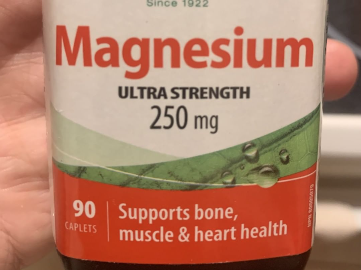
Supplementing with magnesium is reasonable if blood tests prove that there is low magnesium. Supplements with 100-250 mg of elemental magnesium are quite reasonable for 2-3 months but I often start with every other day for 2 weeks to ensure that the patient does not experience diarrhea. Supplements with higher levels of magnesium are not typically recommended. After 3 months, I typically reduce the dose quite significant and recheck levels. Depending on the patient, the old magnesium levels, the new magnesium levels at the end of month 3 and the original suspected reason for the low magnesium, I might either continue at low doses or stop the magnesium altogether.

I have always found this to be an interesting study. I have not found a high proportion of women with hair shedding to have magnesium deficiency but am always on the look out.

Women with high intake of vitamin D may have low magnesium levels as well as other medication users. Low magnesium can give symptoms of muscle pain, fatigue, high blood pressure, irregular heart beats, osteoporosis and mood disorders so certainly we need to be particularly thinking about the possibility of low magnesium levels when this issues are present.


Im general, basic tests for women with hair shedding include:

CBC, TSH, ferritin, 25 hydroxy vitamin D, DHEAS testosterone, AM cortisol, ESR

zinc, magnesium, ANA, creatinine, AST, AST.

Consideration can given to ordering a variety of other tests depending on the exact patient history including syphilis screening, HIV, selenium, mercury and others.


Reference

A Tataru and E Nicoara. Idiopathic diffuse alopecias in young women correlated with hypomagnesemia. J Eur Acad Dermatol Venereol. 2004 May;18(3):393-4.


This article was written by Dr. Jeff Donovan, a Canadian and US board certified dermatologist specializing exclusively in hair loss.
Share This
No Comments

Vitamin D Receptor Polymorphisms in Chronic Telogen Effluvium (CTE)

Certain Vitamin D Receptor Changes May Increase the Risk of Chronic Shedding in Women

The role of vitamin D in human health is the subject of tremendous research interest - and also great controversy and debate. Some experts actually say we are focusing way too much on the role of vitamin D nowadays …. and some say we need to focus more. The same is true for the role of vitamin D in the hair follicle.

It’s clear that vitamin D has some sort of role. Vitamin D binds to the vitamin D receptor (or “VDR”) and together this complex sits on various genes and tells the genes what to do. The vitamin D receptor is expressed in the dermal papilla of the hair follicle as well as in epidermal keratinocytes. VDR expression increases in hair follicles in late anagen and catagen phases. Individuals with mutations in the VDR develop complete hair loss - an indication that vitamin D singling in hairs is important. What is less clear is how vitamin D affects hair loss in adults.

Vitamin D receptor polymorphisms (VDR polymorphisms) refer to subtle changes in the genes coding the vitamin D receptor. These are often one or two nucleotide changes that ever so slightly change the vitamin D receptor gene and/or the way the vitamin D receptor gets manufactured in the body. Studies have focused on whether these subtle changes in the VDR (i.e. VDR polymorphisms) affect various aspects of human health. From colon cancer to heart disease, these VDR receptor polymorphisms are an intensively researched topic.

VDR Receptor Polymorphisms in Chronic Telogen Effluvium

Seliet and colleagues from Egypt set out to determine if two VDR polymorphisms have any role in chronic shedding - or what is typically called chronic telogen effluvium (CTE). They studied 30 women with chronic telogen effluvium and 30 controls who did not have chronic telogen effluvium. The specific VDR polymorphisms that were studied were the Taq1 and the Cdx1 polymorphisms.


Results are shown in the table below. Remarkably, the CC genotype of the Taq1 and then GA genotype of the Cdx1 were associated with increased risks of women having chronic shedding. The highest was for the CC genotype of the Taq1 where a 15 fold increased risk of CTE was found.

Polymorphisms in the VDR. the CC genotype of Taq1 confers a 15 fold increased risk of CTE.

Polymorphisms in the VDR. the CC genotype of Taq1 confers a 15 fold increased risk of CTE.


Conclusion

This work is interesting. It seems that certain polymorphisms increased the risk of hair shedding in women. Growing research over the years has taught us that some polymorphisms affects the stability and activity of VDR mRNA and protein. Furthermore, it does seem that many patients who don’t response well to vitamin D supplementation may in fact have these sorts of vitamin D receptor polymorphisms. For example, studies have shown that women with certain VDR polymorphisms can improve their bone mass with vitamin D supplementation and women with other VDR polymorphisms do not.

Moving forward

A common question I am asked is whether we can just test for these VDR polymorphisms. That answer is ‘not yet’. Testing for these is not something we do routinely …. and it’s still in the realm of ‘research only’. I am also asked if taking more vitamin D is going to help if one actually has these polymorphisms or simply if one has chronic telogen effluvium. We don’t know those answers either. Certainly if one has low vitamin D levels (as measured by the 25 hydroxy-vitamin D test), taking more vitamin D is going to be quite reasonable.

Reference
Seleit I et al. Vitamin D Receptor Gene Polymorphism In Chronic Telogen Effluvium; A Case-Control Study. Clin Cosmet Investig Dermatol. 2019 Oct 8;12:745-750. doi: 10.2147/CCID.S227232. eCollection 2019.


This article was written by Dr. Jeff Donovan, a Canadian and US board certified dermatologist specializing exclusively in hair loss.
Share This
No Comments

The Five Day Modified Hair Wash Test (MWHT)

THE 5 DAY MODIFIED HAIR WASH TEST

The modified hair wash test (MHWT) is an extremely helpful non-invasive test. It is underutilized (or even rarely utilized) by dermatologists mainly because of lack of familiarity, lack of exposure along with the time it takes to interpret the test. It is an extremely powerful technique to differentiate challenging cases of CTE from AGA. The MHWT involves 3 parts: 1) Collecting the Hairs 2) Analyzing the Hairs 3) Interpreting the Results

The patient may perform the first part of the test at home. 


Step 1: Collecting the Hairs

The steps in the MHWT are shown in the diagram below. To perform the MHWT, a patient is instructed to avoid shampooing the hair for 5 days before the date of set test date. On the day of the test, the sink is covered with a gauze. The hair is then shampooed thoroughly and rinsed and rinsed and rinsed again. The hairs trapped in the gauze are collected, dried for 3-4 days and then mailed to the office in the same gauze they were trapped on without moving them off the gauze.

Screen Shot 2019-12-09 at 11.39.12 AM.png


Step 2: Analyzing the Hairs

Step 2 begins when the sample arrives back at the office. Most patients simply mail the gauze and hair back to the office in an envelope. The hairs are then divided according to length into hairs less than 3 cm, hairs 3-5 cm and hairs more than 5 cm. We use the following form to count hairs.

FORM FOR ANALYZING RESULTS OF THE MODIFIED HAIR WASH TEST (MHWT)

FORM FOR ANALYZING RESULTS OF THE MODIFIED HAIR WASH TEST (MHWT)


Step 3: Interpreting the Results

The number of hairs collected in the MHWT can give a good sense of excessive shedding. Results need to be interpreted by a dermatologist who is familiar with the performance and interpretation of this test.

a) Patients with 10% or more of hairs 3 cm or shorter and who shed fewer than 100 hairs are diagnosed as having androgenetic alopecia (AGA).

b) Patients with fewer than 10% of hairs that were 3 cm or shorter and who shed at least 100 hairs are diagnosed as having chronic telogen effluvium (CTE).

c) Patients with 10% or more of hairs that were 3 cm or shorter and who shed at least 100 hairs are diagnosed as having AGA + CTE

d) Finally patients with fewer than 10% of hairs that were 3 cm or shorter and who shed fewer than 100 hairs are diagnosed as having CTE ‘in remission.’


This article was written by Dr. Jeff Donovan, a Canadian and US board certified dermatologist specializing exclusively in hair loss.
Share This
No Comments

The Modified Hair Wash Test (MHWT)

A closer look at the MHWT for Differentiating CTE and AGA

The modified hair wash test (MHWT) is an extremely helpful non-invasive test. It is underutilized by dermatologist mainly because of lack of familiarity and exposure. It is an extremely powerful technique to differentiate challenging cases of CTE from AGA. The patient may perform this test at home. 


Performing the MHWT

To perform the MHWT, a patient is instructed to avoid shampooing the hair for 5 days before the date of set test date. On the day of the test, the sink is covered with a gauze. The hair is then shampooed thoroughly and rinsed. The hairs trapped in the gauze are collected counted and divided into hairs less than 3 cm and more than 5 cm.


Interpreting the MHWT

Patients with 10% or more of hairs 3 cm or shorter and who shed fewer than 100 hairs are diagnosed as having AGA; Patients with fewer than 10% of hairs that were 3 cm or shorter and who shed at least 100 hairs were diagnosed as having CTE; Patients with 10% or more of hairs that were 3 cm or shorter and who shed at least 100 hairs were diagnosed as having AGA + CTE; Finally patients with fewer than 10% of hairs that were 3 cm or shorter and who shed fewer than 100 hairs were diagnosed as having CTE in remission.


This article was written by Dr. Jeff Donovan, a Canadian and US board certified dermatologist specializing exclusively in hair loss.
Share This
12 Comments

Acute and Chronic Telogen Effluvium: How do they differ?

Acute and Chronic TE

There are two common types of telogen effluviums. Both lead to increased daily shedding.

Acute TE

CTE 620

Acute telogen effluvium (ATE) is associated with some type of "trigger" that then leads to massive shedding. Correction of the "trigger" can lead to resolution of the shedding and a return in density back to normal in 9-12 months. Common triggers include thyroid problems, dieting, medications, high stress, low iron and illness in the body.

Chronic TE

"Chronic" telogen effluvium (CTE) is often misdiagnosed and often mixed up with acute telogen effluvium. Chronic TE is associated with shedding too. The actual amount of hair shed is typically less than the worst possible cases of acute TE. CTE can be associated with a "trigger" but less commonly than acute TE. CTE does improve but then shedding occurs again - sometimes in a very unpredictable manner. Resolution can occur but it may take many years. 
This graph shows how density typically changes over time in patients with acute and chronic effluviums.


This article was written by Dr. Jeff Donovan, a Canadian and US board certified dermatologist specializing exclusively in hair loss.
Share This
7 Comments

Density Changes in CTE vs AGA over Time

Chronic Telogen Effluvium: How does density change over time?

Chronic Telogen Effluvium (CTE) and Androgenetic alopecia (AGA) are both commonly encountered diagnoses in women age 40-70 years. They are however, very different conditions. 

CTE-density

AGA: Androgenetic Alopecia

AGA presents with hair thinning and sometimes increased daily shedding as well. The loss of hair is sometimes just frontal in location or the crown but can be diffuse (all over). A key to the diagnosis is recognition of the progressive reduction in the caliber (diameter) of hairs. 


CTE: Chronic Telogen Efflvuium


Patients with CTE can appear to have a similar story. Many have a sudden onset of shedding. The shedding is diffuse. The temples may be particularly affected with reduced density to a much more significant degree than seen in AGA. Reduced hair caliber (miniaturization) is not a feature of CTE. CTE has periods where shedding appears to slow considerably or even stop. When one follows these conditions for many years there is a realization of another important difference: Density in CTE reduces initially but then plateaus and does not reduce further. Density in AGA continues to drop off over time. These points are illustrated in the graph.


This article was written by Dr. Jeff Donovan, a Canadian and US board certified dermatologist specializing exclusively in hair loss.
Share This
No Comments

Chronic Telogen Effluvium (CTE)

Misdiagnoses common with CTE

CTE

Chronic telogen effluvium (CTE) is an interesting and frequently misdiagnosed hair shedding condition. Many patients with androgenetic alopecia, acute telogen effluvium and even alopecia areata are diagnosed as having chronic telogen effluvium.


How does CTE present?


Most patients with true CTE are 40-65 and present with sudden onset of increased hair shedding that fluctuates in intensity. Some days there is alot of shedding. Some days very little. Many patients have scalp pain (trichodynia) which may correlate with the shedding episodes. Patients with CTE often appear to have good hair density to an outsider which makes the condition frustrating for the patient. A careful history and exam can confirm the diagnosis in many cases. Follicular miniaturization is not a feature unless genetic hair loss is present too. A hair collection or scalp biopsy is useful in more challenging cases.


This article was written by Dr. Jeff Donovan, a Canadian and US board certified dermatologist specializing exclusively in hair loss.
Share This
3 Comments

Can stress cause hair loss?

Stress and Hair Loss

It is possible that stress can cause hair loss, although it does not happen to everyone. High levels of stress can trigger an increased amount of hair shedding. The hair shedding is typically experienced 2-3 months later at its peak but is highly variable. Some shed one month later and some 3. Even the same person can experience great variability in how they shed. One stressful event triggers shedding 4 weeks later yet another stressor causes a delay of 3 months.

The diagram above shows a typical stress - shedding response. For some, a high level intense stress in February will trigger a shed sometime starting in April and peaking in May/June. For reasons that are not clear, this stress-shedding cycle does not occur in everyone.

Stress may play a role in other hair conditions. In my opinion, high stress may accelerate androgenetic alopecia a slight bit. Stress can make scarring alopecia much more itchier. I do believe stress has a major role in frontal fibrosing alopecia- with many patients reporting extremely high stress at the time of disease onset.


This article was written by Dr. Jeff Donovan, a Canadian and US board certified dermatologist specializing exclusively in hair loss.
Share This
No Comments

Standardized Hair Collections

Identifying AGA and TE

Telogen effluvium (TE) and androgenetic alopecia (AGA) are common, especially among women. There are many ways to differentiate a shedding disorder (TE) from AGA - and some women have both. 

A clinical examination of the scalp, a biopsy and a so called "hair collection" are three methods to evaluate a patient's diagnosis. Exactly which one I use depends on the specific clinical situation. Certainly not everyone with hair loss needs a biopsy and not everyone needs to perform a hair collection.

There are many different ways to perform a hair collection. Rebora studied the use of the 5 day hair collection, where shampooed hairs are trapped on a gauze 5 days after shampooing. The collected hairs are divided into three groups: telogen vellus hairs (less than 3 cm), intermediate hairs (3-5 cm) and long hairs (more than 5 cm). The presence of more than 10 % non broken hairs 3 cm or less is suggestive of the diagnosis of androgenetic alopecia (AGA).


Reference


Distinguishing androgenetic alopecia from chronic telogen effluvium when associated in the same patient: a simple noninvasive method.
Rebora A, et al. Arch Dermatol. 2005.


This article was written by Dr. Jeff Donovan, a Canadian and US board certified dermatologist specializing exclusively in hair loss.
Share This
No Comments

Hair Shedding and the "Exogen" Phase

Exogen Phase

There are various phases of the hair growth cycle that you may have heard of such as anagen, catagen and telogen. Anagen is the growing phase. Catagen is the transitional phase. Telogen is the resting phase where hairs stop growing. At the end of the telogen phase, hairs shed from the body- and end up in our brushes, combs, and shower drains.

So what is the "exogen phase"? Well, for years it was thought that once a hair is ready to be shed, it simply leaves that scalp when a hair underneath pushes it out. We know now that is untrue. A hair can of course leave the scalp when enough tug is given to it. However, the departure of a hair from the scalp is now recognized to be a highly regulated process which is known as "exogen." Therefore, hairs are not simply pushed out of the scalp - the process is tightly regulated.

This picture shows the scalp of a patient with a telogen effluvium (hair shedding disorder). Upright regrowing hairs (URG) are seen. In addition, a telogen hair (also called a club hair) can also be seen. This hair has officially been shed from the patient's scalp. It is nested amongst the existing hair. At the time of the next patient's next shampooing or brushing it will likely be removed completely from the scalp.


This article was written by Dr. Jeff Donovan, a Canadian and US board certified dermatologist specializing exclusively in hair loss.
Share This
No Comments

What kind of hairs are going down my drain after shampooing?

Hair Shedding: What am I seeing?

What kind of hairs typically go down the drain after shampooing one's scalp? Well, in nearly everyone these are hairs known as "telogen hairs."

Telogen hairs are hairs that have a long history. They were previously tightly rooted in the scalp and had spent many years growing (at which point they were called anagen hairs). But after years of growing without even a moment of rest, anagen hairs retire and become known as telogen hairs - and then drop out of the scalp. Telogen hairs lack a root sheath around the ends.


This article was written by Dr. Jeff Donovan, a Canadian and US board certified dermatologist specializing exclusively in hair loss.
Share This
No Comments

The Pull Test

What really is normal?

The Hair Pull Test: 3 is abnormal

Telogen effluvium is a form of hair loss where patients experience increased daily hair shedding. Instead of losing 40 or 50 hairs per day, patients with “TE” lose 80 to up to 600 hairs per day. A ‘pull test’ has traditionally been one of the methods that hair specialists are taught to perform when examining the scalp. To perform the test, 60 hairs are lightly grasped between the thumb and index finger and gently pulled upwards. Removal of more than 10 % of the hairs in the bundle (i.e more than 6 hairs) has been traditionally viewed as a positive pull test. 


McDonald and colleagues from Ottawa, Canada performed a study revisiting this issues of what exactly constitutes a normal pull test and what limits should be set for abnormal. They studied 181 otherwise healthy individuals. The authors showed that for the vast majority of individuals, a pull test of 60 hairs extracts 0,1 or 2 hairs (97 % or more have 2 or less). The average was 0.44 hairs indicating that many individuals have no hairs removed. Interestingly, the date the patient last washed their hair, did not influence the pull test result nor did the frequency of brushing the hair. 
This is one of my favourite studies of the year. It is simple and elegant and answers a lot important questions. I have long abandoned the “6 hair rule” for the pull test, and frequently have told the dermatology residents and trainees that work with me that even a few hairs coming out is abnormal. I’m grateful for this well conducted study and it has renewed my interest in the pull test.


Reference


McDonald et al. Hair pull test: Evidence-based update and revision of guidelines. Journal of the American Academy of Dermatology 2017; 76: 472


This article was written by Dr. Jeff Donovan, a Canadian and US board certified dermatologist specializing exclusively in hair loss.
Share This
No Comments

Blogs by Topic


Share This
-->