h1.qusth1 { display: none !important; }

QUESTION OF THE WEEK

Dr. Donovan's Articles

QUESTION OF HAIR BLOGS

Filtering by Category: Trichoscopy


TrichoCare 2020 Comes to an End

TrichoCare Education together with the Association of Registered Trichologists put on Excellent 3 Day Program

I had the privilege of being the keynote speaker these past three days at TrichoCare Education’s 2020 conference. The conference was sponsored by the Association of Registered trichologists.

Trichocare 2020

On Monday, June 1 , 2020 I spoke about the training of the hair specialist and the key educational foundations as well as key attitudes that go into producing outstanding hair professionals. On Tuesday, June 2, 2020 I spoke about thee medial treatments for hair loss including medical treatments for endogenetic alopecia, telogen effluvium and alopecia areata. On the day 3 finale, I spoke about scalp trichoscopy or the use of hand held devices to image the scalp with greater detail and precision. I reviewed a 3 step approach to performing trichoscopy.

It was a wonderful meeting and I was honoured to be invited.  Trichocare Education is committed to helping raise standards in the hair and beauty profession through the provision of education and innovation in hair science and technology. The Association of Registered Trichologists (ART) is a membership organization for qualified trichologists and those studying trichology.


This article was written by Dr. Jeff Donovan, a Canadian and US board certified dermatologist specializing exclusively in hair loss.
Share This
No Comments

Scalp Trichoscopy: Completely Wonderful but Complete with Its Own Set of Limitations !

Can I just buy a USB trichoscope and figure out my own diagnosis ?

Trichoscopy is a wonderful diagnostic tool. Trichoscopy refers to the use of some sort of handheld device for viewing the scalp with higher magnification. These devices are widespread - they range in price from $ 35 for a pretty reasonable USB microscopy to $ 1,500 for a hand held device to $ 15, 000 for a video dermatoscope.

There are quite a few misconceptions that the pubic has about these devices.


1. Will trichoscopy tell me the diagnosis?

That answer is no. One can buy a USB device, plug it in and see beautiful pictures on the screen. But what does it mean? That requires an expert! It takes a few weeks to become reasonably good at trichoscopy and then a few years to become an expert. The USB trichoscope device does not give a print out that reads “you have androgenetic alopecia” or “you have telogen effluvium.”

Consider a useful analogy. If my air conditioner breaks down, I can certainly get out my tool box and open up the back of the air conditioner and see inside. But unless I known what I’m looking for, the process is not that useful and I will not know what’s wrong with the air conditioner (I can assure you based on my experience with doing this exact task).


2. If the trichoscope won't tell me the diagnosis, can’t I just email the doctor the pictures and he can tell me the diagnosis ?

I don’t like really ever answering two “no” answers in a row , but this answer is also no. We’re commonly asked this question. We have many people who ask us if they can just send in photos they have obtained with their own trichscope. These photos are not helpful UNLESS I have the entire story of the patient’s hair loss and have reviewed their blood tests and know absolutely everything about them. Then these trichoscopic images are a major bonus! It’s true that I can be pretty sure what’s going on by their photos - but not 100 % sure. Doesn't one want to be 100- % sure or at least as close to 100 % sure as possible?

The mistake people make is thinking trichoscopy is “everything.” They think to themselves that all I need to do is take pictures of my scalp of find some clinic to take trichoscopy pictures of my scalp and I’ll know what’s going on! That’s wrong, wrong wrong ….and that’s where I see people run into problems time and time again. Trichsocopy is wonderful but it’s only part of the puzzle. As an aside, some people also make the similar mistake of thinking that their blood test results are “everything.” They think to themselves that all I need to do is get to my doctor and get some blood tests and I’ll know what’s going on! That’s also not a correct approach. One needs the entire story and the chance to see the scalp in it’s entirely.

Although I’m sure I sound like a broken record, I’d like to remind the reader that the ideal way to diagnose hair loss is using what I termed the ”Diagnostic S.E.T.” I refer to these as the diagnostic “set” because theses 3 aspects all go together. These 3 items include:

1) the patient’s story

2) the findings uncovered during the process of the scalp examination (sometimes including trichoscopy, pull test, clinical exam, card test, etc)

3) the results of relevant blood tests. 

The first letter of each of the three words 1) story, 2) examination and 3) tests spell out the word “S.E.T.” - again a helpful reminder of how the information obtained from reviewing each of these 3 aspects helps solidify a proper diagnosis.

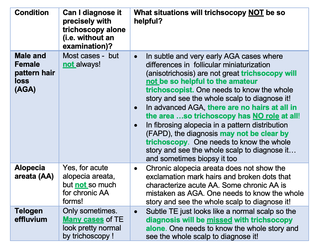
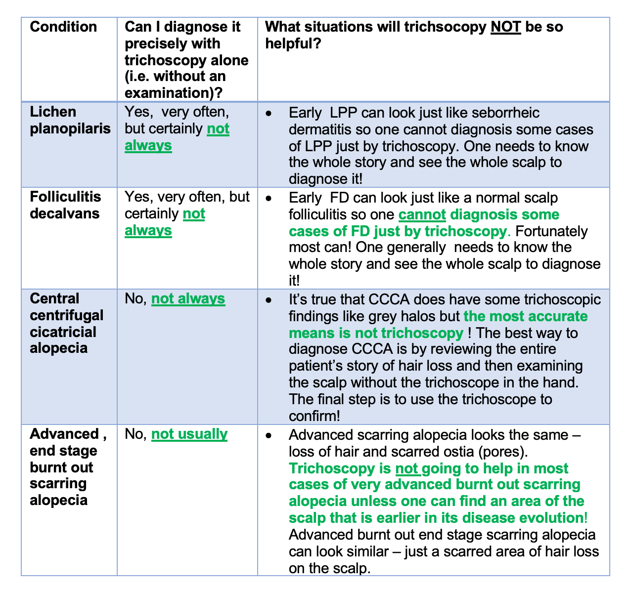
In summary, I can diagnose so many conditions with trichoscopy - but there are so many situations that I can not.

Let’s take a look at some situations where trichsocopy has it’s limitations.


EXAMPLE 1: TRICHOSCOPY IN THE NON SCARRING ALOPECIAS

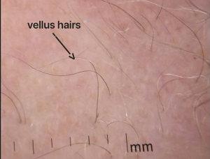
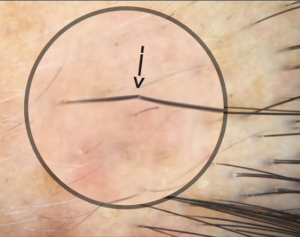
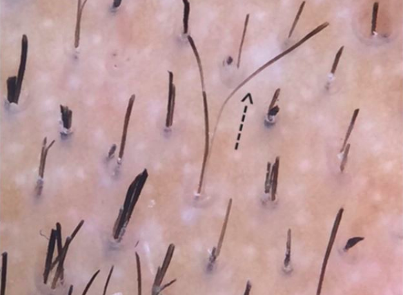
Trichoscopy is completely wonderful. It helps me tremendously in the diagnosis of many non scarring alopecia. Most cases of androgenetic alopecia can be diagnosed with trichoscopy but not all! In fact, unless one is very experienced with trichscopy, the early cases of AGA are going to be very challenging to diagnose by trichscopy because there is just not enough miniaturization that has developed yet. So, if a patient buys a trichoscope and sees that their is not much miniaturization, can they conclude they don’t have AGA? No.

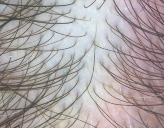
Most cases of acute alopecia areata can be diagnosed with trichoscopy. This is certainly one area where trichoscopy is very helpful. But in cases of advanced AA and some cases of alopecia areata incognito, all that might be seen is miniaturization of hairs. It can be difficult to render the diagnosis from trichscopy alone. So how do we diagnose it? Listen to the patient’s story!

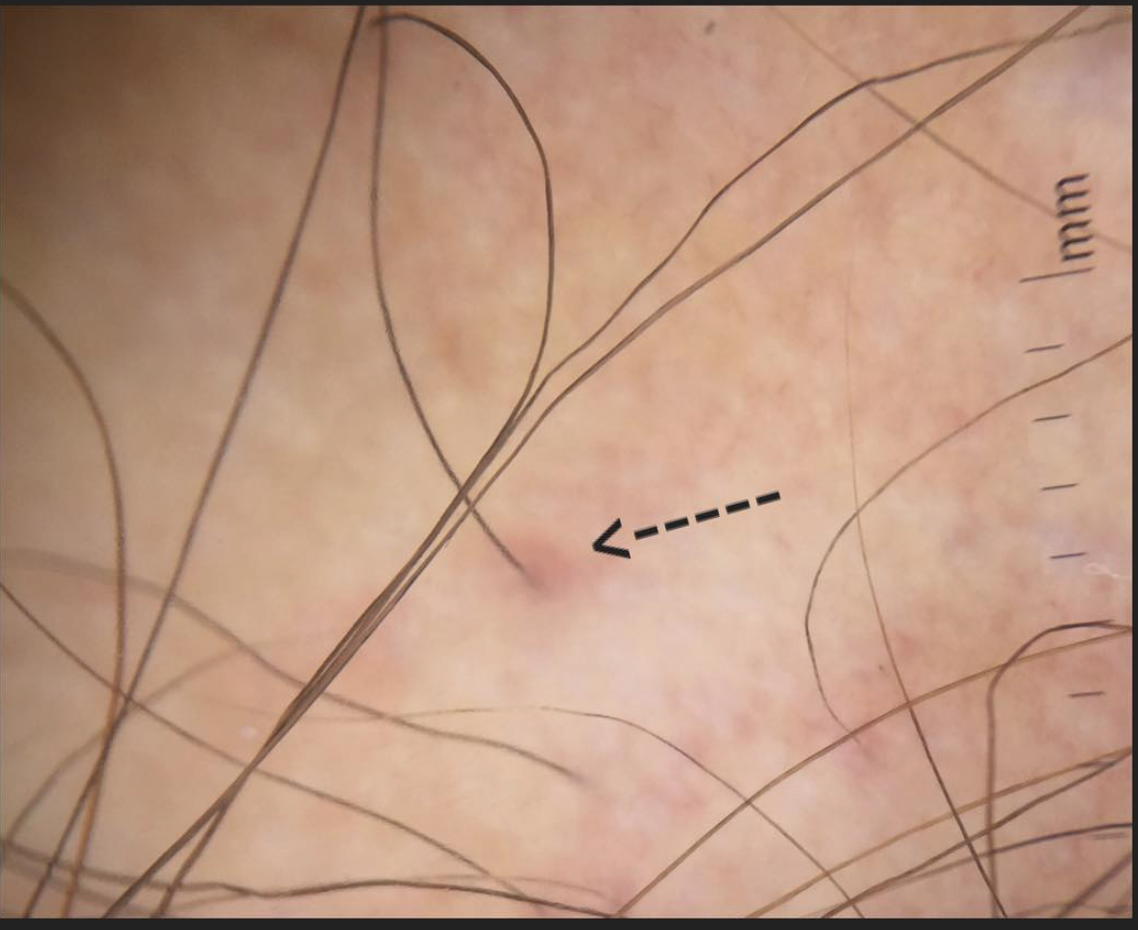
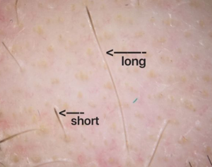
Telogen effluvium (TE) refers to a type of hair shedding and is one of the more common diagnoses in women. Guess what? Telogen effluvium has NO definitive specific diagnostic trichoscopic signs ! Yikes! it’s true that the presence of many upright regrowing hairs can be a tip off from trichoscopy that the diagnosis of TE might be present - but it’s not specific. If a person thinks they are going to diagnose their TE by buying a trichoscope, they are wrong.

trichoscopy in TE- limitations


EXAMPLE 2: TRICHOSCOPY IN THE SCARRING ALOPECIAS

Trichoscopy is completely wonderful. It helps me tremendously in the diagnosis of many scarring alopecia. In fact, the use of trichoscopy has massively reduced my need to perform scalp biopsies. That said, one needs to be aware that some cases of early lichen planopilaris can’t be confidently diagnosed with trichoscopy - the scalp looks just like seborrheic dermatitis! Some cases of early folliculitis decalvans look just like regular ordinary folliculitis !

So does trichosopy help in all these subtle and early forms of these diseases? - no ! It gets me thinking but usually a biopsy is needed to confirm these challenging diagnoses.

Let it be heard though - a good majority of scarring alopecia cases can be diagnosed with trichosopy. Just not all!

As for central centrifugal cicatricial alopecia (CCCA), the best way to diagnose this condition is simply to look at the scalp! Trichoscopy can help but there are not a great number of classic trichscopic signs for CCCA.

trichoscopy scarring alopecia


FINAL SUMMARY

Many patients want to get blood tests because they think that the blood tests will provide the entire answer the diagnosis. Many patients want to buy a trichoscope (USB dermatoscope) because they feel the trichoscope will provide the answers.

We must always remember that the confident diagnosis of hair loss from from use of the diagnostic SET - all comments from the patient’s story, scalp examination,, trichoscopic examination and blood tests go into figuring out the exact cause.


This article was written by Dr. Jeff Donovan, a Canadian and US board certified dermatologist specializing exclusively in hair loss.
Share This
No Comments

Trichoscopy of Telogen Effluvium (TE) - A Closer look at Kenogen Phase and Yellow Dots

Trichoscopy of Telogen Effluvium - Kenogen Phase & Yellow Dots

For a period of time after a hair is shed from the scalp, it appears that the hair follicle opening sits empty. Of course, it is not truly empty because deep underneath the scalp follicular machinery is preparing yet again to manufacture a brand new hair. The period of time between a shed hair and the emergence of a new hair is called “kenogen”. Kenogen phase is typically quite short (1-2 months). The duration that a follicle remains in kenogen increases in patients with some types of hair loss. For example, in some cases of androgenetic alopecia, hairs have been found to remain in kenogen for up to 1 year.

The “sudden” appearance of hair follicle openings that are empty and lack follicles must also prompt one to consider a diagnosis of telogen effluvium.

Yellow dots in telogen effluvium signify hairs in kenogen phase. This finding is a non specific finding of telogen effluvium.

Yellow dots in telogen effluvium signify hairs in kenogen phase. This finding is a non specific finding of telogen effluvium.


Sometimes we see upright regrowing hairs by dermoscopy as a sign of telogen effluvium but sometimes the dermatoscopic findings are extremely non specific and the only finding by trichoscopy is empty pores. It’s possible to have large numbers of hairs in kenogen in advanced stages of androgenetic alopecia but this is uncommon in the early stages. The finding of many empty pores is atypical of the early stages of androgenetic alopecia. The patient in this trichoscopic image androgenetic alopecia as one of the diagnoses. Variation in the caliber of hairs is easy to spot. However the large number of empty follicles is unexpected prompting one to consider that hairs have recently shed - as in a telogen effluvium. Causes include stress, low ferritin levels, thyroid dysfunction, crash diets, medications and internal illness. In this patient, a 49 pound weight loss over 2 months together with low ferritin levels and extremely intense and sudden stressful life events caused the patient’s androgenetic alopecia to suddenly “appear worse.


This article was written by Dr. Jeff Donovan, a Canadian and US board certified dermatologist specializing exclusively in hair loss.
Share This
No Comments

Trichoscopy of Folliculitis Decalvans

Key Trichoscopic Signs of Folliculitis Decalvans

Folliculitis decalvans (FD) is a scarring alopecia which causes permanent hair loss. Patients develop red, itchy scalps that often contains pimples. Bacteria such as Staphylococcus aureus can sometimes be isolated when swabs are taken from these pimples.

A number of “trichoscopic” or “dermatoscopic” signs are suggestive of folliculitis decalvans including some I have shown here: (1) perifollicular “tubular” scaling, (2) compound follicles containing 6 or more hairs, (3) linear fibrotic bands and the (4) red “strawberry ice cream” color.

(1) Perifollicular “tubular” scaling

Trichoscopy (Scalp Dermoscopy) of Folliculitis Decalvans, Image 1. tubular scaling. The scale rides up higher on the hair shaft in folliculitis decalvans than in lichen planopilaris.

Trichoscopy (Scalp Dermoscopy) of Folliculitis Decalvans, Image 1. tubular scaling. The scale rides up higher on the hair shaft in folliculitis decalvans than in lichen planopilaris.

(2) Compound Follicles (Containing 6 or more Hairs)

Trichoscopy (Scalp Dermoscopy) of Folliculitis Decalvans, Image 2. “Compound follicles” are follicles containing more than 6 hairs emerging from a single pore.

Trichoscopy (Scalp Dermoscopy) of Folliculitis Decalvans, Image 2. “Compound follicles” are follicles containing more than 6 hairs emerging from a single pore.

(3) Linear fibrotic bands

Trichoscopy (Scalp Dermoscopy) of Folliculitis Decalvans, Image 3. Linear Fibrotic bands indicate a pattern of scarring associated with the typical starburst scaling.

Trichoscopy (Scalp Dermoscopy) of Folliculitis Decalvans, Image 3. Linear Fibrotic bands indicate a pattern of scarring associated with the typical starburst scaling.

(4) Red “Strawberry ice cream” Color.

Trichoscopy (Scalp Dermoscopy) of Folliculitis Decalvans, Image 4. Strawberry Red Color.

Trichoscopy (Scalp Dermoscopy) of Folliculitis Decalvans, Image 4. Strawberry Red Color.


Folliculitis decalvans (FD) vs Lichen planopilaris (LPP).

Folliculitis decalvans can resemble lichen planopilaris at first glance. However, it does have many differences. Compared to LPP, FD is more likely to have pustules, is more likely to bleed, is more likely to showing compound follicles or “tufting” and is more likely to have tubular scaling the climbs up the follicles (as in this image) and more likely to have these linear fibrotic bands too. Treatment for FD has been discussed in other posts but includes antibiotics, isotretinoin as well as other treatments. 


This article was written by Dr. Jeff Donovan, a Canadian and US board certified dermatologist specializing exclusively in hair loss.
Share This
No Comments

Lichen planopilaris: Classic trichoscopic findings

Classic trichoscopic findings of LPP

Classic trichoscopy of active lichen planopilaris, an immune mediated scarring alopecia is shown below.

Classic trichoscopic image from a patient with active lichen planopilaris (LPP)

Classic trichoscopic image from a patient with active lichen planopilaris (LPP)

There is redness and scale around hairs (called perifollicular erythema and perifollicular scale). Some hairs are twisted (called pili torti). The areas of scalp devoid of hairs are no longer red as the immune system has destroyed hairs in that area and has since left the area. Treatments discussed in other posts as in the following link.

Treatments for LPP: What is available?


This article was written by Dr. Jeff Donovan, a Canadian and US board certified dermatologist specializing exclusively in hair loss.
Share This
No Comments

Scalp Itching in Psoriasis: Don't Forget about "Psoriasitch"

Scalp Itching in Psoriasis Affects Quality of Life

Psoriasis is an immune disease that affects multiple organs and structures. Dermatologists treat skin psoriasis on a daily basis. Rheumatologists treat psoriatic arthritis on a daily basis. 
The scalp is involved a large proportion of patients with psoriasis. In fact, nearly two thirds of patients with psoriasis have scalp psoriasis and it is often the first area where the disease shows up. 

Having scalp psoriasis affects people’s quality of life. Scalp itching is often present and the itching can be troublesome for many. In fact, when I teach other physicians about scalp psoriasis I always encourage them to remember the word “psoriasitch.” It’s a completely made up and completely invented word but it helps clinicians remember the often debilitating effects of the itching that comes with having psoriasis. Not only do we need to remember the flaking and redness and sometimes hair loss that goes with it - but we absolutely need to address the itching these patients live with. And so I call it “psoriasitch.”

The mechanisms that lead to itching are actually quite complex for patients with psoriasis and involve parts of the immune system, nervous system, endocrine system and blood vessels. The mast cell, a key cell normally involved in “allergic responses” in the body, seems to be a key cell type involved in generating itching in patients with psoriasis.

Trichoscopic image from a patient with psoriasis. The patient was extremely itchy. Scalp itching impacts qualify of life and must be addressed by practitioners.

Trichoscopic image from a patient with psoriasis. The patient was extremely itchy. Scalp itching impacts qualify of life and must be addressed by practitioners.


A wide variety of traditional anti-psoriasis agents can reduce itching. Topical steroids, vitamin D analogues, methotrexate, retinoids, and the newer biologics can reduce inflammation and reduce itching. But whether other non traditional strategies that affect the nervous system, endocrine system, blood vessels can also help stop itching is the subject of intense research interest worldwide.

The image here is a trichoscopic image from a patient with scalp psoriasis. The scale is white in contrast to the yellow of seborrheic dermatitis. There is scale around hairs (“perifollicular scale”) and certainly other conditions such as lichen planopilaris could be considered.


This article was written by Dr. Jeff Donovan, a Canadian and US board certified dermatologist specializing exclusively in hair loss.
Share This
No Comments

Challenge Case of Compound Follicles - Final Diagnosis Lichen planopilaris

The Case of Compound Follicles

“Compound follicles” are follicles with 6 or more hairs coming out of a single pore. Occasionally, follicles with 4,5 and rarely 6 hairs coming out of a single pore can be found as an isolated finding- especially towards the more posterior regions of the scalp. But large groupings of hairs like this are rare with advancing age and in fact - the presence of many areas containing compound follicles should prompt the clinician to consider that the patient might have a scarring alopecia. Compounding occurs when 2 or more adjacent follicles “fuse” together. They do not occur because a hair follicle suddenly makes more hairs. Compound follicles are more common in neutrophilic scarring alopecias like folliculitis decalvans (FD) than in lymphocytic scarring alopecias like lichen planopilaris (LPP). Nevertheless, occasional follicles with 5 and 6 hairs can be found in LPP…. as we’ll see in the case below.

I’ve included below a schematic diagram showing the typical findings in lichen planopilaris (LPP) and folliculitis decalvans (FD). Lichen planopilaris typically has no compounding although rarely it’s true that we can see it ….as we’ll seen in the case below. Compounding is more common in folliculitis decalvans.

compound follicles


Generally speaking, the tendency in LPP is for hairs to be destroyed and follicles to contain fewer and fewer hairs over time rather than to contain more follicles in them.

Case

case 1


The case was a patient who presented with redness in the scalp and a loss of hair density. Trichosocpy of the scalp is shown in the photo above. The arrow points to a follicle with 6 hairs coming out of a single pore. There were not many other features in this photos that suggested LPP such as perifollicular scale or perifollicular erythema or pili torti or scarring.

Below is a more typical photo of lichen planoplaris. As you’ll likely agree, the above photo doesn’t quite look like the only below. The typical photo of LPP has scale around hairs. Furthermore, most of the hairs either come out of the pores in groups of 2 or just one hair or no hair at all !

Typical trichoscopic image of lichen planopilaris. Perifollicular scale (white scare around hairs) is evident.

Typical trichoscopic image of lichen planopilaris. Perifollicular scale (white scare around hairs) is evident.


Occasional hairs in our patient’s case had a thicker scale than expected in LPP which prompted me to also consider whether this could be “starburst” scaling of FD. A “hint” of crusting is present but there are no pustules. Redness is interfollicular. A typical trichoscopic image of FD is shown below:

Typical trichoscopic image of folliculitis decalvans. Starburst scale is seen around hairs and compound hairs (hairs with more than 6 hairs coming out of a single follicular opening) are clearly evident.

Typical trichoscopic image of folliculitis decalvans. Starburst scale is seen around hairs and compound hairs (hairs with more than 6 hairs coming out of a single follicular opening) are clearly evident.


A biopsy was be done to evaluate for the possibility of scarring alopecia. The biopsy returned showing lichen planopilaris with no features of folliculitis decalvans. The biopsy also showed that there was a perifollicular inflammatory infiltrate of lymphocytes together with lichenoid change (death of keratinocytes) in the outer root sheath. Perifollicular fibrosis was seen along with loss of sebaceous glands.

FINAL DIAGNOSIS: LICHEN PLANOPILARIS. 

Comment on Case

This was a nice example of a case that was atypical. Not all patients with lichen planopilaris have a typical presentation. Most however have scalp symptoms (like itching or burning or tenderness). Most have redness of some sort in the scalp. Most of scale around hairs. This patient had a bit of subtle redness and not really that much in the way of symptoms. The patient had some unusual compounding by trichoscopy which was the tip off that something might not be right. The biopsy confirmed the diagnosis of lichen planopilaris. Compounding is not a typical feature of LPP but certainly can be seen from time to time. It’s usually not a feature seen in all regions of the scalp and usually the compounding is limited to less than 7 hairs. Compound hairs containing 10, 15 our 20 hairs are almost never seen in lichen planopilaris (LPP) but can be seen very commonly in folliculitis decalvans (FD).

The patient was started on topical steroids and steroid injections together with hydroxychloroquine. The patient will be seen back in 3 months to review response to treatment. Blood tests will be needed monthly for three straight months for CBC, AST, ALT while starting hydroxychloroquine. An eye examination will be needed within 6 months. Clinical photos and trichoscopic photos were taken at the first visit and wil be compared to photos taken a the 3 months follow up. The hope is that redness will be reduced and that that patient’s perception of increased shedding will be reduced. I will monitor over time if more hair loss occurs. Regrowth may or may not occur in scarring Alopecias and this is not a main goal. The goal of treatment is to stop it from getting worse.


This article was written by Dr. Jeff Donovan, a Canadian and US board certified dermatologist specializing exclusively in hair loss.
Share This
No Comments

Vellus Hairs in Alopecia Areata: Sensitivity, Activity, Severity

AA-vellus-sensitivity.png

Vellus hairs are short, thin hairs are commonly seen in patients with alopecia areata. These hairs tend to be seen in patients with more severe and active disease. 
This photo shows vellus hairs in a patient with advanced alopecia areata involving 85 % of the scalp.


This article was written by Dr. Jeff Donovan, a Canadian and US board certified dermatologist specializing exclusively in hair loss.
Share This
3 Comments

Alopecia Areata: Broken Hairs

Hair Breakage

hair breakage.png

Alopecia areata is an autoimmune disease that affects hairs and nails. Inflammation deep under the skin in a region of the hair follicle known as the bulb leads to the production of weak hair follicles that break easily. Hair breakage is commonly seen in active alopecia areata. The photograph here shows a hair follicle that is about to break. Within hours the hair will likely break off at the site demarcated by the arrow. Treatment of alopecia areata can reduce inflammation and lead to the production of stronger hairs that do not break. 


This article was written by Dr. Jeff Donovan, a Canadian and US board certified dermatologist specializing exclusively in hair loss.
Share This
No Comments

Secondary Hair Casts: Psoriasis

What are hair casts?

hair casts.png

Hair casts are thin, elongated, cylindrical concretions that encircle the hair shaft. Hair casts range in size from 2-7 mm and can be easily dislodged. The term was coined by Kligman in 1957.

Hair casts (sometimes called “pseudonits”) can be easily differentiated from true “knits" because they slide along hairs when grabbed with the fingers. They are usually asymptomatic and particularly common in young women.

Hair casts are said to be "primary" in nature when not associated with an underlying scalp disorder and "secondary" when associated with an underlying disorder. Common secondary causes include psoriasis, seborrheic dermatitis, pemphigus and traction alopecia and scarring alopecia. Many other causes are possible too including hair sprays and deodorants.

Hair casts are thought to represent material from both the internal root sheath and the external root sheath.

The photo here shows casts in a patient with psoriasis.


This article was written by Dr. Jeff Donovan, a Canadian and US board certified dermatologist specializing exclusively in hair loss.
Share This
No Comments

Scalp Psoriasis: Many Variations

Many Variations

scalp-psoriasis-many.png

Dermoscopy of Scalp psoriasis. There are many variations in how scalp psoriasis appear. It can be red to pink and scaly white to scaly silver. Psoriasis needs to be differentiated from a range of inflammatory conditions such as seborrheic dermatitis, dandruff, scarring alopecia and various infectious causes.


This article was written by Dr. Jeff Donovan, a Canadian and US board certified dermatologist specializing exclusively in hair loss.
Share This
No Comments

Perifollicular erythema in FFA

Redness around hair follicles: Perifollicular eythema

Frontal fibrosing alopecia or "FFA" is a scarring alopecia (scarring hair loss condition) that most commonly develops in women 45-65. It causes permanent hair loss. The cause is not know although a mix of immune based mechanisms and hormonal mechanics are likely to contribute. The condition can be asymptomatic - and many patients have no itching, burning or pain.


FFA Disease Activity: How do we know it's' active or not

PFE

How do we know whether a given patient's FFA is active? Undoubtedly, the absolute best way is with a photo. If a patient's photo changes over a period of monitoring (6 months or 12 months) the FFA is active by definition.

However, photographs don't capture subtle changes in activity. To accomplish this examination by "dermoscopy" is helpful. In this photo, slight redness around the hairs can be seen. We call this "perifollicular" erythema. (note peri means "around"). In 2013, Spanish researchers Toledo- Pastrana and colleagues published an article in the International Journal of Trichology examining dematoscopic features of FFA. Of 79 patients examined, 66 % showed perifollicular erythema. In patients with active disease, perifollicular erythema was present in 95 % of patients.


Conclusion


Perifollicular erythema is an important sign to look for in FFA. It indicates disease activity and a high likelihood of further hair loss in the patient.


Reference


Toledo- Pastrana et al. Perifollicular erythema as a trichoscopy sign of progression in frontal fibrosing alopecia. Int J Trichoscopy 2013; 5: 151-3.


This article was written by Dr. Jeff Donovan, a Canadian and US board certified dermatologist specializing exclusively in hair loss.
Share This
No Comments

Dissecting Cellulitis: Early Features

Early Stages of DSC

Dissecting Cellulitis (DSC) is a relatively rare scarring alopecia. Men are affected much more than women and affected patients are frequently young males in their 20s.

dsc2

The condition beings with discharge of pus and sometimes blood from the scalp. It can also be quite itchy. The beard, underarm (axilla) and groin can be affected with similar discharge. A key feature of diagnosis is the presence of sinus tracts or "tunnels" underneath the skin. Small thin vellus hairs are also seen in the affect area.

The primary treatment of DSC is isotretinoin although antibiotics, zinc, dapsone, colchicine, and TNF inhibitors can be used.


This article was written by Dr. Jeff Donovan, a Canadian and US board certified dermatologist specializing exclusively in hair loss.
Share This
No Comments

Pseudopelade of Brocq

ppb2.png

Pseudopelade of Brocq

Pseudopelade of Brocq (PPB) is a scarring alopecia. It causes permanent hair loss. The cause is unknown.

In contrast to lichen planopilaris, there is little to no scale around hair follicles. The areas may be pink-colored when active. Treatment is similar to lichen planopilaris including use of topical steroids, steroid injections, topical calcineurin inhibitors, oral methotrexate, oral doxycycline, oral hydroxychloroquine and others.


This article was written by Dr. Jeff Donovan, a Canadian and US board certified dermatologist specializing exclusively in hair loss.
Share This
No Comments

Alopecia Areata

Shaved scalp: Exclamation mark hairs

AA shaved scalp.png

Many patients with advanced alopecia areata shave their scalp. For some, this allows a wig to fit better. For others, especially men, the shaving is done to reduce the appearance of hair loss. 
Even with a shaved scalp, it is sometimes possible to tell if a patient's alopecia areata is active or not. This is especially true if exclamation mark hairs can be seen. "Exclamation mark" (arrow) hairs are easy to identify with a magnifying device. They are 3-5 mm in size and wide at the top and narrow at the bottom. They signal disease activity and the need for more aggressive treatment if hair loss is to be stopped.

Other features can also be seen on a shaved scalp including yellow dots (and hair follicles lacking a hair follicle) and hair follicles with just a single hair coming out (rather than in groups of 2 and 3 haired follicles).


This article was written by Dr. Jeff Donovan, a Canadian and US board certified dermatologist specializing exclusively in hair loss.
Share This
No Comments

Eccrine Glands: White Dots

White Dots

eccrine glands.png

The eccrine glands are a type of sweat gland. They are abundant on the scalp (and even more abundant on the palms and soles). The eccrine gland openings can be more readily seen via trichoscopy (dermoscopy) in darker colored skin than lighter colored skin. This photo shows an arrow pointing to an eccrine gland opening. The variation in the thickness of hairs that can be seen in this image indicates an underlying diagnosis of androgenetic alopecia as well. 


This article was written by Dr. Jeff Donovan, a Canadian and US board certified dermatologist specializing exclusively in hair loss.
Share This
No Comments

EXCLAMATION MARK HAIRS IN ALOPECIA AREATA

 Variations In Size

mark hairs.png

Exclamation mark hairs are hairs that are quite short. Most exclamation mark hairs are 4-6 mm in length but rarely they can be longer depending on the amount of inflammation under the scalp beneath that specific hair. Here in this photo, we can see two exclamation mark hairs of different lengths - one is 3 mm and the other is nearly 15 mm in length. Exclamation mark hairs are seen in alopecia areata, trichotillomania and a few other conditions as well. They are important signs in alopecia areata as they indicate disease activity


This article was written by Dr. Jeff Donovan, a Canadian and US board certified dermatologist specializing exclusively in hair loss.
Share This
No Comments

Female Pattern Hair Loss


Major and Minor Criteria

fphl.png

Female androgenetic alopecia is common. By the age of 50, well over 1/3 of women will have androgenetic alopecia (AGA)- also known as female pattern hair loss (FPHL). This type of hair loss causes thinning in the frontal and mid scalp. The sides and back may also be affected but generally to lesser degrees than the front for most women. Traditionally, the diagnosis of androgenetic alopecia has been made based on the finding of reduced density in the frontal scalp compared to the back of the scalp and the clear demonstration via dermoscopy that there is a variation in the diameter if more than 20% of hair follicles. This is known as anisotrichosis.

In 2009, Dr Rudnicka and colleagues proposed a series of major and minor criteria for diagnosing FPHL.


FPHL MAJOR CRITERIA

(1) ratio of more than four empty follicles in four images (at 70-fold magnification) in the frontal area

(2) lower average thickness in the frontal area compared to the occiput

(3) more than 10% of thin hairs (<0.03 mm in diameter) in the frontal area.


FPHL MINOR CRITERIA

(1) increased frontal to occipital ratio of single-hair pilosebaceous units

(2) vellus hairs

(3) peripilar signs.


Remarkably, the presence of two major criteria or of one major and two minor criteria allow diagnosis FPHL with 98% specificity.


Reference

Rakowska A et al. Int J Trichol. 2009;1:123–30.


This article was written by Dr. Jeff Donovan, a Canadian and US board certified dermatologist specializing exclusively in hair loss.
Share This
No Comments

Trichoscopy of Hair Dyes

Hair Dye Colors the Openings of Hair Follicles

hair dye

Hair dye can sometimes alter the up close appearance of the scalp. Hair dyes can deposit on the scalp given the appearance of hyperpigmentation. In addition, the hair follicle openings or “pores” can take up pigment simulating the appearance of “dots.”

Hair dye is generally safe for most patients with hair loss. However one must be aware that freshly dyed hair may take on a slightly altered appearance. This photos shows brown dots coating hair follicles in a patient with frontal fibrosing alopecia (FFA).


See also

Trichoscopy of Hair Dyes


This article was written by Dr. Jeff Donovan, a Canadian and US board certified dermatologist specializing exclusively in hair loss.
Share This
2 Comments

Trichoscopy of Late DLE

Late DLE: Features


Screen Shot 2018-03-04 at 10.54.42 PM.png

Discoid lupus (DLE) is an autoimmune condition affecting the scalp and skin. It can cause permanent hair loss in affected individuals. About 5% develop systemic lupus erythematosus, an autoimmune condition with the potential to affect many organs of the body. Late scalp lesions of DLE show hyperpigmentation, white structureless areas and telangiectatic vessels,


This article was written by Dr. Jeff Donovan, a Canadian and US board certified dermatologist specializing exclusively in hair loss.
Share This
No Comments

Blogs by Topic


Share This
-->