h1.qusth1 { display: none !important; }

QUESTION OF THE WEEK

Dr. Donovan's Articles

QUESTION OF HAIR BLOGS


What Drugs Cause Alopecia Areata?

The authors of a new study set out to determine drugs implicated in the development of alopecia areata (so-called drug-induced AA). Ezemma et al. performed a systematic review using PubMed to search for all published cases of drug-induced AA.

The authors found sixty-six articles (55 case reports, 9 case series, and two retrospective studies) for their systematic review. These 66 articles included 102 patients. The average age was around 40 years, and approximately half of the patients were female (53.9%).

What drugs are implicated in Drug-Induced AA?

Thirty drugs were found to be associated with the development of drug-induced AA. Monoclonal antibodies caused seventy-six percent of drug-induced AA cases. The TNF-alpha inhibitors and dupilumab comprised most of these cases. Overall, TNF inhibitors were responsible for 47.0% of all patient cases and dupilumab 12.7%.

Adalimumab was the most common TNF inhibitor implicated in drug-induced AA. (18.6% of all cases). Other TNF inhibitors included infliximab (14.7%), etanercept (11.7%), and golimumab (1.9%)

The TOP Causes of Drug-Induced AA

Here is the list of top culprits in drug-induced AA:

  1. Adalimumab (18.6% of all published cases) - TNF inhibitor

  2. Infliximab (14.7% of all published cases) - TNF inhibitor

  3. Dupilumab (12.7% of all published cases) - anti-IL-4/13

  4. Etanercept (11.7% of all published cases) - TNF inhibitor

  5. Alemtuzumab (5.8% of all published cases) - monoclonal anti-CD52 antibody

  6. Hepatitis C therapy (ribavirin, interferon-alpha) (3.9% of all published cases)

  7. Cyclosporine (2.9% of all published cases) - calcineurin immunosuppressive agent

  8. Belimumab (2.9% of all published cases) - monoclonal AB; inhibitor of B-lymphocyte stimulator (BLyS)

  9. Cladribine (1.9% of all published cases) - antimetabolic chemotherapeutic

  10. Golimumab (1.9% of all published cases) - TNF inhibitor

  11. Haloperidol (1.9% of all published cases) - antipsychotic

  12. Pembrolizumab (1.9% of all published cases) - checkpoint inhibitor, anti-PD1

  13. Rifampicin (1.9% of all published cases) - antimicrobial

  14. Oral retinoid (1.9% of all published cases) - vitamin A derivative

Epidemiology of Drug-Induced AA

Overall, patchy AA was the most common presentation. This was seen in 70.5% of drug-induced AA cases.

The average time to onset of AA was 11.7 months, so this was not always seen immediately.

Interestingly, most patients had no family history of AA (93.1%).

What is the effect of stopping the culprit drug?

Forty-eight patients discontinued the drug thought to cause AA, and nearly all patients had regrowth regardless of additional treatment.

What if patients chose not to stop?

Forty-two patients continued the drug, and most did not have regrowth unless they underwent additional treatment. However, the vast majority (75%) of patients who continued the implicated drug and received treatment had hair regrowth.

Comment

This is interesting and provides some beneficial information. The implicated drugs are similar to those reported in Ravipati et al. (see link) but not the same. The most common drug in Ravipati is the taxanes.

REFERENCE*

Ezemma O et al. Drug-induced alopecia areata: A systematic review. J Am Acad Dermatol. 2024 Jan;90(1):133-134. doi: 10.1016/j.jaad.2023.05.022. Epub 2023 May 18.


This article was written by Dr. Jeff Donovan, a Canadian and US board certified dermatologist specializing exclusively in hair loss.
Share This
No Comments

Drug Induced Alopecia Areata : A Closer Look at the FAERS Database

Introduction

Medications have the potential to trigger many diseases. This includes hair loss. It’s always important to pay close attention to drugs that cause or trigger a specific disease because this provides important clues to the underlying mechanisms governing the disease pathogenesis.

The FDA Adverse Events Reporting System (FAERS): A Powerful Tool to Track Post-Marketing Side Effects

The Food and Drug Administration (FDA) launched a publicly available database that allows individual patients, healthcare professionals, and manufacturers to report adverse events caused by medications.

This data is essential to better understanding “real-world” side effects. By real-world, I mean side effects outside a well-controlled, highly monitored clinical trial.

There are now over 26 million side effects reported on the FDA database.

Ravipati A et al, 2024

The authors of a new study set out to review the FAERS database to understand better medications associated with alopecia areata.

The authors found 1,331 AA cases reported on FAERS since 1997. Reports are increasing over time. Approximately 75.6% of AA reports were in females, whereas 24.4% were in males. Most cases were in patients ages 18-64, but there are pediatric patients in the database and patients over 65.

Top 10 Medications Associated with AA in FAERS

The following are the ten medications:

  1. 29.2% of AA cases were seen with docetaxel (a taxane chemotherapy drug)

  2. 8.3% were with adalimumab (a tumour necrosis factor (TNF) inhibitor)

  3. 4.7% with trastuzumab (monoclonal antibody; HER2 inhibitor cancer therapy)

  4. 4.4% with cyclophosphamide (alkylating agent- used in cancer treatment and autoimmune dz)

  5. 4.3% with infliximab (a tumour necrosis factor (TNF) inhibitor)

  6. 4.1% with carboplatin (alkylating agent- used in cancer treatment

  7. 3.4% with dupilumab (monoclonal antibody, blocks IL4 and IL13)

  8. 2.7% with levonorgestrel (progestin)

  9. 2.6% with etanercept (a tumour necrosis factor (TNF) inhibitor)

  10. 2.6% with from pertuzumab (monoclonal antibody; HER2 inhibitor cancer therapy)

A Closer Look at the Top 10

Monoclonal antibodies accounted for 6 out of the top 10 drugs. 3 of the top 10 medications were chemotherapeutic agents, including two HER2 receptor inhibitors and carboplatin.

Among the monoclonal antibodies, males were more likely to report AA when taking adalimumab (OR: 1.79, P = 0.04) and dupilumab (OR: 2.56, P = 0.03).

Other less common reported causes in the FAERS (rank order 11-30)

11. methotrexate (antimetabolite used in cancer therapy and autoimmune disease)

12. levocetirizine (antihistamine)

13. secukinumab (interleukin-17A (IL-17A) receptor inhibitor)

14. montelukast (leukotriene receptor antagonist - used in allergy treatment)

15. Olopatadine (selective histamine H1 antagonist and mast cell stabilizer)

16. prednisolone (corticosteroid)

17. tacrolimus (calcineurin inhibitor immunosuppressant)

18. doxorubicin (anthracycline chemotherapeutic drug)

19. fexofenadine (antihistamine)

20. tofacitinib (JAK 1/3 inhibitor)

21. levothyroxine (thyroid medication)

22. azathioprine (antiproliferative and immunosuppressive agent)

23. ustekinumab (monoclonal antibody, anti IL-12/23 inhibitor)

24. sertraline (selective serotonin reuptake inhibitor (SSRI)

25. simvastatin (cholesterol medication)

26. leflunomide (disease-modifying anti-rheumatic drug)

27. paclitaxel (taxane chemotherapeutic)

28. dexamethasone (corticosteroid)

29. certolizumab (a tumour necrosis factor (TNF) inhibitor)

30. Ribavirin (nucleoside analogue, used in hepatitis C treatment)

Comment

This is an interesting paper that highlights the new world of drug-induced AA. Monoclonal antibodies will increasingly be a part of our therapeutic toolbox for many diseases, so it will be important to understand how this changes over time. Of the 26 million side effects in FAERS, just 1400 are related to AA. It could be that this is underreported. However, drug-induced AA is still likely a minor contributor to AA.

REFERENCE

Ravipati A et al. A cross-sectional analysis of medications used by patients reporting alopecia areata on the FDA adverse events reporting system. Int J Dermatol. 2024 Apr;63(4):497-502. doi: 10.1111/ijd.17014. Epub 2024 Jan 12.


This article was written by Dr. Jeff Donovan, a Canadian and US board certified dermatologist specializing exclusively in hair loss.
Share This
No Comments

A Meta-Analysis of the Effects of Smoking on AGA

Over 1 billion people in the world smoke - which here is defined as the consumption of nicotine-containing tobacco. Smoking is felt to be a risk factor for both the development of AGA and the worsening AGA. The subject, however, remains somewhat controversial.

Several studies have examined the relationship between smoking and androgenetic alopecia. To date, there are almost a dozen such studies.

Mosley and Gibbs, 1996

The Mosley and Gibbs study of 1996 often gets referenced when it comes to smoking and hair loss studies. This was one of the first reports that meant to address the relationship between smoking and hair loss meaningfully. The authors studied 606 patients (268 male and 338 female) aged 30 and over who were visiting a surgical outpatient department over the three months of the study. Patients who were smokers had a nearly two-fold greater risk for balding than non-smokers (odds ratio 1.93 (95% confidence interval [CI]: 1.13–3.28). Interestingly, in that same study, smokers were 4.4 times more likely to have greying of hair than non-smokers, implicating smoking in the greying of hair, too.

Gupta et al. 2024

The authors systematically searched the literature for published studies with suitable data better to understand the relationship between smoking and hair loss and then conducted a meta-analysis. In total, the authors found eight studies to include in their meta-analysis.

Results

The authors made several key observations in their study:

  • The authors found that “ever smokers” were more likely than “never smokers” to develop AGA (pooled odds ratio (OR) = 1.82, 95% confidence interval (CI): 1.55–2.14).

  • The odds of developing AGA were significantly higher in men who smoke ten or more cigarettes per day compared to men who smoke up to 10 cigarettes per day (pooled OR = 1.96, 95% CI: 1.17–3.29).

  • For men with AGA, the odds of disease progression (from mild stages to more advanced stages) were significantly higher among ever-smokers than in never-smokers (pooled OR = 1.27, 95% CI: 1.01–1.60).

  • There was no significant association that could be found between smoking intensity (less than 20 cigarettes per day vs 20 or more) and disease progression.

REFERENCES*

Gupta AK et al. A meta-analysis study on the association between smoking and male pattern hair loss. J Cosmet Dermatol. 2024 Apr;23(4):1446-1451. doi: 10.1111/jocd.16132. Epub 2024 Jan 4.

Dai X et al. Evolution of the global smoking epidemic over the past half century: strengthening the evidence base for policy action. Tob Control. 2022 Mar;31(2):129-137. doi: 10.1136/tobaccocontrol-2021-056535

Kavadya T and Mysore V. Role of Smoking in Androgenetic Alopecia: A Systematic Review. Int J Trichology. 2022 Mar-Apr; 14(2): 41–48.

Su LH and Chen T H-H. Association of Androgenetic Alopecia with Smoking and Its Prevalence Among Asian Men. Archives of Dermatology 2007 143; 1401-1406.

Mosley JG and Gibbs AC. Premature grey hair and hair loss among smokers: a new opportunity for health education? British Medical Journal 1996; 313: 1616.

Fortes C, Mastroeni S, Mannooranparampil TJ, Ribuffo M. The combination of overweight and smoking increases the severity of androgenetic alopecia. Int J Dermatol. 2017;56:862–7. [PubMed: 28555720]

Salem AS, Ibrahim HS, Abdelaziz HH, Elsaie ML. Implications of cigarette smoking on early-onset androgenetic alopecia: A cross-sectional study. J Cosmet Dermatol. 2021;20:1318–24. [PubMed: 32946667]

Park SY, Oh SS, Lee WS. Relationship between androgenetic alopecia and cardiovascular risk factors according to BASP classification in Koreans. J Dermatol. 2016;43:1293–300. [PubMed: 27028221]

Gatherwright J, Liu MT, Gliniak C, Totonchi A, Guyuron B. The contribution of endogenous and exogenous factors to female alopecia: A study of identical twins. Plast Reconstr Surg. 2012;130:1219–26. [PubMed: 22878477]

Vora RV, Kota RK, Singhal RR, Anjaneyan G. Clinical profile of androgenic alopecia and its association with cardiovascular risk factors. Indian J Dermatol. 2019;64:19–22. [PMCID: PMC6340244] [PubMed: 30745630]

Gatherwright J, Liu MT, Amirlak B, Gliniak C, Totonchi A, Guyuron B. The contribution of endogenous and exogenous factors to male alopecia: A study of identical twins. Plast Reconstr Surg. 2013;131:794e–801e.


This article was written by Dr. Jeff Donovan, a Canadian and US board certified dermatologist specializing exclusively in hair loss.
Share This
No Comments

The Epidemiology of Alopecia Areata in Germany

Below is a link to an article I wrote on the epidemiology of alopecia areata in Germany. The full article is on the Donovan Hair Academy website.

Eczema, Lupus, Urticaria and Psoriasis Top List of AA Comorbidities in Germany

REFERENCE

Augustin M et al. Epidemiology of alopecia areata and population-wide comorbidities in Germany: analysis of longitudinal claims data. Br J Dermatol . 2024 Feb 16;190(3):374-381. doi: 10.1093/bjd/ljad381.


This article was written by Dr. Jeff Donovan, a Canadian and US board certified dermatologist specializing exclusively in hair loss.
Share This
No Comments

Peripilar Sign in AGA: Is it possible we have it all wrong?

Introduction

It is increasingly clear that androgenetic alopecia (i.e. male and female genetic hair loss or “balding) is an inflammatory condition. Even though the scalp typically looks non-inflamed, scalp biopsies show that inflammation is present just a few millimetres beneath the scalp in an area known as the isthmus and infundibulum. Studies have shown that inflammation occurs relatively early in the course of androgenetic alopecia. This inflammation is believed to facilitate the progressive miniaturization of hair follicles.


Although the scalp usually looks fairly normal and non-inflammatory in patients with androgenetic alopecia, evaluation of the scalp via biopsy may also show features that suggest there is inflammation under the scalp.


The brown peripilar sign (BPPS) was proposed as a trichoscopic sign in patients with AGA. The PPS is defined as the presence of a brown halo, roughly 1 mm in diameter, at the follicular ostium. It is thought to represent the inflammation beneath the scalp in patients with AGA. Let’s look at one of the original 2004 studies that led to the current understanding of the BPPS and then at a new study that calls this all into question!

Brown peripilar sign (arrow) in a patient with androgenetic alopecia (AGA)


The 2004 Deloche Study

In 2004, Deloche and colleagues from France studied 40 patients with androgenetic alopecia. The researchers showed that the brown discolouration around hairs seen with dermoscopy correlated nicely with inflammation under the scalp when evaluated by biopsy. They called this the peripilar sign (PPS) and proposed that it was an early sign of AGA. They found it present in 90% of males and 86% of females with AGA.

“Peri” means around, and pilar means hair. The peripilar sign is also known by many other names, including the “brown peripilar sign (BPPS)” and “peripilar halo.”


Abdalla D et al. 2024

Authors from Egypt set out to study the significance of the peripilar sign (PPS) in a cohort of patients with AGA and to evaluate whether it is associated with perifollicular inflammation.

The authors recruited 100 patients with trichoscopically confirmed AGA. The study group included 87 (87%) females and 13 (13%) males. Patient ages ranged from 16 to 67, and the duration of hair loss ranged from 0.16 to 20 years.

Peripilar signs were present in 50% of the 100 cases studied. Scalp biopsies were performed on 22 patients with PPS and 23 patients without PPS.

Interestingly, perifollicular inflammation on biopsy was present in AGA patients with PPS as well as in those without PPS. PFI was present in 82.2% of all studied biopsies. Inflammation was rated as mild in most patients (89.2%). The inflammation surrounded the infundibulum in 100% of cases. No significant difference was identified between those with and without PPS regarding the amount of inflammation.

The peripilar sign was more often encountered in patients with skin type III (p=0.001). Patients without the PPS had fewer yellow dots. Histopathologically, biopsies from patients with the peripilar sign were significantly associated with absent melanophages (p=0.011).


Comment

This is an interesting study that questions the meaning of the peripilar sign. The authors propose that the peripilar sign in AGA is not linked to perifollicular inflammation. They feel it is a dark colour in those with lighter skin types.

Further studies are needed to verify and extend our understanding of the BPPS.

The authors wonder whether UV radiation is somehow affecting the melanocytes.


REFERENCES*

Abdalla D et al. Peripilar Sign in Androgenetic Alopecia: Does It Really Indicate Peripilar Infiltrate? Dermatol Pract Concept . 2024 Jan 1;14(1):e2024096. doi: 10.5826/dpc.1401a9


Deloche C et al. Histological features of peripilar signs associated with androgenetic alopecia. Arch Dermatol Res. 2004.


This article was written by Dr. Jeff Donovan, a Canadian and US board certified dermatologist specializing exclusively in hair loss.
Share This
No Comments

New Study Adds Support to Notion that COVID19 Can Trigger Alopecia Areata

COVID-19 infection can trigger several autoimmune diseases, including rheumatoid arthritis, lupus, and many more. (For further review, see the article “Autoimmune Disease Risk Increased After COVID19 Infection.) There is growing evidence that COVID-19 infection can trigger AA or flare AA in those who already have the disease.

  • A 2023 study by Lim et al. showed an increase in both alopecia areata and alopecia totalis following COVID infection and a reduced risk in those who were vaccinated

  • A 2023 study by Tesch  et al. et al. showed an increase in both alopecia areata following COVID infection

Kim et al. 2024

A new study supports the notion that COVID-19 infection increases the risk of all forms of alopecia areata in adults.

The authors conducted a cohort study to investigate the association between COVID-19 and AA. They studied patients between October 8, 2020, and September 30, 2021.

The study comprised 259 369 patients with COVID-19 and 259 369 uninfected controls.

Results

To begin with, the authors found an increased risk of telogen effluvium in the cohort with COVID-19 compared with the uninfected cohort (AHR, 6.40; 95% CI, 4.92- 8.33).

Incidence of AA Increased in COVID-19.

The incidence of alopecia areata in patients with COVID-19 was found to be almost two times higher than in patients who did not have COVID infection (i.e. 43.19 per 10 000 person-years [PY] vs 23.61 per 10 000 PY)

Incidence of both Patchy AA and AT/AU Increased in COVID-19.


The authors studied the incidence of mild and severe alopecia areata separately. They showed that the incidence of all forms of alopecia areata increased with COVID-19 infection.

For patchy AA. The incidence of patchy AA was 35.94 per 10,000 PY in patients with COVID-19 and 19.43 per 10,000 PY in controls.

For AT/AU. The incidence of alopecia totalis and unversalis was 7.24 per 10,000 PY in patients with COVID-19 and 4.18 per 10,000 PY in controls.

Adult Patients and Females at Higher Risk

The incidence of AA after COVID-19 was significantly increased in all groups older than 20 years. In this study, the risk of AA was not increased in children and adolescents following COVID-19. There was a higher risk among both females and males, with a higher risk in females than males.


The Prevalence of Alopecia is Increasing in post-pandemic Korea

The authors noted that the incidence and prevalence of AA and AT/AU remained constant from 2006 to 2015. They point out that the prevalence has now increased following the COVID-19 pandemic.


REFERENCES**

Kim J-S et al. Risk of Alopecia Areata After COVID-19. JAMA Dermatol. 2024 Feb 1;160(2):232-235. doi: 10.1001/jamadermatol.2023.5559.

Christensen RE, Jafferany M. Association between alopecia areata and COVID-19: a systematic review.JAAD Int. 2022;7:57-61. doi:10.1016/j.jdin.2022. 02.002 3.

Birkett L, Singh P, Mosahebi A, Dhar S. Possible associations between alopecia areata and COVID-19 vaccination and infection. Aesthet Surg J. 2022; 42(11):NP699-NP702. doi:10.1093/asj/sjac165


Kutlu Ö, Aktaş H, İmren IG, Metin A. Short-term stress-related increasing cases of alopecia areata during the COVID-19 pandemic. J Dermatolog Treat. 2022;33(2):1177. doi:10.1080/09546634.2020.1782820


Kim J, Hong K, Gómez Gómez RE, Kim S, Chun BC. Lack of evidence of COVID-19 being a risk factor of alopecia areata: results of a national cohort study in South Korea. Front Med (Lausanne). 2021;8:758069. doi:10.3389/fmed. 2021.758069


This article was written by Dr. Jeff Donovan, a Canadian and US board certified dermatologist specializing exclusively in hair loss.
Share This
No Comments

Gut Microbiome Changes in Androgenetic Alopecia: The Good and the Bad

Specific Gut microbiota Alterations Linked to AGA

The gut microbiome refers to the array of organisms that live in our bowels. It’s clear that the health of the gut microbiome is extremely important for human overall health. An altered gut microbiome can lead to disease.

A recent study sought to identify changes in the microbiome associated with androgenetic alopecia and those felt to help prevent AGA.

Risk Factors for AGA included the genus Olsenella, genus Ruminococcaceae UCG-004, and genus Ruminococcaceae UCG-010

Preventive factors for AGA included the family Acidaminococcaceae and genus Anaerofilum, along with the genus Ruminiclostridium

Comment:

This was an interesting study. In the coming years, we’ll hear much more about our microbiome and how various changes influence human health.

REFERENCE*

Fu H et al. Roles of gut microbiota in androgenetic alopecia: insights from Mendelian randomization analysis. Front Microbiol. 2024 Apr 2:15:1360445. doi: 10.3389/fmicb.2024.1360445. eCollection 2024.


This article was written by Dr. Jeff Donovan, a Canadian and US board certified dermatologist specializing exclusively in hair loss.
Share This
No Comments

Does minoxidil cause hair greying? A new study suggests the answer is 'yes'

26% of Patients Felt Minoxidil Causing Greying

Authors from Saudi Arabia set out to evaluate whether minoxidil can affect hair greying. They used two surveys: one given to patients and one given to dermatologists.

Patient Survey

The patient survey comprised 453 patients. 56.7% of patients were aged 18-24 and mostly female. 26% of patients felt that minoxidil caused hair greying, and 14.8% noticed other colour changes (such as yellow, orange, and light brown). Hair colour changes occurred within the first five months in about one-third of women, but many had more delayed colour changes. Patients using minoxidil for a longer duration and patients with a family history of greying were more likely to report hair greying.

Dermatologist Survey

In the second surgery, 57 dermatologists completed the survey. 60% of dermatologists reported that they observed hair greying after minoxidil use. 17% of dermatologists who saw grey hair felt that minoxidil was responsible for the greying of hair.

Conclusion

Overall, the authors proposed that prolonged use of minoxidil can cause hair greying, especially in those with a family history of greying.

I really enjoyed reading this study! Every day, I’m faced with numerous questions from patients. A common question is, “Do you think minoxidil is responsible for my greying of hair?” This study suggests that it very well could be!

It’s interesting to me that the product monograph for Rogaine states, “some patients have experienced changes in hair colour and/or texture with ROGAINE MINOXIDIL TOPICAL 2% SOLUTION, Men’s ROGAINE FOAM 5% or Women’s ROGAINE FOAM 5% use.”

What’s even more interesting to me is that the product monograph errs on the side of extreme safety in advising patients to stop using the product if this occurs.

This is not always practical, as some patients will lose hair if they stop! What the product monograph should say if truly the company feels patients should stop is “We advise patients to stop using the product if hair colour changes occur and accept the fact that they may experience hair shedding and hair loss from stopping”

More studies are needed to better understand this phenomenon and determine whether the current era of extreme popularity of oral minoxidil use will lead to more people noticing grey hair.

REFERENCE*

Alhayaza G et al. Topical minoxidil reported hair discoloration: a cross-sectional study. Dermatol Reports. 2023 Aug 10;16(1):9745. doi: 10.4081/dr.2023.9745. eCollection 2024 Mar 12.


This article was written by Dr. Jeff Donovan, a Canadian and US board certified dermatologist specializing exclusively in hair loss.
Share This
No Comments

Topical Minoxidil: Not Great for Patchy AA, but not too Bad Either!

Topical Minoxidil Inferior to Betamethasone dipropionate in Treatment of Patchy Alopecia Areata

I enjoyed reading a study from Pakistan comparing topical betamethasone dipropionate and topical minoxidil in the treatment of patchy alopecia (less than 50% loess). This non-randomized study involved 100 patients—50 received topical betamethasone dipropionate twice daily, and 50 received topical minoxidil twice daily. The study lasted four weeks.


The authors showed that topical betamethasone dipropionate was superior to topical minoxidil in helping people regrow hair. 74 % of patients in the betamethasone dipropionate group achieved a 50 % or more improvement in their hair, compared to 42% of patients in the minoxidil-only group. However, the point that must not be missed in this study is that topical minoxidil still helped!


Conclusion

The study's conclusion is, of course, that topical betamethasone dipropionate twice daily is better than topical minoxidil 5% twice daily for patchy alopecia areata.

What I liked in this study is something that is ignored and pushed aside - that topical minoxidil still performed reasonably well! It was not as good as topical betamethasone dipropionate, but it had a respectable performance.

Let’s face it—topical betamethasone dipropionate and topical minoxidil are both pretty safe over a short period, like four weeks. But you can’t keep rubbing on betamethasone dipropionate forever. Prolonged use can thin the skin and even contribute to side effects if too much is used. One must take breaks every now and then if betamethasone dipropionate is going to be used.

Topical minoxidil, on the other hand, is reasonably safe for continued use. It does not thin the skin, and it is widely available and does not require a prescription in many countries.

One must always remember that topical minoxidil still has a role in the alopecia areata treatment toolbox!


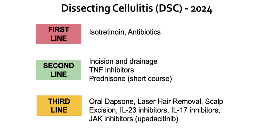
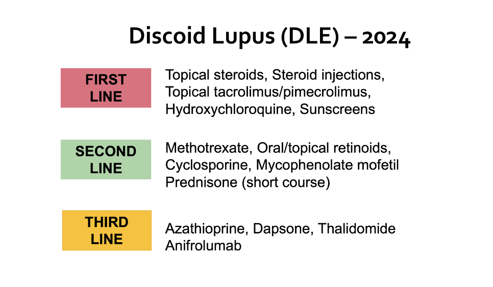
Here are my first line, second line and third line options for the treatment of alopecia areata


REFERENCES

Aslam S et al. Comparison of Efficacy of Topical Betamethasone Dipropionate and Topical Minoxidil in Patients With Alopecia Areata. Cureus . 2024 Mar 16;16(3):e56282. doi: 10.7759/cureus.56282. eCollection 2024 Mar.


This article was written by Dr. Jeff Donovan, a Canadian and US board certified dermatologist specializing exclusively in hair loss.
Share This
No Comments

The Benefits of Topical Minoxidil for Facial hair

Topical Minoxidil helps Facial hair Growth

Many males value good facial hair growth. Growth patterns are strongly genetically determined. There is a tremendous amount of information online about using topical minoxidil to enhance facial hair. Despite all these anecdotal reports, few studies have been done. An important study to note was a 2016 study by Ingprasert et al. This was a randomized study of 48 males using 3 % minoxidil for facial hair growth (…to review, click on the link).


Shokravi and  Zargham, 2024

In a new study, the authors present a case showing the potential benefits of topical minoxidil for facial hair. What was unique in this report was that the patients were identical twin males. One used minoxidil, and one did not. The twins had similar facial hair density before using minoxidil.


TWIN 1: used ¾ cap topical 5% minoxidil foam for over a year on the beard and mustache area

TWIN 2: did not use topical minoxidil


Results in Treated Twin

The study's authors described that the twin using minoxidil had improvements in facial hair that were not seen in the untreated twin. New, finer, lighter-colored hairs were noted by month 1. A modest increase in facial hair density was noted by month 2. The treated twin reported shedding of the hairs in the beard area approximately three months after initiating minoxidil.

Overall, after 16 months of use, the minoxidil-treated twin had a greater hair count and density in both the beard and mustache areas.


Photos show the differences in facial hair ten days after shaving.

SIDE EFFECTS

There were no major side effects.

Minimal mild side effects with foam minoxidil use included:

  • Mild skin dryness

  • hypertrichosis on the ears and forehead

  • An increase in body hair (chest, abdomen, forearms, and legs)


Interestingly, the patient initially used minoxidil solution but switched to the foam due to DRY FLAKY SKIN.


CONCLUSION

This study concluded that topical minoxidil is one strategy to consider for males wishing to improve facial hair growth.


REFERENCES*

Shokravi A and  Zargham  H. Facial hair enhancement with minoxidil-an off-label use. SAGE Open Med Case Rep. 2024 Feb 23:12:2050313X241231490. doi: 10.1177/2050313X241231490. eCollection 2024.

Ingprasert S et al. Efficacy and safety of minoxidil 3% lotion for beard enhancement: A randomized, double-masked, placebo-controlled study. J Dermatol . 2016 Aug;43(8):968-9. doi: 10.1111/1346-8138.13312. Epub 2016 Feb 19.


This article was written by Dr. Jeff Donovan, a Canadian and US board certified dermatologist specializing exclusively in hair loss.
Share This
No Comments

Patients Using Dupilumab Have an Increased Risk of Being Diagnosed with Cutaneous T Cell Lymphoma More Often

Dupilumab and CTCL: What does it mean for our alopecia areata patients?


I’ve been increasingly prescribing dupilumab, the IL4/13 antagonist, in the management of some patients with alopecia areata and atopic dermatitis. Dupilumab is a drug often used for atopic dermatitis but has the potential to help hair growth in a small proportion of patients with alopecia areata. Safety seems reasonably good. Blood tests are not needed for most, and it’s reasonably easy to administer.

We’ve talked about the role of dupilumab in alopecia areata in prior articles.

But then there’s the lymphoma studies. Studies keep coming out showing a similar trend: if you are prescribed dupilumab, you have an increased chance you’ll be diagnosed with a specific skin lymphoma known as cutaneous T cell lymphoma. It’s not a massively increased risk, but it’s an increased risk.

I’ll review a new study in a moment, but let’s talk about this subject!

Challenges in the CTCL Data

Let’s face it; the data is challenging to decipher. On the one hand, some feel the lymphomas associated with dupilumab are not really lymphomas at all. Boesjes CM et al., in 2023, showed that dupilumab treatment can cause a reversible and benign ‘lymphoid reaction’ that mimics a CTCL but is not CTCL.

On the other hand, there are more and more and more reports showing a massive “unmasking” of CTCL following the initiation of dupulimab therapy. The cases showing that some of the patients treated with dupilumab end up dying are a stark reminder of how important this issue is.

Espinosa et al. 2020

In 2020, Espinosa et al. described four patients given dupilumab for presumed atopic dermatitis and 3 for use off-label to treat pruritic cutaneous T-cell lymphoma. (In cutaneous T-cell lymphoma, IL-13 is overexpressed and regulates cell proliferation, so it was initially felt it could be helpful to block tumour progression.)

All patients treated with dupilumab had disease progression. The four patients initially presumed to have atopic dermatitis subsequently received a diagnosis of cutaneous T-cell lymphoma. The three patients with existing cutaneous T-cell lymphoma progressed to Sézary syndrome while receiving treatment, and two died.

Jfri A et al. 2023

Jfri et al. systematically reviewed twenty-three dupilumab-associated CTCL cases from 11 sites.

The median (range) age was 58 (27-74). Atopic dermatitis onset was mostly in adulthood in 19 (82.61%) cases and childhood-onset in 4 (17.39%) cases. In 13 patients (56.52%), skin biopsies done before starting dupilumab were not diagnostic of MF/SS. In 5 patients, there were atypical features for AD (epidermotropism, folliculotropism, lichenoid inflammation).

The mean time between the start of dupilumab and biopsy-proven cutaneous T-cell lymphoma was 7.5 months. Eleven had advanced disease (Stage IIB-IV) at diagnosis. Personal/family atopic history, a major criterion for clinical AD diagnosis, was found in only 4 of 23 cases. Several patients initially diagnosed with atopic dermatitis had features that are not typical of AD, including indurated plaques, nodules, poikiloderma, and sparing of flexural sites.

When looking back at these cases, the authors found that there were two cases where it was clear that the patient had atopic dermatitis to begin with. These are 2 cases with a long-standing diagnosis of AD since childhood, and biopsies done before starting dupilumab showed features consistent with AD. Of those 19 patients with adult-onset atopic dermatitis, only five patients had clinical presentations and pre-dupilumab biopsies truly consistent with AD. In many cases, it was really not possible to exclude that patients had skin lymphoma before they started therapy.

Hasan et al. 2024

In a new study, Hasan and colleagues used a large database to compare the incidence of CTCL in those who used dupilumab compared to those who never used dupilumab.

This was an extensive database. Data was extracted from 60 healthcare organizations, encompassing 22,888 AD patients who were prescribed dupilumab and 1,115,235 AD patients who were not prescribed dupilumab,

The cohort of AD patients who used dupilumab had an increased risk of CTCL (OR 4.1003, 95% CI 2.055-8.192). The risk was not increased for other cutaneous or lymphoid malignancies, like non-Hodgkin lymphoma or leukemia. Most (27/41) cases of CTCL were diagnosed more than one year after dupilumab use, suggesting that perhaps these are not simply unmasked cases.

So does dupilumab Cause or UnMASK Skin Lymphoma?

It’s pretty clear that if you use dupulimab for atopic dermatitis, you are at increased risk of being diagnosed with a type of skin rash that causes everyone to worry. That is something that is undebatable. It’s also clear that if you have a skin rash before starting dupilumab and it does not respond to treatment with dupilumab or gets worse, it needs to be biopsied to make sure one is not missing a diagnosis of CTCL.

We don’t yet have evidence that patients who use dupilumab for alopecia areata who do not have atopic dermatitis develop skin lymphoma or lymphoid reactions.

What is debatable is whether dupilumab actually causes a lymphoma or lymphoid-like reaction or whether patients had this skin malignancy to begin with, and dupilumab simply encouraged it to come out. Atopic dermatitis and cutaneous T-cell lymphoma can certainly mimic each other, and a widely held concern is that some of the cases of CTCL with dupilumab were simply cases that were first misdiagnosed as atopic dermatitis.

Most studies in the literature conclude their study with the unsatisfying sentence that goes something like “These data raise the question as to whether the affected patients were first misdiagnosed as having atopic dermatitis and the CTCL was unmasked by dupilumab or whether cutaneous T cell lymphoma is truly is an adverse effect of treatment with dupilumab. Other studies end with comments like lymphomas developing with dupilumab might be unrelated to the drug use itself, but long-term follow-up data of large patient cohorts is necessary to rule out such a possibility. Even the discussion section of the paper by Hasan et al. starts out with, “It is unknown if dupilumab causes CTCL or unmasks CTCL.”

For now, it’s clear that patients with atopic dermatitis who are considering dupilumab need one (or more) skin biopsies

  1. Their atopic dermatitis starts in adulthood (instead of childhood)

  2. They have a diagnosis of atopic dermatitis but don’t have a strong family history

  3. They have significant body surface area (erythroderma) - also need flow cytometry

  4. They don’t have involvement of the flexures

  5. They have atypical features like nodules or poikiloderma

Patients who start dupilumab and develop new rashes or worsening of existing rashes also need a low threshold to have biopsies.

I’m following this field closely!


REFERENCES*

Hasan I et al. Dupilumab therapy for atopic dermatitis is associated with increased risk of cutaneous T cell lymphoma: a retrospective cohort study. J Am Acad Dermatol. 2024 Apr 6:S0190-9622(24)00566-8. doi: 10.1016/j.jaad.2024.03.039. Online ahead of print.

Boesjes CM et al. Dupilumab-Associated Lymphoid Reactions in Patients With Atopic Dermatitis. JAMA Dermatol . 2023 Nov 1;159(11):1240-1247.


Espinosa ML et al. Progression of cutaneous T-cell lymphoma after dupilumab: Case review of 7 patients. J Am Acad Dermatol . 2020 Jul;83(1):197-199. doi: 10.1016/j.jaad.2020.03.050. Epub 2020 Mar 27.


Harada K et al. The effectiveness of dupilumab in patients with alopecia areata who have atopic dermatitis: a case series of seven patients. Br J Dermatol. 2020 Aug;183(2):396-397.


Poyner EFM et al. Dupilumab unmasking cutaneous T-cell lymphoma: report of a fatal case. Clin Exp Dermatol. 2022 May;47(5):974-976. doi: 10.1111/ced.15079. Epub 2022 Feb 9.

Du-Thanh A et al. Lethal anaplastic large-cell lymphoma occurring in a patient treated with dupilumab. JAAD Case Rep. 2021 Oct 12:18:4-7. doi: 10.1016/j.jdcr.2021.09.020. eCollection 2021 Dec.

Jfri A et al. Diagnosis of mycosis fungoides or Sézary syndrome after dupilumab use: A systematic review. J Am Acad Dermatol . 2023 May;88(5):1164-1166. doi: 10.1016/j.jaad.2022.12.001. Epub 2022 Dec 5.


This article was written by Dr. Jeff Donovan, a Canadian and US board certified dermatologist specializing exclusively in hair loss.
Share This
No Comments

Frontal Fibrosing Alopecia in India: A Reminder of a Truly World-Wide Epidemic

First Report of FFA Cases in North-East India

Several months ago, I had a discussion with a several colleagues. We spoke about FFA and the role of stress, diet, cosmetics, family history, hormones. Basically … everything!

Not too long into the discussion, a point was made by my colleague: “I don’t think we’re seeing much FFA in India!” Another made the point that if this is indeed true that there must be important dietary and genetic reasons that need exploration.

A new report in the Indian Journal of Dermatology reminds us that while FFA in India might be a little less frequent than some pockets of the world, it’s probably understudied - and by no means absent from India!

Authors from North-East India, in Meghalaya state, reported 21 patients with FFA that they saw at their center over the period 2013-2023. 19 were female and the average age was 48 years. 28% had lichen planus pigmentosus of the skin. Only 10 % had androgenetic alopecia and only 10 % had classic LPP.


Comment

This was interesting to see. In this small world we live in with global travel and global products and expanding ingredients, pollution, cosmetics, etc, one can only imagine that FFA must be increasing in most part of the world.

More studies are needed but I congratulate these authors!


REFERENCE

Verma S et al. A Retrospective Study of Frontal Fibrosing Alopecia from North-East India. Indian J Dermatol. 2023 Nov-Dec;68(6):598-602. doi: 10.4103/ijd.IJD_290_23. Epub 2024 Jan 9.


This article was written by Dr. Jeff Donovan, a Canadian and US board certified dermatologist specializing exclusively in hair loss.
Share This
No Comments

1 in 20 Drugs that Get Approved Will Eventually Be Removed From Market

When a new drug comes to market, I’m pretty excited. After all, it’s proof of something I fundamentally believe in : good science can create better treatments and help more people lead fuller lives.

Whenever a new drug comes to market, I also remember something I learned many years ago: about 5 % of the time, the drug you are cheering about is going to eventually be removed from the market because of the harm it caused someone.


Why are drugs removed?

Drugs are removed from the market for several reasons. The top 4 reasons seem to be hepatotoxicity, cardiac disorders hypersensitivity and kidney disease nephrotoxicity. Of course, these are not the only reasons.

According to Craveiro et al, it tends to be the case report and spontaneous reporting that triggers the steps that ultimately lead to removal of a drug - rather than some piece of evidence from a well designed clinical trial. This is important for all of us to remember. Yes, completing that side effect reporting form does make a difference.


How long does it take to get a drug off the market?

A study in Canada by Lechin found that if a drug is going to be removed from the market, then 50% will be gone by 3.5 years and 50% will stay on longer than 3.5 years. A 2020 study by Craveiro NS et al. showed the delay could be even longer. In their study, the average time (rather than median) between the introduction of a drug and its withdrawal due to safety reasons was 20.3 years (SD±13.8).


Comment

Yes, some of the drugs we prescribe are going to eventually be found to harm people seriously enough that they get removed from the market. I’m hoping that number drops lower and lower and lower below 5 %. I still remember the day in 2009 that I learned that Raptiva was removed from the market. I still remember the day in 2009 that I learned that a drug I had prescribed (Raptiva) for psoriasis was removed from the market. The FDA approval of Raptiva was based on initial studies in approximately 2,700 patients - most received the drug for only a few months. Fast forward 6 years to 2009, after over 46,000 patients had received efalizumab. The world came to learn that 3-4 patients developed a rare and serious disease known as progressive multifocal leukoencephalopathy (PML) after using Efalizumab. 3 died. The only way that these side effects came to be realized was through reporting of side effects “after” the drug was released: doctors and patients saw the side effect and reported it regardless of whether they felt it was truly linked or not. Over time, it became clear it was linked.


The next 20 years will see a tremendous number of new treatments in our field. It’s going to be incredible. I’m exited to be part of this expansion of options for our patients.


But approximately 5 % of the time, we’re going to have it all wrong.


REFERENCE

Joel Lexchin. How safe are new drugs? Market withdrawal of drugs approved in Canada between 1990 and 2009. Open Med. 2014; 8(1): e14–e19. Published online 2014 Jan 28.

Craveiro NS et al. Drug Withdrawal Due to Safety: A Review of the Data Supporting Withdrawal Decision. . Curr Drug Saf. 2020;15(1):4-12. doi: 10.2174/1574886314666191004092520.


This article was written by Dr. Jeff Donovan, a Canadian and US board certified dermatologist specializing exclusively in hair loss.
Share This
No Comments

Mice, Men and Evolving The Story of Topical Rosemary For Hair Loss

Topical Rosemary Helps Males and Mice with Hair Loss: Anything Else is Simply A Guess


There is a lot of excitement around the use of topical products containing Rosemary oil for hair loss. The reality is that we’re still not really sure how to use rosemary and we’re still not sure it truly helps all that much. A new study created some “buzz” as it showed rosemary helped grow hair in mice. This study is yet another study in the growing list of studies pointing to a favourable role for rosemary. Let’s take a look at the 2015 study that created quite a bit of interest in rosemary oil and then the new study in mice. We’ll come to see that we still don’t really know if rosemary helps that much.


Panahi et al 2015

This was a randomized controlled study of 100 male patients with androgenetic alopecia. (No females were included in the study - this is strictly a study of males). 50 males applied minoxidil 2 % (not 5 %) twice daily and 50 males applied rosemary oil twice daily. They did this for 6 months.

At the end of the 6 months, hair density increased very slightly from 138.4 hairs per cm2 to 140.7 hairs per cm2 in the minoxidil group and 122.8 to 129.6 in the rosemary group. 24 % of males in the minoxidil group felt they had mild improvement and 38 % in the rosemary group felt they had mild improvement. Nobody in either group felt they had moderate improvement. There were no changes in the perception of greasy hair or dandruff but both groups reported more scalp itching. The itching was greater for minoxidil users than rosemary users.

All in all, the conclusion was if you are male and willing to use rosemary oil twice daily, it may help your hair a little bit. Improvements will be very mild and not any better than 2% minoxidil. Likely 5 % minoxidil is better than rosemary oil given than we know 5 % minoxidil is better than 2 % minoxidil We can’t comment on the effectiveness in females as that was not studied here.


Begum A et al. 2023

A new study in mice showed that a 1 % rosemary lotion was just as helpful as 2 % minoxidil in promoting hair growth in mice. Hair was removed from a 3 cm2 hair from mice using a depilatory cream. Then mice were divided into 3 groups. One group received rosemary lotion on the hairless area twice daily. One group received 2 % minoxidil lotion twice daily and one group receive water (control) twice daily for 30 days. After 30 days, hair regrowth was studied. All in all, hair regrowth was pretty similar in mice treated with 2 % minoxidil and 1% rosemary - and far superior to water


Conclusion

All in all, rosemary is proving itself to have some use in hair regrowth. It’s a pretty mild growth stimulator at best and needs to be applied twice daily. It’s probably not as good as 5 % minoxidil. Just all all treatments, this will need to be done forever in the setting of androgenetic alopecia. We know that rosemary helps males and mice - but we have zero evidence it does anything for females. So, you and I are just guessing if we think it helps women with hair loss. I imagine that it does but that’s just a guess. Rosemary oil can irritate so we’ll need to warn patients about this.


REFERENCES

Panahi Y, Taghizadeh M, Marzony ET, Sahebkar A. Rosemary oil vs minoxidil 2% for the treatment of androgenetic alopecia: a randomized comparative trial.Skinmed. 2015 Jan-Feb;13(1):15-21.

Begum A et al. Evaluation of Herbal Hair Lotion loaded with Rosemary for Possible Hair Growth in C57BL/6 Mice. Adv Biomed Res. 2023 Mar 21:12:60.


This article was written by Dr. Jeff Donovan, a Canadian and US board certified dermatologist specializing exclusively in hair loss.
Share This
No Comments

Spironolactone: Should we also be checking zinc?

A study from Japan examined risk factors for zinc deficiency. The researchers used a study population of 13,100 patients who had reliable data available for zinc levels. Interestingly, associations with zinc deficiency were noted among older adults, males, and inpatients.

Following multivariate analysis, after adjusting for age and sex, there were significant associations with comorbidities, including pneumonitis (adjusted Odds Ratio (aOR) of 2.959), decubitus ulcer and pressure area (aOR 2.403), sarcopenia (aOR 2.217), COVID-19 (aOR 1.889), and chronic kidney disease (aOR 1.835).

What was particularly interesting to me as a hair specialist were the associations with certain drugs. Patients using the following medications were most likely to develop zinc deficiency: spironolactone (aOR 2.523), systemic antibacterials (aOR 2.419), furosemide (aOR 2.138), antianemic preparations (aOR 2.027), and thyroid hormones (aOR 1.864).

Comment

Zinc deficiency can cause a number of issues in humans. Globablly, zinc deficiency is said to affect 2 billion people.

Hair specialists need to know about zinc deficiency given that low zinc can sometimes cause hair loss or at least impact how hair grows.

Spironolactone was surprisingly a top drug in the list of drugs linked to zinc deficiency. More studies are needed to determine if hair loss patients using spironolactone are at risk for zinc deficiency and if we really should be checking zinc levels more often than we tend to do so now.


REFERENCE


Hirohide Yokokawa et al. Demographic and clinical characteristics of patients with zinc deficiency: analysis of a nationwide Japanese medical claims database. Sci Rep. 2024 Feb 2;14(1):2791. doi: 10.1038/s41598-024-53202-0.


This article was written by Dr. Jeff Donovan, a Canadian and US board certified dermatologist specializing exclusively in hair loss.
Share This
No Comments

Baricitinib (Olumiant) Now Approved in Canada for Alopecia Areata

on Friday Jan 26, 2024, Health Canada approved baricitinib for the treatment of severe alopecia areata. This makes it the second approved JAK inhibitor in Canada. Ritlecitinib was approved in early December 2023. Baricitinib is sold under the name Olumiant and Ritlecitinib is sold under the name Litfulo. Our DonovanMedical youtube channel has extensive discussion of both drugs.

VIDEO DISCUSSING RITLECITINIB

VIDEO DISCUSSING BARICITINIB


This article was written by Dr. Jeff Donovan, a Canadian and US board certified dermatologist specializing exclusively in hair loss.
Share This
No Comments

Androgenetic Alopecia: What works?

Article from Cutis Now Published

I appreciated the opportunity to be invited by the journal Cutis to publish on androgenetic alopecia. This article can be downloaded in the following link

Donovan, J. Androgenetic Alopecia: What works? Cutis 2024.

For more information visit the Cutis website.


This article was written by Dr. Jeff Donovan, a Canadian and US board certified dermatologist specializing exclusively in hair loss.
Share This
No Comments

Year End Challenge 2023

Year End Challenge: Myocarditis Following Adenoviral Infection

In keeping with our year end tradition, the following situation was posted on our social medial channels. Participants had the opportunity to guess what was going on and how to help David.

CHALLENGE CASE

David is your healthy 28 year old male patient. He is on 5 mg of oral minoxidil and 0.5 mg dutasteride for treating hair loss.  He leaves a message on your office answering machine on new year’s eve wanting to know what he should do to help his feet swelling and dizziness. There is nobody in the office to take his phone call but he decides to leave a voicemail message.

David says on his message that he has been on oral minoxidil for 9 months and dutasteride for 2 years and absolutely loves his results. He is terrified to stop these drugs but feels they are now starting to cause major problems. He feels these drugs are delaying his recovery from a recent illness and maybe making everything worse.

He tells you in his message that he had a respiratory tract infection 4 weeks ago after attending a family reunion celebration. At the time, David says he had high fever, cough, runny nose. After a few days of feeling sick, David said he felt better for the next 7 days. David says he started feeling so much better that he went out to a party with friends about 1 week ago. He left the party early as he was tired. 

David says things have now worsened to the point where he needs to call you at the office. He continues to be very tired. He tells you has now had 7 straight days of heart burn. He says it started soon after going out drinking and partying with friends last week. He now has mild chest pain and you can hear him coughing on the answering machine message. He has gotten quite dizzy today and feels a bit short of breath. All in all, David feels oral minoxidil is somehow too strong for him and wonders if he should return back to using 2.5 mg. He tells you he notices a sock line so there may be ankle swelling.  He has  not examined it too closely as it’s not very comfortable to be moving around and bending too much.

He tells you that when he first started oral minoxidil 9 months ago at 2.5 mg he had no problems at all. He says he did have problems for a few days when he increased the dose up to 5 mg and he specifically remembers having dizziness and a chest tightness at that time. This lasted only a few days and he has been fine since. He likes oral minoxidil. He feels dizziness is somehow coming back and the mino pill is acting stronger than it really is. 

David says that his two sisters and his mother and 97 year old grandfather were also sick after attending the family reunion 4 weeks ago. He says everyone has completely recovered.  He generally considers himself very healthy so this is all confusing as to why he is not better yet.

David says his friend uses oral minoxidil and had similar symptoms of chest tightness and ankle swelling and needed to reduce the dose to 3.75 mg. His friend had to stop dutasteride because of feeling low energy so he wonders if dutasteride is somehow also causing problems.

David concludes his message by thanking you for listening to his long message and closes by asking “What should I do?”

What would you advise David?

ANSWER TO CHALLENGE CASE

This is a case example of viral myocarditis from the pre-COVID era. The cause here was adenovirus.

Myocarditis refers to inflammation of the heart layer known as the myocardium. Patients with myocarditis can present with a variety of symptoms.  

Most cases of myocarditis are identified in young adults  20-40 with males affected more often than females. Children and adolescents can also have myocarditis and sometimes have a serious course.

Myocarditis must also be considered in patients with shortness of breath, dizziness and especially loss of consciousness. It’s not the only disease that causes these symptoms of course.

Symptoms such as shortness of breath, fatigue and ankle swelling are concerning as they indicate possible heart failure.


Signs and Symptoms of Viral Myocarditis: What do patients experience?

It’s important to realize that there are no unique and specific symptoms that tell a doctor that a patient has myocarditis. Rather, it’s the constellation of symptoms that puts myocarditis on the list. Some patients with myocarditis feel chest tightness or a squeezing sensation.


Adults with viral myocarditis often have a 1-2 week history of a flu-like illness. Patients may have chest pain and shortness of breath. Fatigue, lower blood pressure, fever, tachycardia may be present.  Dizziness and general lightheadedness and even sycope are sometimes present. This may be  due to issues with the conduction system. There can be palpitations and flutters and in some cases even serious life threatening arrythmias.

If there is also pericarditis, then the patient may feel worse leaning back and feel better leaning forward.

Myocarditis can be caused by infections, vaccines, drugs. In the case of viral myocarditis, the patient develops symptoms a few days to weeks after a flu-like illness.  

Most cases of myocarditis are mild and improve with measures that help the heart function and treat conduction problems and rhythm issues.


What causes myocarditis?

Often the cause of myocarditis can’t be determined. In North America and Western Europe, viral infections are the most common identified causes of myocarditis. These include coxsackie virus, echo virus, human herpesvirus 6, parvovirus 19, adenovirus, enterovirus, cytomegalovirus, Epstein Barr virus, and influenza. COVID 19 is now a common cause of myocarditis as well. In some part of the world, bacteria and HIV are important causes.

 Drugs, toxins and autoimmune disease are among other causes.


How is myocarditis diagnosed?

There is no unique blood test that indicates myocarditis. Usually a patient with chest discomfort and shortness of breath will have several tests done in the emergency setting including troponin levels (a test of heart muscle damage), ECG, chest x ray and sometimes tests such as echocardiography. An elevated troponin without evidence that the patient is having a heart attack is often a tip off that myocarditis may be happening.

MRI is increasing used in diagnosing myocarditis. Only rarely is a heart muscle biopsy performed. This is a particularly risky test.

Myocarditis vs pericarditis: Are there differences in symptoms?

Myocarditis is inflammation of the myocardium (middle of the heart wall) where pericarditis is inflammation of the pericardium (a thin sac surrounding the heart). About one-third of patients with myocarditis also have pericarditis. Myocarditis and pericarditis have similar symptoms in many ways so there is a lot of  overlap. But there is some overlap.  It’s often said that pericarditis tends to  be more likely to severe chest pain, while myocarditis is more likely to cause fatigue and shortness of breath. There is overlap of course. The two can be better differentiated with ECG, clinical examination and MRI. In 50 % of cases of myocarditis, the exact cause is never found. In pericarditis, this is as high as 90 % of cases.

How is viral myocarditis treated?

Viral myocarditis is generally treated in what’s called a supportive manner. The patient’s blood pressure, heart rate, heart function is controlled using medications. This may be done in hospital or intensive care (ICU) setting in some cases. Some patients are very sick and require advanced ICU care.  NSAID and steroids are sometimes used in treatment. Colchicine is sometimes used in some cases of pericarditis.

Many cases of mild myocarditis and pericarditis resolve with supportive care.


This article was written by Dr. Jeff Donovan, a Canadian and US board certified dermatologist specializing exclusively in hair loss.
Share This
No Comments

WELCOME TO THE NEW EBHF PARTICIPANTS!

EBHF: 110 participants from 33 countries for 20 months .... with one main goal: … to develop a new level of expertise in the diagnosis and treatment of hair loss!

Just one week to go until I welcome 100 incredible hair loss specialists for a 20 month journey through the Evidence Based Hair Fellowship (EBHF) training program. Participants meet weekly online for a challenging but fun program that stimulates the brain to think and problem solve like an expert.

The journey starts off with 7 weeks of some of the most important material - the basic principles of hair loss diagnosis and treatment. We then journey through “blocks” of 1-2 month duration dedicated to subjects such as androgenetic alopecia, telogen effluvium, alopecia areata, scarring alopecia, hair shaft disorders, psychodermatology, traction alopecia, contact dermatitis, cancer, dysesthesias, hair transplantation and pigmentation. We discuss hair loss in children and infants and adults and pregnancy and older age.

We challenge ourselves each week with quizzes and tough cases and assignments.

In Aug 2025, we reach the finish line with a new set of skills to last a lifetime !

I am looking forward to meeting this amazing group of EBHF participants!


This article was written by Dr. Jeff Donovan, a Canadian and US board certified dermatologist specializing exclusively in hair loss.
Share This
No Comments

Patients with Scarring Alopecia Are At Risk for Cardiovascular Disease

Males with Primary Scarring Alopecia are At Particularly Increased Risk

Authors of a new study set out to determine if patients with primary scarring alopecia are at increased risk to develop cardiovascular disease, coronary artery disease or stroke. The authors from Korea performed a nationwide longitudinal cohort study of 406,016 patients including 7,986 with primary scarring alopecia, 78,590 with non scarring alopecia and 319, 440 controls without hair loss. Patients were from the National Health Insurance Service (NHIS) database from 2013 to 2020. Patients were followed up until Dec 31 2020 or until they had a cardiovascular disease event or died.

The scarring alopecias studied included pseudopelade of Brocq (PPB), lichen planopilaris/frontal fibrosing alopecia (LPP), folliculitis decalvans (FD), dissecting cellulitis (DC). A category called cicatricial alopecia unspecified (CAU) was also included in this study.

The mean age of patients was 36.3 years, and 65.4% were men. Patients with PCA tended to have more underlying diseases and higher body mass index and FSG levels than controls.


 Patients with Scarring Alopecia have increased Risk for Heart Disease

After fully adjusting for potential confounders (like age, sex, household income, smoking, alcohol intake, physical activity, systolic blood pressure, fasting serum glucose, total cholesterol, and Charlson comorbidity index), patients with PCA had an increased risk of CVD (aHR 1.18; 95% CI 1.01-1.38) and CHD (aHR 1.26; 95% CI 1.02-1.55) compared to controls


Folliculitis Decalvans Patients

Patients with FD had an elevated risk of CVD (aHR 1.29; 95% CI 1.04-1.61) and stroke (aHR 1.39; 95% CI 1.05-1.84) compared to controls.


Lichen planopilaris/FFA Patients

Patients with LPP (aHR 1.93; 95% CI 1.07-3.49) had an increased coronary heart disease risk compared to controls.


Risk by Sex

When evaluated by sex it was found that males with PCA had a much greater risk of CVD, CHD and stroke compared to females. In fact, the risk was mainly in males


Conclusion

All in all, the authors found that patients with PCA had an increased CVD risk compared with controls without alopecia. In particular, among the subtypes of PCA, FD or LPP was significantly associated with an elevated risk of CVD, CHD, or stroke. Dissecting Cellulitis and Pseudopelade of Brocq did not appear to have any associated risk.

It’s not clear exactly why patients with scarring alopecia have this risk. It has been hypothesized that abnormal lipid metabolism might be a common link. It is well known that lipid metabolism dysregulation may be an important etiology for PCA . 

These data are important as it suggests that we need to think more carefully about how to help patients with PCA reduce their risk of CVD. This may be particularly important in males with scarring alopecia who are at greater risk.


REFERENCE

Kim SR et al. Association of Primary Cicatricial Alopecia with Subsequent Cardiovascular Disease. J Invest Dermatol. 2023 Nov 19:S


This article was written by Dr. Jeff Donovan, a Canadian and US board certified dermatologist specializing exclusively in hair loss.
Share This
No Comments

Blogs by Topic


Share This
-->