Blue Sclera and Iron Deficiency: An Often Forgotten Sign
Blue Sclera: A Well Recognized but Often Forgotten Sign of Iron Deficiency
The concepts surrounding iron deficiency is important for hair loss specialists to understand. Low ferritin levels, especially levels less than 20 ng/mL are often associated with hair shedding (telogen effluvium). Levels less than 15 ng/mL are quite likely to give shedding. Although low ferritin levels are best diagnosed with a blood test, several clinical parameters can provide hints that iron deficiency anemia might exist.
POSSIBLE FINDINGS ON HISTORY IN PATIENTS WITH IRON DEFICIENCY ANEMIA
From the history, patients who report fatigue, headaches, shortness of breath, difficulty concentrating, weakness, dizziness, paresthesias, depression, anxiety, irritability restless legs syndrome, cold hands and feet, and cravings for non food items like clay or dirt or paper (pica), or craving of ice (pagophagia) sore or smooth tongue, brittle nails should alert the clinician to at least consider iron deficiency. These symptoms can be associated with other conditions as well, so they are by no means specific for iron deficiency.
POSSIBLE CLINICAL FINDINGS IN IRON DEFICIENCY ANEMIA.
Clinical examine findings in someone with iron deficiency include spoon shaped nails, rapid heart rate, atrophic glossitis (smooth tongue), angular cheilitis (cracking of corners of lips), aphthous ulcers, conjunctival pallor (when hemoglobin drops below 8.0 g/dL or 80 g/L) and pale skin. As we will see in a moment, blue sclera are also on the list.
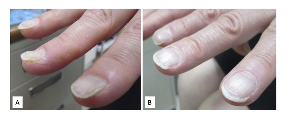
Spoon shaped nails or “koilonychia” in a patient with iron deficiency anemia. From Yoshiharu Taguchi, Shutaro Takashima, Kortaro Tanaka. Koilonychia in a patient with subacute iron-deficiency anemia. Intern Med. 2013;52(20):2379. Used with creative commons license
LAB FINDINGS IN IRON DEFICIENCY ANEMIA
Lab findings in a patient with iron deficiency include low ferritin (often under 20). A patient with iron deficiency anemia has low ferritin and low hemoglobin (under 12 g/dL or 120 g/L).
Other finding are seen in patients with iron deficiency anemia include:
reduced hematocrit (often under 33 %)
low MCV (usually well under 80 and maybe 65-75)
increased RDW (especially well above 15 and maybe 16-17)
reduced serum iron (around 60 with mild iron deficiency; may be under 40 with severe iron deficiency anemia)
increased total iron binding capacity (TIBC) - often above 300 with mild iron deficiency and above 450 with iron deficiency anemia
low transferrin saturation (30 in mild iron deficiency and under 20 in more severe iron deficiency anemia)
low MCH (often less than 27)
low MCHC (especially under 32)
Kano et al 2022
Dr Kano from Japan presented a nice article in the Cleveland Clinic Journal of Medicine of a 46-year-old woman presented with a 3-month history of progressive fatigue as well as dyspnea (shortness of breath). She had a history of uterine fibroids with heavy menstrual cycles. Physical examination revealed facial and conjunctival rim pallor, blue sclera with conjunctival rim pallor, and koilonychia (spoon shaped nails).
Laboratory tests were hemoglobin 4.0 g/dL (reference range 12–16), hematocrit 16.7% (37–47), mean corpuscular volume 54.8 fL (80–98), serum ferritin 0.8 ng/mL (24–307), and transferrin saturation 2.8% (20%–50%). Iron deficiency anemia was diagnosed.
The patient received a blood transfusion and began taking iron supplements. Her uterine fibroids were treated with oral gonadotropin-releasing hormone antagonists. Three months later, her symptoms and physical findings, including the blue sclera had resolved, and her hemoglobin and ferritin levels had normalized.
Blue Sclera and Iron deficiency
Blue sclera is a common and useful finding of iron deficiency but is often overlooked. In 1908, Sir William Osler first described a blue discoloration of the sclera as a symptom of anemia in iron-deficient undernourished adolescent females
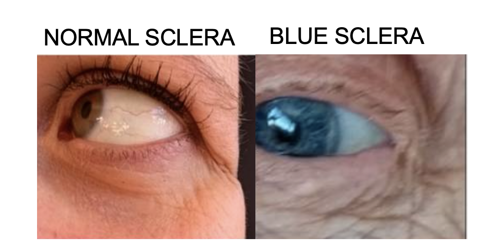
Lobbes H et al. Computed and Subjective Blue Scleral Color Analysis as a Diagnostic Tool for Iron Deficiency: A Pilot Study. J Clin Med. 2019 Nov; 8(11): 1876. Image used with creative commons license
In a 1986 publication in the Lancet, Kalra et al reported that the finding of blue sclera was more common in patients with iron deficiency anemia (87%) than in those with other types of anemia (7%). The presence of blue sclerae was unaffected by age, sex, or colour of iris. In adult patients, blue sclera is reported to have an 87% to 89% sensitivity and 64% to 94% specificity for iron deficiency anemia and iron deficiency.
In 1971 Pope tested the predictive value of blue sclerae in undiagnosed chronic iron deficiency in otherwise healthy individuals attending an outpatient clinic. Of the 1889 patients screened, blue sclerae were observed in 41 patients. Of the 41 patients, 34 (83%) had evidence of past or present iron deficiency.
What conditions are associated with blue sclera?
Blue sclera is seen in other conditions. It has been reported in osteogenesis imperfecta, rheumatoid arthritis, myasthenia gravis, inherited disorders of connective tissue and collagen disorders (e.g. Ehlers-Danlos syndrome, pseudoxanthoma elasticum), incontinentia pigmenti, high myopia, minocycline therapy and long-term steroid therapy but appears relatively rare in these conditions. Its pathogenesis is thought to involve thinning of the collagen fibers of the sclera due to iron deficiency, which allows the bluish color of the underlying uvea to become visible.
In vitro studies have shown that fibroblasts in culture do not synthesize collagen when iron is not available. It is therefore hypothesised that collagen synthesis is impaired in patients with iron deficiency resulting in thin sclerae through which the choroid can be seen, making the sclerae appear blue.
Main Reference
Kano Y. Blue sclera: An overlooked finding of iron deficiency. Cleve Clin J Med. 2022 Oct 3;89(10):549-550.
Additional References
Pope FM. Blue sclerotics in iron deficiency. Lancet. 1971;2:1160
Kalra L, Hamlyn AN, Jones BJ. Blue sclerae: a common sign of iron deficiency? Lancet 1986; 2(8518):1267–1269. doi:10.1016/s0140-6736(86)92688-7
This article was written by Dr. Jeff Donovan, a Canadian and US board certified dermatologist specializing exclusively in hair loss.