Does Fibrosis in the Bulge Region Drive Miniaturization?
New Study Shows Markers of Fibrosis are Upregulated During The Process of Miniaturization
Since 1990, it’s been pretty clear that ‘bulge’ region of the hair follicle is of utmost importance. This region of the hair follicle is home to multipotent stem cells that are important to regenerate hair follicles as well as to regenerate sebaceous glands and the epidermis. It’s increasing clear that the bulge portion of hair follicles (HFs) plays an important role in the pathogenesis of androgenetic alopecia (AGA.) In fact, it’s thought that the failure of bulge stem cells to convert to progenitor cells is key in how AGA develops. Fibrosis may participate in this process as prior studies have shown that fibrosis related genes are overexpressed in the bulge portion of hairs that are undergoing miniaturization.
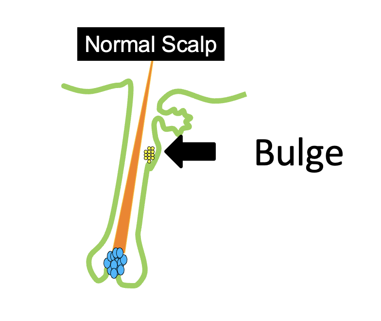
The bulge region is home to stem cells. The failure of stem cells to become progenitor cells contributes to AGA.
Li et al, 2022
A new study from China set out to examine how fibrosis changes during the process of miniaturization. The researchers obtained 300 follicular units from 10 study participants (30 follicular units each person). Hairs were taken from the frontal scalp and divided into three groups: early miniaturized hairs were those 80-100 micrometers in diameter, intermediate miniaturized were 60-80 micrometers in diameter and advanced miniaturized hairs were less than 60 um in diameter.
Spindled Cells Appear in Intermediate to Advanced AGA
Hematoxylin-eosin (H&E) staining showed that most of hair follicles in the mid and late AGA stages had some “spindled cells” appearing in the bulge region. In fact, about 90 % of hairs had these cells present compared to only 12 % of hair follicles that were in an early stage of AGA or 4-5 % of control hairs that weren’t undergoing balding.
Fibrosis Markers Gradually Appear in Intermediate to Advanced AGA
The authors studied fibrosis markers including vimentin, fibronectin, HSP47, and S100A4. These markers were expressed at low levels in early AGA but gradually increased during progression to mid stage AGA and late stage AGA.

Conclusion
The authors proposed that fibrosis of the bulge portion is positively correlated with the miniaturization of AGA-affected HFs. The authors hypothesize further that fibrosis of the bulge portion might cause dysfunction of stem cells and ultimately inhibit hair growth. The authors leave us with the interesting comment that drugs that can inhibit fibrosis might have future potential to reverse hair miniaturization during AGA development.
Listen to Full Article on Dr. Donovan’s Evidence Based Hair Podcast

REFERENCE
Li K et al. Association of fibrosis in the bulge portion with hair follicle miniaturization in androgenetic alopecia. J Am Acad Dermatol 2022; Jan;86(1):213-215.
This article was written by Dr. Jeff Donovan, a Canadian and US board certified dermatologist specializing exclusively in hair loss.