New Report Highlights Clinical and Trichoscopic Features of Folliculotropic MF Involving the Scalp
6 Key Trichoscopic Features of Folliculotropic MF
Authors of a new study set out to evalaute the clinical and trichoscopic features in 18 patients with folliculotropic MF. Their 18 patient included 11 female and 8 male. Patients had a mix of disease stages including stage 1a to IIIa but 50 % of patients were stage 1a. In 77.8% of patients, the scalp was first site of disease
Most folliculotropic mF lesions involving the scalp presented as: inflammatory or non-inflammatory patches or plaques (72.2%). However, in about one-quarter of patients, there was a generalized alopecia (27.8%).
6 Key Trichoscopic Features of Follicular MF involving the Scalp
The authors highlighted 6 of the most common features of FMF of the scalp
1.single hair (83.3%)
2.dotted dilated vessels (77.8%)
3.broken-dystrophic hairs (66.7%)
4.vellus hairs (61.1%)
5.spermatozoa-like pattern vessels (55.6%)
6.yellow dots (55.6%).
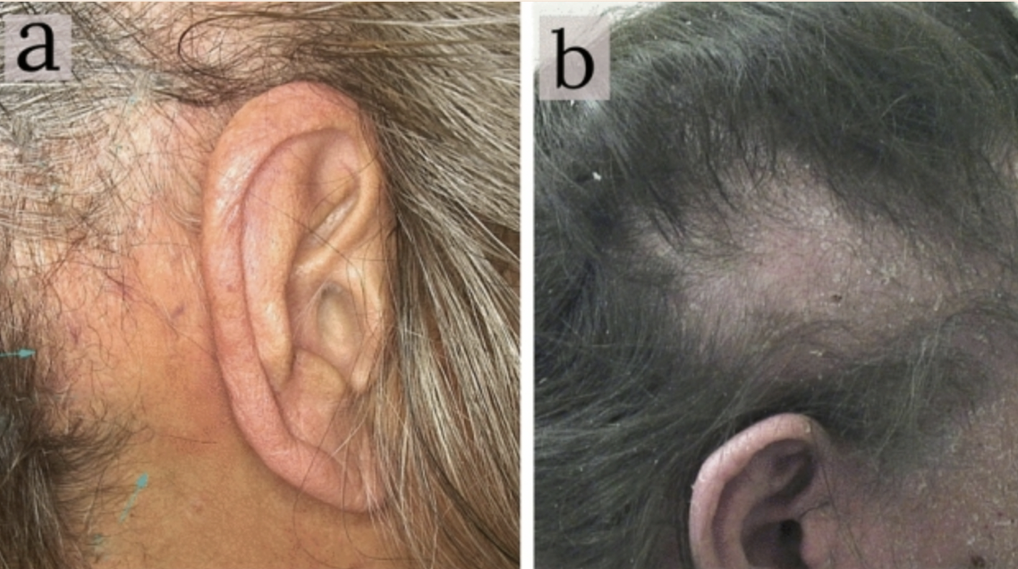
Images from Gallo et al. Image in “A:” shows red patch behind the ear and image in “B” shows scaly patchy. Source Gallo G et al Clinical and trichoscopic features in 18 cases of Folliculotropic Mycosis Fungoides with scalp involvement. Scientific Reports. 2021 May 18;11(1):10555. used with creative commons license
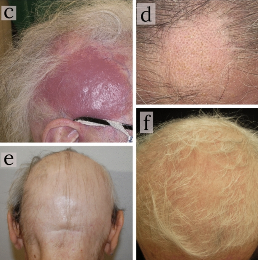
Images from Gallo et al. Image in “C” shows a red nodular lesion in late FMF. Image in “D” shows a non inflammatory pattern mimicking alopecia areata. Image in “E” shows generalized alopecia which is a pattern seen in 25% of patients. Image in “F” shows generalized alopecia. Source Gallo G et al Clinical and trichoscopic features in 18 cases of Folliculotropic Mycosis Fungoides with scalp involvement. Scientific Reports. 2021 May 18;11(1):10555. used with creative commons license
To Read more about CTCL, visit our prior articles
“CTCL: A Short Overview for Hair Specialists”
”Scarring Alopecia due to Follicular MF”
REFERENCE
Gallo G et al Clinical and trichoscopic features in 18 cases of Folliculotropic Mycosis Fungoides with scalp involvement. Scientific Reports. 2021 May 18;11(1):10555.
This article was written by Dr. Jeff Donovan, a Canadian and US board certified dermatologist specializing exclusively in hair loss.