Occipital Fibrosing Alopecia: Another Variant of FFA
Occipital Fibrosing Alopecia
Frontal fibrosing alopecia (FFA) is a scarring alopecia that typically affects the front of the scalp. After all, that’s how the name came about when it was first given its name in 1994.
However, we know the name given to this particular condition is not really so ideal. Perhaps it’s too late to change. It serves us fairly well for now. (Scarring alopecia of the tiny baby hairs just doesn’t sound as nice nor quite as professional).
Non-Frontal Frontal Fibrosing Alopecia
Some patients with FFA have only loss of eyebrows at the time of diagnosis and no loss of frontal scalp hair. Is it still FFA? Yes, although clearly it’s a reminder that we probably need a better term for the condition. Some patients with FFA have only loss of leg or body hair at the time of diagnosis. Is it still FFA? Yes, although clearly it’s a reminder we probably need a better term for the condition.
Some patients with FFA have only loss of hair at the back of the scalp at the time of diagnosis. Is it still FFA? Yes. Some would choose to call this occipital fibrosing alopecia (OFA). It’s probably best to call it frontal fibrosing alopecia, occipital fibrosing alopecia variant ( FFA-OFA).
Occipital Fibrosing Alopecia (OFA): Another Non-Frontal Frontal Fibrosing Alopecia
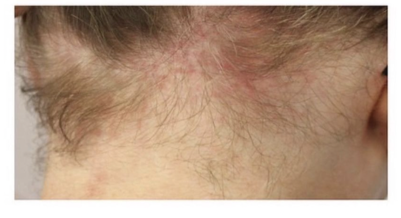
A patient with frontal fibrosing alopecia affected the occipital scalp.
Velaso-Martinez et al, 2021
If you don’t know about the Velasco Martinez study of 2021, it’s likely time now that you come to know this study now. This was a study of 14 women with occipital fibrosing alopecia. The women ranged in age from 42 to 65 years of age. There were 4 Caucasian and 10 Latin American women. 5 women arrived at the appointment specifically concerned about hair loss at the back of the scalp. However, 9 women made an appointment because they were concerned with diffuse shedding rather than occipital hair loss.
In 14 of 14 patients, hair loss was noted in the occipital hairline region. In 14 of 14 patients, the frontal hairline appeared normal. Eyebrows were normal in 10 of 14 patients. Trichoscopy showed loss of follicular openings, loss of vellus hair along with perifollicular scaling. Pathology showed perifollicular fibrosis with lichenoid infiltrates at the level of the isthmus.
Comment
Frontal fibrosing alopecia, occipital fibrosing alopecia variant (FFA-OFA) is a diagnosis to be on the lookout for. I think that once we start more consistently introducing this condition into our diagnostic language and into our diagnostic toolbox we’re going to pick up more and more cases in the world. We’ll also hand out more diagnoses of OFA that really aren’t OFA. That’s inevitable too. This can be a challenging condition to diagnosis sometimes .
I must point out that early cases of OFA are challenging to diagnose (i.e. before the clinical presentation is well developed). Early stages of OFA can mimic traction alopecia and even diffuse androgenetic alopecia in women affecting the back of the scalp Even variants of psorasis can easily fool you!
They key in my mind when making a diagnosis of OFA is to:
a) Make sure one has taken a really good history. Does a diagnosis of traction alopecia even make sense? Has there been loss of eyebrow or body hair? Is there a family history of scarring alopecia? Is there a personal history of thyroid disease? Is there a personal history of other autoimmunity? Is there a history of early menopause?
b) Make sure one has performed a really good physical examination, including trichoscopy. Is there evidence of FFA in the frontal hairline? Is there loss of eyebrows? Does trichoscopy in the back show loss of vellus hairs, perifollicular casts and perifollicular erythema. In my opinion, perifollicular casts are far far less common in OFA than classic FFA so one should not use this as a deciding factor to sway a decision. What I tend to see in OFA is some perifollicular erythema and marked distortion of vascular patterns including telangiectasias and dilated venous structures. Whether this is due to atrophy is not clear.
c) Take a scalp biopsy. In my view, one needs to lower the threshold a bit for taking biopsies in rare variants of most hair loss conditions. If one is going to convince generations of health care providers that will assume care in the future, it’s important to fully secure a diagnosis. We need to lock in this diagnosis! The last thing I want is for the patient to head off to another provider and for the provider to say “sure doesn’t look like FFA to me.”
One must keep in mind that the pathology of very early FFA and OFA is challenging. One must really have a good conversation with the pathologist in equivocal cases (on the fence types cases). There is not always perifollicular fibrosis on biopsy. There is not always lichenoid change of the terminal hairs. The two consistent finings are atrophy of sebaceous glands and some inflammation around vellus hairs.
d) Take repeated (serial) photos of all areas of the scalp. Photos are critically important to follow this condition. Hair must be pulled out of the way to expose the hairline. Photos of the occipital scalp, frontal scalp, eyebrows, mid scalp must be followed over time. If FFA is not clear on day 1, the diagnosis most likely will become clear on day 101, 301 or 1001. Photos are key.
REFERENCE
Martinez-Velasco et al. Occipital fibrosing alopecia. Int J Dermatol . 2021 Feb;60(2):e44-e45.
This article was written by Dr. Jeff Donovan, a Canadian and US board certified dermatologist specializing exclusively in hair loss.