Tinea Capitis in Infants: An Uncommon Age Group
Tinea Capitis in Infants: 2 Rare Cases
Tinea capitis is a fungal infection of the scalp caused by dermatophyte species. Tinea capitis is common in children, especially those 5-10 years of age. It is less commonly diagnosed in those under 1 years of age. A new report highlights 2 interesting cases of tinea capitis in infants under 1 year of age.
PATIENT 1 (10 months, female)
The first patient the authors presented was a 10 month old girl. A 10 % KOH mount showed endothrix species although a culture sent off to the lab did not grow any organism. Trichoscoppy showed perifollicular scaling, hair dust, comma hair, corkscrew hair, and horseshoe hair. The girl was treated with 2% miconazole cream and fluconazole 30 mg twice a week for 8 weeks with complete resolution of the lesions.
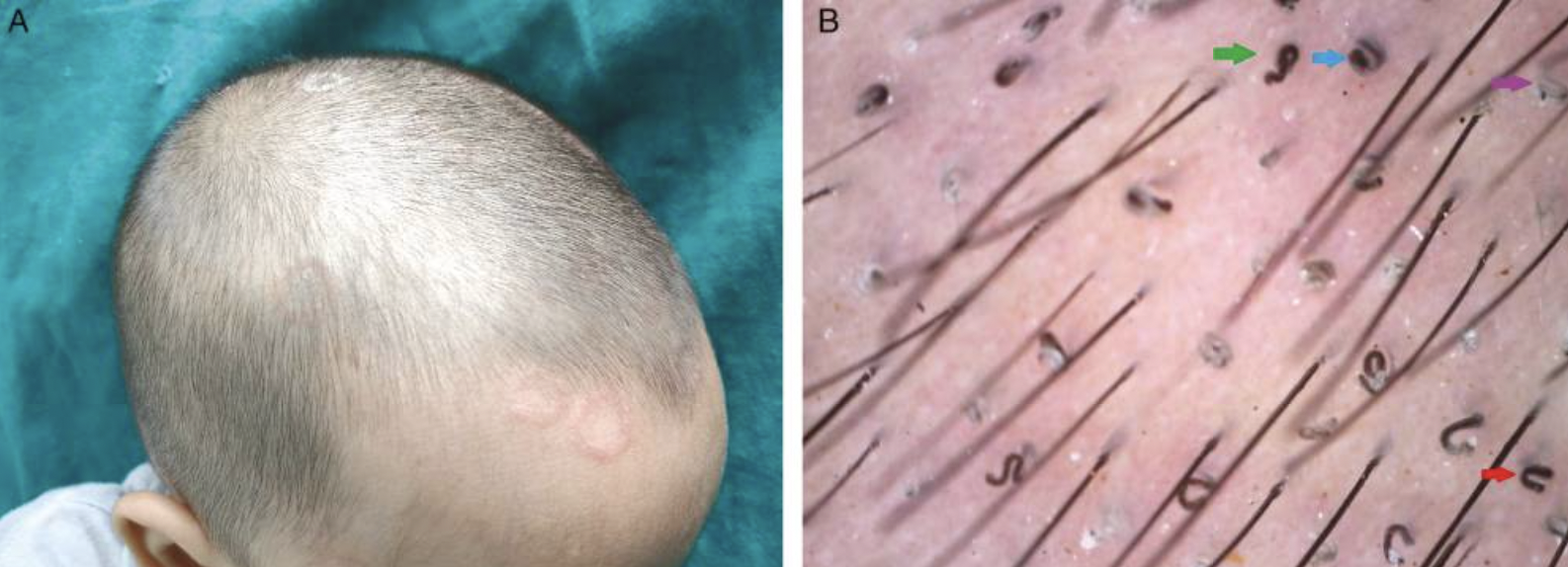
10 month old presenting with scaly patches. Trichoscopy shows perifollicular scaling (violet arrow), comma hair (blue arrow), corkscrew hair (green arrow), and horse-shoe hair (red arrow). Source. Kumar P and Pandhi D. Role of Trichoscopy in the Management of Tinea Capitis in Two Infants: A Case Report. J Cutan Aesthet Surg. Oct-Dec 2021;14(4):443-445. This is an open access journal, and articles are distributed under the terms of the Creative Commons Attribution-NonCommercial-ShareAlike 4.0 License, which allows others to remix, tweak, and build upon the work non-commercially, as long as appropriate credit is given and the new creations are licensed under the identical terms.
PATIENT 2 (3 months, female)
The second patient the authors presented was a 3 month old girl. presenting with gray patch tinea capitis. A 10% KOH mount revealed ectothrix variant. Similar to the first patient, there was no growth was seen in Sabouraud dextrose agar (SDA) culture media. Her trichoscopy showed perifollicular scaling, comma hair, corkscrew hair, and prominent telangiectasia. The patient was treated with oral fluconazole 20 mg twice a week and ketoconazole shampoo for 6 weeks.

3month old presenting with scaly patches. Trichoscopy shows perifollicular scaling (violet arrow), comma hair (blue arrow), corkscrew hair (green arrow), and prominent telangiectasia (white arrow). Source. Kumar P and Pandhi D. Role of Trichoscopy in the Management of Tinea Capitis in Two Infants: A Case Report. J Cutan Aesthet Surg. Oct-Dec 2021;14(4):443-445. This is an open access journal, and articles are distributed under the terms of the Creative Commons Attribution-NonCommercial-ShareAlike 4.0 License, which allows others to remix, tweak, and build upon the work non-commercially, as long as appropriate credit is given and the new creations are licensed under the identical terms.
Summary
I liked this report for a couple of reasons. It reminds us of just how uncommon tinea capitis is in those under 1 year. The medical literature contains under 100 such reports. Most cases of tinea capitis are in those 3-14 and the vast majority in fact are in those 5-10 years of age.
Second, I also liked this report because it reminds us all of the importance of trichoscopy in diagnosing tinea capitis. Although the gold standard for mycological diagnosis is fungal culture, such results can take 4 weeks or more for the fungal colonies to form and for the lab to be able to identify morphological features. Trichoscopy is extremely valuable tool to assist with the diagnosis.In 2020, the team of Anna Waśkiel-Burnat and colleagues set out to review all the articles published in medical journals related to the “trichoscopy” of tinea capitis.
This is an incredibly helpful paper. They showed that the most common findings in tinea capitis are
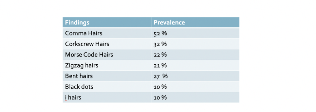

The authors calculated the sensitivity, specificity, positive predictive value and negative predictive value of a all the main specific findings in tinea capitis:

The third reason I liked this paper is it reminds us how best to treat tinea in infants. There are no formally FDA approved options! Everything is off label !! All of the treatments have been described but there is far less experience treating tinea capitis in infants!
REFERENCE
Kumar P and Pandhi D. Role of Trichoscopy in the Management of Tinea Capitis in Two Infants: A Case Report. J Cutan Aesthet Surg. Oct-Dec 2021;14(4):443-445.
This article was written by Dr. Jeff Donovan, a Canadian and US board certified dermatologist specializing exclusively in hair loss.