QUESTION OF THE WEEK

Dr. Donovan's Articles

QUESTION OF HAIR BLOGS

Filtering by Category: All 2018 Questions


Treatments for Scalp Dysesthesias

Question:

I have been to over 7 dermatologists and several other doctors as well. I have been told I have a diagnosis of ‘scalp dysesthesia.’ For almost two years, I have had severe scalp burning and hair loss. My doctors put me on several benzodiazepines but it makes my shedding worse. I have had no luck.

Do you have any suggestions?

Answer

Thank you for your question. The scalp dysesthesias are a group of conditions that are associated with marked scalp symptoms in the absence of any underlying obvious disease of the scalp. When hair loss is present together with the scalp symptoms one must strongly consider the possibility that the scalp symptoms could be due to the hair loss condition as well or perhaps even solely from the hair condition. 

There are many causes of scalp dysesthesias rather than a single cause. Cervical spine disease, neurological disease, depression are some of possible the underlying causes. It seems that patients worsen with stress. When hair loss is present one must always consider whether a primary scalp issue is the reason for the symptoms. A full review of a patient’s medical history as careful examination of the scalp is necessary to rule out primary causes such as scarring alopecia, psoriasis, seborrheic dermatitis, telogen effluvium. Blood tests for vitamin, mineral and hormonal abnormalities are needed and if any doubt exists a biopsy may be needed as well. Patch testing may be needed if allergic contact dermatitis is considered. 

There are many possible treatments for scalp dysesthesias including antidepressants (SSRI, tricyclic antidepressants), oral gabapentin, and oral prebagalin (Lyrica). Topical agents such as topical gabapentin or topical TKAL (ketamine, amitriptyline, lidocaine) can be considered as well. 


There are a variety of non-pharmacological techniques that can be used to combat the scalp symptoms that patient’s with scalp dysesthesias experience as well. These include:

1. Ice packs or Cool water

Ice packs, frozen peas and cool towels are useful for many individuals with challenging scalp syndromes. These are safe to use provided they are not too cold and not left on too long. 

2. Apple cider vinegar

Apple cider vinegar rinses are helpful for individuals with many different scalp syndromes including itching, burning and pain. Most often the apple cider vinegar is diluted 1:4 in water and applied to the scalp for 5-10 minutes before rinsing off. 

3. Witch hazel

Several herbal ingredients are proposed to have an anti-irritant tendency and can be helpful in scalp pain syndromes. These include chamomile (Marticaria chamomilla), heart seed (Cardiospermum halicacabum), peony (Paeonia lactiflora), and the virginian witch hazel (Hamamelis virginiana). Witch hazel in particular has received great attention. We generally recommend application of pure witch hazel with a cotton ball for periods of 5-10 minutes before rinsing off. Many patients find relief from these agents. 


4. Allergen free shampoos 

Although contact allergy must be considered in patients with scalp symptoms, a variety of allergen free shampoos can be considered even in the absence of any evidence of a true scalp allergy. A list of helpful low allergen shampoos is provided in the link below

ALLERGEN FREE SHAMPOOS

5. Vitamin C

Vitamin C or ascorbic acid occasionally helps some individuals with scalp nerve and pain syndromes. The dose is 500 mg daily. 

6. Low level laser therapy. 

Low level laser therapy (LLLT) involves the application of red light therapy to the scalp. The treatments were originally designed for use in androgneetic alopecia but have helped many patients with scalp dysesthesia. Some patients, however, find that the warmth of these devices makes their scalps feel worse. Therefore LLLT is not helpful for everyone. 

7. Menthol 

Products with menthol have a cooling effect and help some patients. Many of our paitents have found relief from a tea tree oil menthol – peppermint product from Holista products. These are available on amazon as well. Ingredients include denatured alcohol, water, glycerin, Melaleuca alternifolia (Tea Tree) leaf oil, menthol, Mentha piperita (Peppermint) oil. We recommend 3-4 sprays up to three times daily


8. Relaxation, medication, yoga and tai chi

Many of our patients have experienced tremendous benefit from the practice of a variety of techniques. 

Conclusion and Summary

I would need to examine your scalp and obtain a great deal more information to determine how best to proceed. The fact that you have hair loss makes it important to make sure there is not one or more hair conditions present as well. As mentioned above, many hair and scalp disorders are associated with scalp symptoms. If this is the case, it may or may not be appropriate to actually use the term scalp dysesthesia.


Blood tests are essential in anyone with scalp symptoms. Typical tests to consider include CBC, TSH, ferritin, zinc, ANA, ESR. Other blood tests may be important as well depending on the precise story. In your case, as well as with all patients with scalp dysesthesia, a scalp biopsy and consideration of patch testing needs to be discussed. These are certainly not appropriate for everyone but they must be considered. Evaluation for potential cervical spine disease (with x-ray +/- magnetic resonance imaging) and a detailed specialist evaluation for potential neurological, rheumatological

Share This
2 Comments

Finasteride Use and Breast Cancer in Women

Question:

I read your recent article discussing the risk of breast cancer in spironolactone users. Do women using finasteride place themselves at increased risk of breast cancer?

ANSWER:

Thanks for the question. We received quite a few inquiries last week when I posted this question:

Does Using Spironolactone Increase the Risk of Breast Cancer?

This week, I’d like to address the evidence for another group of antiandrogens known as the 5 alpha reductase inhibitors. This includes finasteride and dutasteride. To date, there is no good evidence to support the notion that use of oral anti-androgens such as finasteride or spironolactone are associated with an increased risk of breast cancer in women either in the general population or in women at increased risk of breast cancer. For finasteride, studies in women have never been done and we rely entirely on studies in males at low risk. For women, only a few studies have looked at breast cancer risks in spironolactone users. These studies have not suggested an increased risk of cancer in spironolactone users who are at low risk for breast cancer. 

 We do not have data on the risk of breast cancer in users of anti-androgens at highest risk. If the patient or her treating physicians feels that her risk of breast cancer may be higher than current estimates then the drug might not be used given that we have no information about the risks in these high risk groups.  I will first address what is known at present about the risk of breast cancer from finasteride and then address how a patient may come to get a better estimate of risk.  References for all the studies discussed are provided at the end.


Finasteride and Breast Cancer Risk: No studies in Women

For finasteride-related risk, the best means we have in the present day of addressing this question is by looking at the risk of breast cancer in men using finasteride and extrapolating the data the best we can to estimate the potential risk in female users.  We do not have studies in women. Male breast cancer is a rare condition with a lifetime risk of 0.1 %. In men, its behavior is similar to breast carcinoma in postmenopausal women.  So while studying male breast cancer and extrapolating the information to female breast cancer is not ideal, it is the best method we have in the present day.

There have been case reports and clinical trial results that suggested that treatment with 5ARIs may be associated with male breast cancer. Most studies to date however, suggest that it is not. All data needs to be taken into context with all available data to date. It should be noted that a warning label has been placed on finasteride packaging in many countries until this issue is further evaluated. An evidence review by the United Kingdom’s (UK) national drug agency resulted in a finasteride drug warning label for breast cancer in the UK and Canada and initiation of an FDA safety probe for all 5ARIs in 2010.


It is impossible to ascertain risk of cancer from the original short duration clinical trials. In other words, one is not going to get a sense of the risk of breast cancer by looking up a journal article about a 1 or 2 year study with finasteride. It’s far too short of a period. This is because such typical clinical trials are neither large enough nor have long enough follow-up to identify male breast cancer cases in men who use finasteride. The best type of studies we have at present are observational studies where men with breast cancer are compared to men without breast cancer. Such “case-control studies” are an invaluable tool to assess this important question. I will review many such case control studies below. 


A. 2016-2018 PUBLISHED STUDIES ON FINASTERIDE AND BREAST CANCER IN MEN


Meijer and colleagues recently assessed the possible relationship between finasteride and breast cancer by combining nationwide registers in 4 countries (Denmark, Finland, Norway, and Sweden) to assess the potential association between finasteride and male breast cancer.  A cohort of all males with dispensed finasteride (1,365,088 person years) was followed up for up to 15 years for breast cancer, and compared to a cohort of males not receiving finasteride.  An increased risk of male breast cancer was found among finasteride users (IRR = 1.44, 95% confidence interval [95% CI] = 1.11-1.88) compared to nonusers.  The analyses suggested possible ascertainment bias and did not support a clear relationship between dispensed finasteride and male breast cancer.  

Hagberg et al conducted a cohort study with nested case–control analyses using the UK Clinical Practice Research Datalink. 5ARI users did not have an increased risk of breast cancer compared to unexposed men (OR=1.52, 95% CI 0.61–3.80).


B. 2013-2015 PUBLISHED STUDIESON FINASTERIDE AND BREAST CANCER IN MEN

In 2014, Duijnhoven and colleagues in the Netherlands performed acase-control study with data from the United Kingdom Clinical Practice Research Datalink database among all men aged 45 years and older. Cases of men diagnosed with breast cancer were matched to up 10 controls. There were 398 cases were identified and matched to 3,930 controls. The “ever use” of 5-ARIs was associated with an adjusted odds ratio for breast cancer of 1.08 (95 % CI 0.62-1.87) compared to non-users. Increasing cumulative duration of treatment showed no increasing risks. The conclusion here in Duijnhoven’s study was that there was no evidence of an association between short- or long-term treatment with 5-ARIs and the risk for breast cancer in older men.

In 2013, Bird and colleagues in the United States published a cased control study of men age 40 to 85 years old. Here there were 339 breast cancer cases matched to 6,780 controls. There were no statistically significant associations observed between 5α-reductase inhibitors and breast cancer regardless of exposure assessment. Their conclusion was that the lack of an association in our study suggests that the development of breast cancer should not influence the prescribing of 5α-reductase inhibitor therapy.


C. 2003-2012 PUBLISHED STUDIESON FINASTERIDE AND BREAST CANCER IN MEN

In the 2003 PCPT study, Thompson and colleagues published data on 18882 men aged 55 years or older who were randomized to treatment with 5 mg/day finasteride (n = 9423) or placebo (n = 9459) for 7 years. One case of breast cancer was reported as an adverse experience in each treatment group during the study.  In this very large long-term study, an increased incidence of breast cancer in the finasteride group compared to placebo was not observed.

The main study that drew attention to a potential relationship between finasteride and breast cancer was a 2003 study by McConnell. In this study, 3047 patients were randomized to a double-blind, multi-center, placebo-controlled clinical trial for 4-6 years. The 4 different patient groups were administered different drugs: placebo; 8 mg doxazosin; 5 mg finasteride and a combination of 8 mg doxazosin and 5 mg finasteride. Three cases of breast cancer occurred in the finasteride-treated group and 1 case of breast cancer occurred in the combination group. No predisposing factors were identified. Duration of treatment ranged from 1.8 years to 5 years. The occurrence of 4 cases of breast cancer in 3047 patients was considered high considering the normal incidence in the general population of 1 case in 100,000 man-years. Treatment with finasteride appeared in this study to confer 200-fold risk for breast cancer in comparison to patients not receiving the drug.


D. 1996-2002 PUBLISHED STUDIES ON FINASTERIDE AND BREAST CANCER IN MEN


In 1996, Prescription Event Monitoring (PEM) Study was published. This study was conducted by Drug Safety Research Unit (DSRU) and involved a total of 14,772 patients (mostly male) under observation of General Practitioners from 1992–1994. There were 2 reported breast carcinomas. For one of the events, the time to onset from commencement of finasteride treatment was recorded as 5 months, the other was unknown. The PEM study in 1996 concluded overall that that finasteride is acceptably safe when used in accordance with the current prescribing information.However, it is not possible from this study to evaluate the cases of breast cancer and their causal relationship with finasteride, as enough data is not available regarding the 2 events of breast carcinoma.


In 1998, McConnell and colleagues published the Proscar long term efficacy and safety study (PLESS). There were 3040 patients were followed up for a period of 4 years. The patients were randomized in approximately equal proportions to receive either 5 mg finasteride or placebo for up to 4 years. In this study, there were no cases of male breast cancer reported in finasteride-treated subjects, and 2 cases were reported in placebo-treated subjects.

Summary and Conclusion

The evidence to date does not point to an association between finasteride or dutasteride use and breast cancer in women at low risk. It is absolutely critical to keep in mind that the studies I have mentioned above above were conducted in men with low risk of breast cancer and not in women (and not in women who have high baseline risks of breast cancer and not in women who already have breast cancer). Also most of the studies have looked at finasteride rather than dutasteride but of course some of the studies looked collectively at both types of alpha reductase inhibitors. This is important to keep in mind. Many physicians continue to avoid avoid prescribing any type of anti androgen to patients with a history of breast cancer (or at highest risk of breast cancer) given that no such studies have been done. However, for most women, there is no evidence to suggest that their use of finasteride or dutasteride increases their risk of developing breast cancer.

REFERENCES


1.    Hagberg KW, et al. Risk of gynecomastia and breast cancer associated with the use of 5-alpha reductase inhibitors for benign prostatic hyperplasia. Clin Epidemiol. 2017.

2.     Wiebe JP, et al. Progesterone-induced stimulation of mammary tumorigenesis is due to the progesterone metabolite, 5α-dihydroprogesterone (5αP) and can be suppressed by the 5α-reductase inhibitor, finasteride. J Steroid Biochem Mol Biol. 2015.

3.     Meijer M, et al.  Finasteride treatment and male breast cancer: a register-based cohort study in four Nordic countries. Cancer Med. 2018.

4.     Duijnhoven RG, et al. Long-term use of 5α-reductase inhibitors and the risk of male breast cancer. Cancer Causes Control. 2014.

5.    Bird ST, et al. Male breast cancer and 5α-reductase inhibitors finasteride and dutasteride.

J Urol. 2013.

6.    McConnell JD, Bruskewitz R, Walsh P, Andriole G, Lieber M, Holtgrewe HL, et al. The effect of finasteride on the risk of acute urinary retention and the need for surgical treatment among men with benign prostatic hyperplasia. N Engl J Med. 1998;338:557–63.  


7.    McConnell JD, Roehrborn CG, Bautista OM, Andriole GL, Jr, Dixon CM, Kusek JW, et al. The long-term effect of doxazosin, finasteride, and combination therapy on the clinical progression of benign prostatic hyperplasia. N Eng J Med. 2003;349:2387–98 

8.    Thompson IM, Goodman PJ, Tangen CM, Lucia MS, Miller GJ, Ford LG, et al. The Influence of finasteride on the development of prostate cancer. N Engl J Med. 2003;349:215–24.

9.    Wilton L, Pearce G, Edet E, Freemantle S, Stephens sMD, Mann RD. The safety of finasteride used in benign prostatic hypertrophy: a non-interventional observational cohort study in 14,772 patients. Br J Urol. 1996;78:379–84.  

10.  Mackenzie IS, et al. Spironolactone use and risk of incident cancers: a retrospective, matched cohort study. Br J Clin Pharmacol. 2017.

11. Biggar RJ, et al. Spironolactone use and the risk of breast and gynecologic cancers. Cancer Epidemiol. 2013.

12.  Mackenzie IS, et al. Spironolactone and risk of incident breast cancer in women older than 55 years: retrospective, matched cohort study. BMJ. 2012.

Share This
No Comments

Does using spironolactone increase the risk of breast cancer?

Question

I have been prescribed spironolactone for female pattern hair loss but have heard confusing information about whether or not the drug increases the risk of breast cancer. Do women using spironolactone have an increased risk of breast cancer?

Answer

Thanks for the great question. I’ll answer this with some depth but I’ll begin by saying that the most recent well conducted studies do not support an association between breast cancer and the use of spironolactone in women at low risk for the disease.

Concerns about the possibility of an increased risk of cancer from spironolactone date back to 1975. Studies at the time showed that rats ingesting spironolactone (at 25–250 times the exposure dose in humans) for 2 years developed several types of tumors including benign adenomas of the thyroid and testes, malignant mammary tumors, and growths on the liver.

To date, there is no good evidence to support the notion that women using spironolactone are at increased risk for breast cancer. In the most recent 2017 study, McKenzie studied the risk of cancer among users of Spironolactone. The participants were 74 272 patients exposed to spironolactone between 1986 and 2013 using the Clinical Practice Research Datalink from the UK. In this study, there was no increased risk of cancer in spironolactone users. 

In 2013, Biggar published data specifically looking at the risk of breast cancer in female spironolactone users. The researchers used anationwide prescription drug registry between 1995 and 2010 and identified use of spironolactone in a cohort of Danish women (≥20 years old).  After studying 2.3 million women (28.5 million person-years), the authors concluded that with respect to breast, uterus, ovarian and cervical cancer, there is no evidence of increased risk with spironolactone or furosemide use.

In 2012, McKenzie published a study a retrospective cohort study evaluating whether exposure to spironolactone treatment affects the risk of incident breast cancer in women over 55 years of age. The study involved 1,290,625 female patients, older than 55 years and with no history of breast cancer, from 557 general practices with a total follow-up time of 8.4 million patient years.  Although the vast majority of women were using doses under 100 mg, 17.2 % of women in the study were using 100 mg doses and 3.6 % were using 200 mg doses. The data suggested that the use of spironolactone did not increase the risk of breast cancer.


Summary and Conclusion

The evidence to date does not point to an association between spironolactone and breast cancer in women at low risk. These studies above were conducted in women with low risk of breast cancer and not in women at highest risk and not in women who already have breast cancer. This is important to keep in mind. Many physicians continue to avoid avoid prescribing spironolactone to patients with a history of breast cancer (or at highest risk of breast cancer) given that no such studies have been done. However, for most women, there is no evidence to suggest that their use of spironolactone increases their risk of developing breast cancer.

References

Barker DJP. The epidemiological evidence relating to spironolactone and malignant disease in man. J Drug Dev. 1978;1(Suppl 1. 2):22–25.

Biggar RJ, et al. Spironolactone use and the risk of breast and gynecologic cancers.Cancer

Epidemiol. 2013.

Danielson DAN, Jick H, Hunter JR, et al. Nonestrogenic drugs and breast cancer. Am J Epidemiol. 1982;116:329–332. 

Mackenzie IS, et al. Spironolactone use and risk of incident cancers: a retrospective, matched

cohort study. Br J Clin Pharmacol. 2017.

Mackenzie IS, et al. Spironolactone and risk of incident breast cancer in women older than 55

years: retrospective, matched cohort study. BMJ. 2012.


Share This
No Comments

Hair Loss from Relaxers

Question

Can my scalp be treated after severe damage from a hair relaxer?

Answer

Thanks for the great question. This is such an important question and also a very common one. There is a lot to discuss. As you’ll see, the answer to your question is ‘maybe.’ Some patients with hair loss form relaxers will grow back their hair. Some patients do not.

Let’s begin.

When a patient says to me they have hair loss from a relaxer, it’s important to keep in mind that there is not one type of hair loss that they might have. In fact, they might have one or more of many types of hair loss, including hair breakage, inflammatory scarring alopecias, hair loss from chronic inflammation, traction alopecia, telogen effluvium or androgenetic alopecia. Some patients just have one type of hair loss. Others have two or three.

Let’s take a look.

1. Hair breakage (Trichorrhexis nodosa)

Both chemical and heat relaxing of hair can cause breakage of the hair. The hairs simply break off because of the damage to the delicate strand. The heat or chemicals cause “micro tears” in the hair shaft which we call “trichorrhexis nodosa.”

The photo below shows a picture of a hair fibre that has such a tear.

Trichorrhexis nodosa of a hair fiber. This can occur from many agents including heat and chemicals used to relax hair.

Trichorrhexis nodosa of a hair fiber. This can occur from many agents including heat and chemicals used to relax hair.

If trichorhexis nodosa is the only reason for the hair loss, the hair will grow back. The damaged sections may need to be cut off, but the long term prognosis is good. It may take 6-9 months before hair returns back to the way it once was but it will return. Unfortunately, trichorrhexis nodosa as the ONLY and sole reason for a person’s hair loss from relaxers is not common. Ofter there is another reason present as well, and these are discussed below.


2. Telogen Effluvium

Many patients who come to see me with concern about their hair after using a relaxer also have a a diagnosis of telogen effluvium or “TE.” Telogen effluvium is a type of hair loss that occurs when the body feels some type of shock. This can occur from low iron (low ferritin), thyroid problems, anemias, crash diets, weight loss, stress, medications, and illness. Some of these issues such as anemia and low iron levels may make the hair slightly weaker and slightly more susceptible to hair damage. These issues must be addressed fully. For this reason, I always order blood tests for ferritin, 25 hydroxy-vitamin D, TSH, CBC, ANA in all patients who come to see me with concerns about hair loss from a relaxer. Other causes must be fully evaluated.


3. Traction alopecia.

Traction alopecia is a type of hair loss that occurs from the chronic pulling of hair. Patients who use relaxers may be more susceptible to traction alopecia because their hair is subjected to many pulling forces during relaxers and the hair fibers may be weaker. Traction alopecia can occur anywhere on the scalp. The frontal regions near the temple are often a common site of traction.

Treatment for traction alopecia involves stopping the pulling forces that caused the traction in the first place. If traction alopecia is diagnosed and pulling is stopped immediately (within a few months of the new hair care practice), hair might grow back. However, if traction alopecia has been present many months, the hair may not fully return. Long standing traction alopecia is permanent and may even continue to progress once the hair pulling is stopped.

Traction alopecia of the frontal hairline.

Traction alopecia of the frontal hairline.

4. Scarring Alopecia and Chronic Inflammation .

It’s a little known fact but chronic use of relaxers, especially chemical relaxers, can create scalp inflammation. It may not be a type of inflammation that can be seen on the surface but rather a type of inflammation that is occurring deep under the scalp. In some people using relaxers (but certainly not all people), this chronic inflammation triggers the body to also create scar tissue beneath the scalp. The exact mechanism is not clear but micro injury to the skin creates microinflammation and chronic microinflamation may induce scar tissue to form.

This pattern of hair loss from relaxers has been most carefully studied in women with afro-textured hair but likely applies to all hair types. Chronic use of relaxers in women with afro-textured hair may be linked to the development of several types of hair loss including traction alopecia and central centrifugal cicatricial alopecia (CCCA). CCCA often affects the central scalp first. The diagnosis must be caught as early as possible to prevent progress and prevent irreversible loss of hair. Too often women with CCCA are told that their hair loss is simply from a relaxer and it will grow back. CCCA is a cause of permanent hair loss. If there is any doubt, a punch biopsy should be considered to properly evaluate for scarring alopecia. Treatment with agents such as topical steroids, steroid injections, and doxycycline can help stop the disease. Hair growth does not usually occur. A photo of a woman with CCCA is shown below.

Central centrifugal cicatricial alopecia (CCCA) in a woman initially misdiagnosed as having temporary hair loss from a relaxer. The correct diagnosis for this patient was CCCA which causes permanent hair loss.

Central centrifugal cicatricial alopecia (CCCA) in a woman initially misdiagnosed as having temporary hair loss from a relaxer. The correct diagnosis for this patient was CCCA which causes permanent hair loss.

Summary and Conclusion

Thanks again for the great question. Let’s now return to the original question regarding whether or not your scalp can be treated. As we’ve seen above, it really comes down the the exact cause of the hair loss. If the relaxer caused trichorrhexis nodosa, the damaged hair simply needs to be trimmed and hair density will eventually come back. If however, the relaxers have caused traction alopecia, it may or may not come back even if the relaxers are stopped. If the cause is CCCA, the hair is less likely to return and aggressive treatment with various anti-inflammatory medications are needed to stop the inflammation. If there is a telogen effluvium (from low iron for example) that predisposed to some fragility and hair loss, there could be some improvement with iron supplementation and stopping the relaxers as well.


Be sure to see a dermatologists as relaxer related hair loss can be complex sometimes. Blood tests for ferritin, 25 hydroxy-vitamin D, TSH, CBC, ANA might be considered and if any doubt exists, a biopsy might be considered to rule out scarring alopecia.

Share This
No Comments

MTHFR Gene Mutations and Hair Loss: Is there a link?

QUESTION:

Is there a MTHFR gene mutation that can cause hair loss?  There are some people reporting success in getting their hair back after discovering that they have this mutation and taking supplements.

ANSWER

Thanks for the excellent question. The topic of the MTHFR gene is certainly being discussed in many areas of medicine. Before answering your question, I’ll review what is known about the MTHFR gene, what is known about MTHFR mutations, and finally what is known about gene mutations and polymorphisms in hair loss

What is the MTHFR gene?

The MTHFR gene is a stretch of DNA that has all the instuctions for making a protein known as methylenetetrahydrofolate reductase. This protein is actually an enzyme which helps our bodies convert the folate vitamin into a usable form called methylfolate.  When the MTHFR gene has mutations or the gene is altered in some way (i.e. a polymorphism), the enzyme might either work just fine, or not work quite as well, or not work at all. Many polymorphisms in the MTHFR gene are associated with a 40-70 % reduction in the enzyme’s ability to function.

What is known about MTHFR gene mutations?

There are many different ways that the MTHFR gene is coded in human. We call these differences “polymorphisms.” When there is only one common way a gene is made, we use the word mutation to refer to anything that is different than the main way. However, when there are so many ways that the gene can be coded in the population, we refer to the various changes as “polymorphisms.” The proper terminology here is really MTHFR gene polymorphisms and so I’ll call these MTHFR changes polymorphisms rather than mutations.

When it comes to the MTHFR, there are two particular polymorphisms that are studied. The MTHFR 677T allele and the MTHFR 1298C allele. Recent studies by Nefic and colleagues suggested that the frequency of the MTHFR 677T allele in the population was approximately 33% and the frequency of MTHFR 1298C allele was around 38%.

How do we test for the MTHFR polymorphism ?

A variety of labs will do this, including some online sources. Individuals should check with their physician for local testing.

What is the main effect of poorly functioning MTHFR enzymes in patients with the MTHFR polymorphisms?

Individuals with hypo functioning enzymes have increased levels of blood homocysteine and reduced levels of B12 and serum folate.

Is there a link between MTHFR gene mutations (i.e. polymorphisms) and human disease ?

To date, there have been approximately 75 different human diseases and conditions that appear to be altered in patients with MTHFR gene polymorphisms. These range from asthma to heart attacks, to blood clots in the lungs to stroke, depression, cancers and neurological disease.

Anyone can search the PubMed Medical Library on the Topic

Is there a link between MTHFR gene mutations (ie. polymorphisms) and hair loss?


There have been few studies regarding the relationship between various MTHFR gene polymorphisms and hair loss. In fact, to date there has only been one - and that was a study examining the autoimmune condition known as alopecia areata (rather than genetic hair loss). This study included 136 patients affected by alopecia areata and 130 healthy controls. As in most studies, the researchers looked at the MTHFR gene C677T mutation. There were differences observed between the two groups suggesting that the MTHFR gene mutation might play some type of role in this immune condition. How best to treat alopecia in patients with the MTHFR polymorphism was not discussed as it was not the purpose of the research paper.

We don’t really know how best to treat the hair loss associated with various MTHFR gene polymorphism or if there the MTHFR gene polymorphism really has anything to do with the hair loss. Simply taking folic acid pills might not be the answer. Depending on the specific polymorphism, supplementation with methyl folate and B12 may be important to consider.

CONCLUSION

There seems to be a link between various MTHFR polymorphisms and several human diseases. I’d like to say there’s a clear link between changes in the MTHFR gene and hair loss and that everyone with hair loss should be tested. That’s just simply not the case. There is a whole lot of extremely poor and anecdotal evidence online about the relationship between MTHFR polymorphisms and hair loss and also how best to treat hair loss if someone does have these polymorphisms. Here are the main summary points and conclusions:

1) The MTHFR gene is coded very differently from person to person. We call these differences ‘polymorphisms.’ Most people who decide to get their MTHFR gene tested will fine that they don’t have the ‘standard’ template. They have slight changes. One must be very careful to interpret these results as most will find they have some type of change (not just a few but most).

2) If one has a MTHFR polymorphism, they may speak to their physician about the appropriateness of ordering B12, homocysteine and folate levels. The levels of these will determine what therapies are appropriate.

3) To date, these is absolutely no good evidence that MTHFR polymorphisms have a clear link to genetic hair loss, telogen effluvium or scarring hair loss (scarring alopecia). This information, may change over time. It’s certainly possible that if levels of folate or B12 are low enough that negative consequences on hair would result. The main point is that for many with MTHFR polymorphisms, the changes are not severe enough for detrimental effects on hair.

4) To date, there has been only 1 very small study examining the relationship of MTHFR polymorphisms to alopecia areata and suggested a possible weak relationship. The study has never been independently repeated.

5) Routine testing of MTHFR status in a person with concerns about hair loss can not be recommended at this time.

6) To date, there is absolutely no good evidence consuming methylfolate or any specific type of folic acid, or B12 or methyl-B12 or B6 in patients with MTHFR polymorphisms provides benefit to those experiencing hair loss. These supplements may be very important in other conditions however, (for example the benefit of B12 or folate supplementation to lower high plasma homocysteine levels). This information as it relates specifically to hair may change over time. One must of course consider the levels of B12 or folate and take these in context with the help of a physician.

More research is needed.

REFERENCES

Kalkan G, et al. Methylenetetrahydrofolate reductase C677T mutation in patients with alopecia areata in Turkish population. Gene. 2013.

Nefic H, et al. The Frequency of the 677C>T and 1298A>C Polymorphisms in the Methylenetetrahydrofolate Reductase (MTHFR) Gene in the Population. Med Arch. 2018.

Share This
No Comments

What features do you look for in a good biopsy report?

QUESTION

biopsy-reports

QUESTION:

What kind of things do you look for in a biopsy report that tells you it was a good biopsy report?

ANSWER

Thanks for the great question. This is a topic we don’t touch upon all that often and so I’m glad you’ve brought it to attention. Let me begin by saying that biopsies are important for some complex cases of hair loss. They are not needed in every patient. Biopsies can be extremely useful if done properly. They can also be extremely misleading if certain pieces of information are left out. Here are some of the top 10 things I look for when reading a report.


TOP 10 FEATURES OF A BIOPSY REPORT


1. SIZE OF THE BIOPSY SHOULD 4 mm

A punch biopsy is deal and should be 4 mm in size. Too often smaller punches (3 or 2 mm) are used in attempt to limit scars for the patient. While this is a good thought, smaller biopsied provided limited information.


2. THE PATHOLOGIST SHOULD COMMENT ON THE DEPTH OF THE BIOPSY

Ideally, it’s nice to see that the biopsy goes deep enough into the scalp so that the pathologist has a good chance to see what’s happening at the very bottom of the hair follicles and even into the fat. Conditions like alopecia areata and many autoimmune and inflammatory conditions and scarring alopecias (dissecting cellulitis) go quite deep. A biopsy must be deep enough to capture. this.

Many biopsies are not deep enough. Sometimes, there’s just too much bleeding during the biopsy and a physician is afraid to go deeper. Sometimes the punch technique the physician is using is not adequate and the punch is simply not pushed deep enough when taking the sample.


3. HORIZONTAL SECTIONING OF THE BIOPSY SAMPLE WAS CONSIDERED.

A biopsy specimen can be cut side to side (horizontal sections) or up and down (vertical sections). Many labs nowadays will do both. It’s nice to have horizontal sections as this gives the pathologist a lot of information on 12-30 hair follicles. Vertical sections give information on 3-7 hair follicles. I prefer to work with a pathologist who reads horizontal sections as it gives a great deal more information. Most labs perform horizontal sections.


4. THE PATHOLOGIST COMMENTS ON THE PROPORTION OF TERMINAL HAIRS, VELLUS HAIRS, ANAGEN HAIRS AND TELOGEN HAIRS IS GIVEN.

If horizontal sections are used to process the sample, it’s important to know exactly what the pathologist sees. In this regard it’s nice to have information about the proportion of terminal hairs, vellus hairs, anagen hairs and telogen hairs in the biopsy.

1) A high proportion of vellus hairs relative to terminal hairs given a clue to possible androgenetic alopecia. Horizontal sections allow the pathologist to comment on the ratio of terminal hairs to vellus hairs - which is a wonderful clue for diagnosing andrognetic alopecia and in some cases also chronic telogen effluvium. A terminal to vellus ratio less than 4:1 means androgneetic alopecia in most cases and a T:V ratio above 8:1 signifies chronic TE.

2) A high proportion of telogen hairs may also offer information about possible underlying telogen effluvium. For example, normally there are less than 12 % telogen hairs in a biopsy. As the percent of telogen hairs rises above 12-15 % one must also wonder if a telogen effluvium is present. The diagnosis of telogen effluvium is more of a clinical diagnosis than a pathology diagnosis. So, even if the percentage of telogen hairs is less than 12 %, it’s still posisble that a patient has a telogen effluvium.


5. THE PATHOLOGIST COMMENTS ON THE SEBACEOUS GLAND (OIL GLAND) DENSITY

When I read a report, I want to know what’s happening to the sebaceous glands (oil glands). This is often surprising to hear as one would normally imagine one would like to know what’s happening to the hair follicles themselves. I certainly do want to know what’s happening to the hair follicles (see below), but I’d like to know if the sebaceous glands are present, if they appear bigger in the biopsy or if they are reduced. Reduction in the density of sebaceous glands is very much a feature of scarring alopecias. A relative increase in the appearance of sebaceous glands is a feature of many non scarring alopecias such as androgenetic alopecia. This information is sometimes left out of reports and it’s so incredibly helpful to have.


6. THE PATHOLOGIST COMMENTS ON THE PRESENCE OR ABSENCE OF SCAR TISSUE (FIBROSIS)

It’s important to know if there is scar tissue present in the skin. Scar tissue is often referred to as fibrosis, and it’s nice to know if there is scarring around the hairs (perifollicular fibrosis) or more widespread in the skin (interfollicular fibrosis).

This is perhaps the most easily confused part of interpreting biopsies since perifolliclar fibrosis can be seen in both non scarring as well as scarring alopecias. It’s easy to over interpret the presence of perifollicular fibrosis as indicating a scarring alopecia.

For example, perifollicular fibrosis is seen in many biopsies from the non scarring hair loss condition known as androgneetic alopecia (male balding). However, the tip off that a biopsy from a patient with male balding is not a scarring alopecia is the fact that the sebaceous glands are still present and there is no lichenoid inflammation (see below). In lichen planopilaris (one of the scarring alopecias), there is perifollicular fibrosis, lichenoid change and reduction in sebaceous glands.


7. THE PATHOLOGIST COMMENTS ON THE PRESENCE OR ABSENCE OF INFLAMMATION.

Most biopsies have bits of inflammation here and there (sparse inflammation) and some have more Inflammation. When inflammation is present, it’s nice to know whether the pathologist feels it’s mild, moderate or severe. In addition, it’s important to know where the inflammation is found. Is it up high in the skin… or is it in the middle or is it down low at the level of the bulb. In scarring alopecias and androgenetic alopecia, the inflammation is high up in the level of the so called isthmus (fairly close to the skin level). In alopecia areata, the inflammation is quite deep in the skin around the hair follicle bulb.

I also look for the type of inflammation. Most inflammation in biopsies is comprised of a type of white blood cell called lymphocytes. But the presence of other inflammatory cells like lymphocytes, neutrophils or plasma cells might mean different things in different situations.


8. THE PATHOLOGIST COMMENTS ON CELL DEATH

If hair follicle cells in the biopsy are dying this is important. There are two mains ways that cells die - apoptosis and necrosis. These is a specific pattern of cell death in scarring alopecias called “lichenoid inflammation” that gives death of hair follicle keratinocytes. The presence of lichenoid inflammation in a biopsy really points towards a diagnosis of certain scarring alopecias such as lichen planopilaris or frontal fibrosing alopecia. . This information on cell death is desperately needed but too often left out of reports.

9. THE PATHOLOGIST COMMENTS ON WHAT’S HAPPENING ELSEWHERE IN THE BIOPSY

It’s nice to know the biopsy was looked at carefully. Ideally a report should comment on the presence of inflammation in other parts of the biopsy - not only around the hair follicle. This would include the presence of inflammation around blood vessels (called perivascular inflammation) and the presence of inflammation around other gland structures (like eccrine glands). In addition, some biopsies show that the body has produced and left behind extra material in the skin (like mucin for example). All this information is very helpful to know about.


10. THE PATHOLOGIST DECIDES IF ANY SPECIAL STAINS WERE NEEDED AND COMMENTS ON THESE FINDINGS

A variety of special ‘stains’ are available in the world of pathology. These stains are used to help identify specific substances in the skin or specific markers on cells. Special stains are commonly used to identify fungi (PAS stain). Other stains like gram stain may be used for identifying the presence of bacteria. An Alcian blue stain is commonly used to identify a substance in the biopsy known as mucin which may point to come autoimmune processes. Elastic stains are used to identify patterns of scarring.


Conclusion

I don’t usually need a biopsy to make a diagnosis. But if I’m going to do a biopsy, I want the pathology report that comes back to be a good one. I want it to be useful to my patient. I want the report to contain all the features that allows be to be as close to 100 % confident as to what’s going on in the scalp of the patient.

Share This
1 Comment

Seborrheic Dermatitis vs Lichen Planopilaris: Which do I have?

QUESTION

Screen Shot 2018-10-15 at 5.54.06 AM.png

QUESTION:

My doctors can’t decide if I have seborrheic dermatitis or lichen planopilaris. My scalp does feel less itchy and becomes less red with anti dandruff shampoos. However, it also becomes less red and itchy with topical steroids. Overall my shedding has improved after 4 weeks of treatment. Does this information suggest one diagnosis over the other?


ANSWER:

Thanks for the great question. The short answer is that the information provided here does not actually suggest one diagnosis over another. You may have scarring alopecia and you may have seborrheic dermatitis. The key point I would like to make is that you may have both! Up to 50 % of patients with lichen planopilaris have seborrheic dermatitis too. A scalp biopsy can fully answer your question.

Let’s take a closer look at both of these conditions and we’ll see why some patients with lichen planopilaris will benefit from anti-dandruff shampoos and we’ll see why some patients with seborrheic dermatitis benefit from topical steroids (the same ones used to treat lichen planopilaris.)

SEBORRHEIC DERMATITIS
First, seborrheic dermatitis is closely related to dandruff. The exact cause is still being worked out but yeast such as Malassezia may have an important role. Patients with seborrheic dermatitis have many similar (and sometimes identical) symptoms to patients with lichen planopilaris. They have a red, itchy scalp! Seborrheic dermatitis however does not cause scarring for most people and usually only gives minor amounts of hair shedding. (Everything in medicine has exception and seborrheic dermatitis may cause scarring in some cases and may give excessive hair shedding when severe - see previous articles below).

DOES SEBORRHEIC DERMATITIS CAUSE SCARRING?

CAN SEBORRHEIC DERMATITIS TRIGGER SHEDDING?


Seborrheic dermatitis is an inflammatory condition which means there is inflammation in the scalp. Although the standard first line treatment for seborrheic dermatitis is topical anti-dandruff shampoos, treatment with anti-inflammatory agents like topical steroids can help reduce the inflammation which in turn reduces redness and itching. Many patients with seborrheic dermatitis feels better with use of both antidandruff shampoos and topical steroids. In fact, studies have shown that adding topical steroids to a patient’s seborrheic dermatitis treatment plan can greatly help.

To come back to your question for a moment, we would expect seborrheic dermatitis to improve with dandruff shampoos and topical steroids. However, fact that your scalp did improve does not rule out a scarring alopecia as we’ll see next.

LICHEN PLANOPILARIS
Lichen planopilaris is a scarring alopecia that causes patients to experience itching and sometimes burning and tenderness in the scalp. The scalp is typically red. An important difference between lichen planopilaris and seborrheic dermatitis is that lichen planopilaris always associated with scarring. Biopsies of LPP show rings of scar tissue around hair follicles in early stages (called concentric perifollicular fibrosis) and deposits of large bits of scar tissue in the scalp in advanced stages.

Topical steroids are one of many agents used to treat LPP. They help reduce redness and scaling and help the patient feel better too with less itching, burning or pain.

Seborrheic Dermatitis in Patients with LPP: Is is More Common than We Realize?

Seborrheic dermatitis is present in a very large proportion of patients with LPP. In fact, a greater proportion of patients with LPP have seborrheic dermatitis compared to people in the general population. (About 5% of people in the general population have seborrheic dermatitis compared to nearly 50% of patients with LPP). On account of seborrheic dermatitis being so common in LPP, it makes sense that many people with LPP will feel better and gain some relief with use of antidandruff shampoos! The fact that a patient with LPP reports improvement with antidandruff shampoos does not rule out a scarring alopecia. It simply means they may have seborrheic dermatitis too!


Cleveland Clinic 2016 Study of Seborrheic Dermatitis in LPP

In 2016, Berfeld’s group at the Cleveland clinic studied the incidence of seborrheic dermatitis in patients with lichen planopilaris. This study is important to understand and relevant to the above discussion. It was one of the few studies to date which really documented the increased incidence of seborrheic dermatitis in patients with LPP.

The study I am referring to was a retrospective review of 246 patients seen over the period 2004-2015. Interestingly seborrheic dermatitis (SD) was present in 46.2 % of LPP cases. In 27.4 % of cases the SD was found outside the area affected by the LPP. On average the SD was diagnosed 7.8 months prior to the LPP diagnosis. Having SD seemed to delay an actual diagnosis of LPP. Patients with both SD and LPP diagnosis (LPP-SD) received their diagnosis with significantly more delay than patients with LPP who did not have SD (ie LPP). For example, patients with LPP-SD received their diagnose in 7.6 months on average comapred to 2.3 months for LPP alone.

Whether SD actually plays a role in the scarring process as well remains to be determined. It is interesting that there was a greater prevalence of late stage scarring alopecia in ptient with LPP-SD than LPP alone (41.5 % vs 15.7%). Patients with LPP-SD had greater rates of hyperandrogenism compared to patients with LPP alone.

SUMMARY AND CONCLUSION

Thanks again for the great question. One can’t determine if you are more likely to have SD or LPP from the information provided. It would be entirely within the realm of expected for a patient with LPP to improve with topical antidandruff agents since many have seborrheic dermatitis present as well. Likewise, it would be expected that a patient with seborrheic dermatitis would improve with topical steroids because this is an inflammatory disease just like LPP.

A biopsy can help distinguish if lichen planopilaris is truly present or not.


Reference
Ratnaparkhi et al. Association of lichen planopilaris with seborrheic dermatitis l: A retrospective case-control study. Poster 3727. JAAD May 2016.


https://www.aad.org/eposters/view/Meeting.aspx?id=43&c=2

Share This
No Comments

Hair loss after Breast Cancer Endocrine Therapy

QUESTION

eia


QUESTION: Many of us on breast cancer drugs like Letrazole/Zoladex end up with hair loss.  In my case, it occurred after a few months and was associated with an itchy scalp and diffuse shedding of 200+ hairs a day.  Some have become very fine.

If one stops these drugs, how long does it generally take for them to clear out of the system and stop affecting the hair?


ANSWER

Thanks for the excellent and very important question. These types of questions and their answers are important for both patients and their doctors. These issues continue to be very much overlooked in the present day and so your question has broad relevance.

I’ll review the subject broadly and then return to your question.

Endocrine Therapy for Breast Cancer.

The breast cancer drugs you are referring to are broadly classified as ‘endocrine therapy.’ These drugs include three main categories: 1) the selective estrogen receptor modulators (SERMs), 2) aromatase inhibitors (AI) and 3) GnRH agonists. These drugs have shown benefits in the adjuvant and therapeutic setting for premenopausal or postmenopausal women with early-stage or advanced breast cancer. These drugs may be taken in some cases for 5-10 years to reduce the risk of recurrence.


SERMs. The selective estrogen receptor modulators include tamoxifen, raloxifene, and toremifene. These act as competitive inhibitors of estrogen binding to estrogen receptors. From this group, tamoxifen has been the most widely used for adjuvant endocrine therapy and is still the the gold standard for premenopausal patients at risk of recurrence.


AI. By contrast,aromatase inhibitors, including letrozole, anastrozole, and exemestane. These drugs inhibit the enzyme aromatase and thereby suppress plasma estrogen levels. These drugs are now considered the preferred option for adjuvant endocrine therapy in postmenopausal patients. They all appear to be comparable in efficacy and have similar AE profiles that include hot flashes, mood disorders, osteopenia, and arthralgias.


GnRH agonists. Gonadotropin releasing hormone agonists (GnRH) are used for premenopausal women with hormone receptor–positive breast cancer Drugs like leuprolide are prescribed to suppress estrogen production by the ovary and may be combined with other endocrine therapies or chemotherapies.


Breast cancer endocrine therapy induced alopecia (BC-EIA)


Hair loss is also a potential side effect of the medications used as ‘endocrine therapy’ discussed above including the SERMs, AI, and GnRH agonist. The type of hair loss that can occur in patients using these drugs is broadly referred to as “breast cancer endocrine therapy induced alopecia (BC-EIA)

These changes come about because of the effects of hormone changes on the hair follicle - particularly the reduction in blood levels of estrogen (or estrogen related signals that are sent into cells) that accompanies use of these drugs. The hair follicle has a highly responsive endocrine organ. It produces hormones itself and responses quickly to changes in hormones in the body.

In 2013, the a meta-analysis of 13 415 patients in 35 clinical trials revealed an overall incidence of hair loss of 4.4%. The highest incidence in this study was with with the highest incidence in tamoxifen-treated patients (25%). In 2015, Moscetti and colleagues published data showing that about 8 % of 236 women using aromatase inhibitors stopped treatment on account of their hair loss.

HAIR LOSS IN PATIENTS USING ENDOCRINE THERAPY
The short answer to your question is that there can be more than one type of hair loss associated with these types of hormone blocking drugs. The main types of hair loss that are associated with these drugs include 1) acute telogen effluvium and 2) androgenetic alopecia.


Possibility 1: The Hair Loss is From a Drug Induced Telogen Effluvium


Telogen effluvium refer to a type of hair loss whereby the affected patient notices increased daily hair shedding. Instead of finding a few hairs in the shower or sink, the individual finds dozens and dozens or even hundreds. The key point here is that the shedding rate is increased over what is normal. Telogen effluvium from a drug typically occurs 2-3 months after the drug was started and can continue in some cases if the drug is continued. if a decision is made to stop the drug, the shedding can last 6-9 months after the drug is stopped.

Telogen effluvium is not common with tamoxifen. In fact, a 2014 study by Kanti et al did not show any changes in telogen hairs in 17 women using tamoxifen who were followed for 28 weeks. Although uncommon, tamoxifen related hair cycle changes are certainly possible and tamoxifen may have a growth inhibitory effects in some situations. Clearly a pure telogen effluvium is not like to be common but it can occur. A 2010 study by Bhatia et al showed that tamoxifen loaded liposomal topical formulation actually arrested hair growth in mice.

The aromatase inhibitors can also trigger a telogen effluvium. For example, Litt’s Drug Eruption Manual lists telogen effluvium from letrozole as occurring in less than 5 % of users.

Leuprolide, the GnRH agonist sometimes used in premenopausal women with breast cancer, may also cause a telogen effluvium. Litt’s Drug Eruption Manual lists telogen effluvium from Leuprolide as occurring in less than 5 % of users.


Possibility 2: The Hair Loss is From Patterned Alopecia (Androgenetic Alopecia)

The most important concept really understand is the development of androgenetic alopecia in women using endocrine therapy for breast cancer.

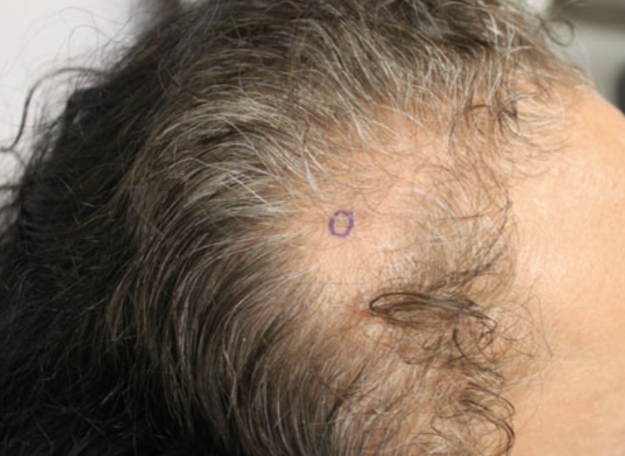
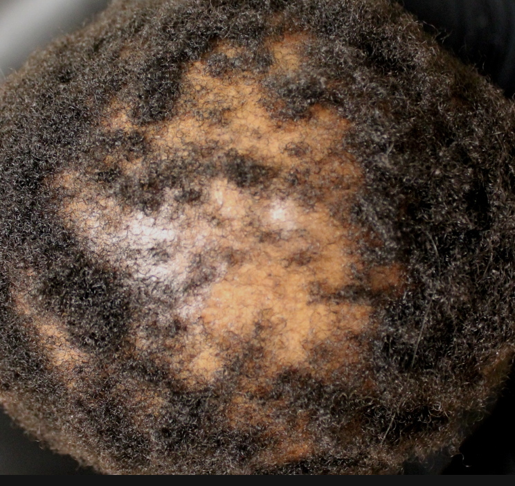
It’s becoming increasingly recognized that many of the hormone blocking drugs used to treat breast cancer can cause a type of hair loss that very much resembles male and female pattern balding (also called androgenetic alopecia and also called female pattern hair loss, FPHL). This type of hair loss is associated with an actual thinning of the hair strands and generally occurs in specific areas of the scalp. Women with FPHL notice that their hair is thinner and finer and this change typically affects the central scalp and crown area. The sides and the back can be affected as well but these areas are usually less affected than then middle and top of the scalp.

The drugs used as ‘endocrine therapy’ for breast cancer can cause a type of hair loss that very much resembles FPHL. It may be that women with breast cancer with underlying susceptibilities to FPHL (based on their family history or genetics) are more likely to develop this type of hair loss when they are prescribed endocrine therapy.

This type of hair loss is important to identify because it has a different course than the telogen effluvium discussed above. FPHL that develops from endocrine therapy does not improve over time if the drug is continued. Even with stopping of the drug, the hair density may not revert back to the original density. With stopping, however, the rate of hair loss may slow down or even stop.

Let me introduce you to a few important studies.


STUDY 1: Park et al 2014

In 2014, a group in Korea reported five cases of pattern alopecia in female patients who are undergoing anticancer hormonal based therapy (with aromatase inhibitors or selective estrogen receptor modulators) for the prevention of recurrence of breast cancer after surgery. This type of patterned alopecia developed after the full recovery of global hair loss of the entire scalp due to previous cytotoxic chemotherapy. The authors proposed that the androgen-estrogen imbalance caused by the drugs was thought to be the reason for the onset of pattern alopecia in the patients.


STUDY 2: Freites-Martinez A, et al 2018

in 2018, Freitas-Martinez and colleagues performed a retrospective cohort study of 112 patients with BC-ETIA. Alopecia was attributed to aromatase inhibitors in 75 patients (67%) and tamoxifen in 37 (33%). Severity was grade 1 in 96 of 104 patients (92%), and the pattern was similar to androgenetic alopecia.


STUDY 3: Gallicchio L et al. 2013

Gallicchio’s study showed that hair thinning was common with aromatase inhibitors and the chances of developing hair loss was not dependent on the age of the patient nor whether they had received chemotherapy in the past. The study was a survey-based study including a total of 851 female patients with breast cancer receiving aromatase inhibitors. 34% reported hair loss or hair thinning during their last month of therapy, and these hair changes were independent of previous chemotherapy and age.


Possibility 3: The Hair Loss is From A Different Reason Altogether

In patients who have hair loss after breast cancer treatment, one must consider a number of possibilities and keep an open mind. A number of possible reasons for hair loss after breast cancer include:

1) hair loss in the form of a telogen effluvium from the stress associated with illness

2) hair loss in the form of a telogen effluvium from one or more surgeries and the anesthetics involved in those surgeries

3) hair loss from chemotherapy, either a temporary or permanent form

4) hair loss from another issue such as a thyroid disorder, poor diet, cancer associated weight loss, or another medication used as part of treatment.


Treatment of Breast cancer endocrine therapy induced alopecia (BC-EIA)

The ideal treatment protocol for BC-ETIA remains to be determined. Hair loss with these endocrine therapies is known to have a significant effect on quality of life and this makes it imperative to develop strategies to address the hair loss.

Stopping the drug is not always an option as doing so may put the patient and highly increased risk of breast cancer recurrence. Such discussions about stopping require a thorough review by the oncologist and dermatologist.

Minoxidil may be one option for treating BC-ETIA. Freitas-Martinez and colleagues showed in 2018 that after treatment with topical minoxidil, moderate or significant improvement in alopecia was observed in 37 of 46 patients (80%). Low level laser may also be an options but this remains to be fully evaluated. Traditional anti-androgen options for addressing androgenetic alopecia in women such finasteride and spironolactone are usually not recommended for women who have been diagnosed with breat cancer.


Summary

Your question is and excellent one and I would encourage you to speak with your doctors about these issues I have raised here. Letrozole is an aromatase inhibitor and Zoladex is a GnRH type analogue. Both of these, as reviewed above, are part of what is termed endocrine therapy. While I can’t comment on whether what you have described truly fits the definition of breast cancer endocrine therapy induced alopecia (BC-EIA), certainly your description would suggest this.

Together with your physicians you can decide whether to continue these treatments or change treatments and whether to now begin specific hair loss treatments such as minoxidil. The data to date would suggest a reasonably good chance of improvement with minoxidil. In some patients minoxidil is combined with low level laser in attempt to further stimulate growth.

We don’t yet know if women with hair loss from one type of endocrine therapy are more or less susceptible to hair loss from another type of therapy. For example, we don’t yet know if women with hair loss from an aromatase inhibitor like Letrozole are less likely to have hair loss if they switch to tamoxifen. It would seem reasonable to conclude that the effects on the hair could be different.

Even with stopping a drug that may have caused BC-EIA hair may or may not improve. Some reduced shedding may occur if a culprit drug was stopped. But the pathways that were triggered often remain triggered to some degree which means the thinning does not revert back to normal in most people. With treatment of course, there can be an improvement.

Thank you again for the question.

Reference

Bhatia A, et al. Tamoxifen-loaded liposomal topical formulation arrests hair growth in mice. Br J Dermatol. 2010

Freites-Martinez A, et al. Endocrine Therapy-Induced Alopecia in Patients With Breast Cancer. JAMA Dermatol. 2018 Jun 1;154(6):670-675. doi: 10.1001/jamadermatol.2018.045

Gallicchio L et al. Aromatase inhibitor therapy and hair loss among breast cancer survivors. Breast Cancer Res Treat. 2013;142(2):435-443.

Kanti V, et al. Analysis of quantitative changes in hair growth during treatment with chemotherapy or tamoxifen in patients with breast cancer: a cohort study. Br J Dermatol. 2014.

Moscetti L, et al. Adjuvant aromatase inhibitor therapy in early breast cancer: what factors lead patients to discontinue treatment? Tumori. 2015;101(5):469-473.

Park J, et al. Pattern Alopecia during Hormonal Anticancer Therapy in Patients with Breast Cancer. Ann Dermatol. 2014.

Sagger V et al. Alopecia with endocrine therapies in patients with cancer. Oncologist. 2013;18(10):1126-1134.


Share This
No Comments

Why isn't my hair loss improving despite improving my iron?

QUESTION

iron levels

I was told that my hair loss was from my low iron levels. However, after working hard for the past 6 months to bring my ferritin levels up from 23 to 55…… I am still not seeing any improvement with my hair at all. Is my hair loss related to iron or not?


Answer

Thanks for the question. It’s certainly a possibility that a person’s iron levels are related to their hair loss. It’s just that they are not implicated as often as most people think. For every one patient I meet with whose lower iron levels are truly related to their hair loss, there are 6 or 7 others where the lower iron levels don’t really seem to be playing role. It’s common to hear stories from patients that they were told their low iron is the reason for their hair loss. Many such patients spend months trying to improve their iron only to find that their hair density has not improved even after correcting their iron.

The short answer is that the lower a person’s ferritin is - the more likely it’s related to the hair loss they are experiencing. It’s a scale from “very likely related” when the ferritin is down below 15 to very like unrelated. I often think in terms of the following table:

ferritin

With a ferritin of 23 you described, there is a good chance it will help. But it’s far from 100 %. In fact, as you’ll see in the studies I discuss below, almost one half of people in the general population with ferritin levels of 23 will have no hair loss problems.

Hypoferritinemia without anemia (HWA): Is it consistently implicated ?

Ferritin is a measure of iron storage levels in body. In order to get a sense of a patient’s iron status, we measure “ferritin” levels rather than iron. Males tend to have higher ferritin levels than females. Premenopausal women tend to have lower ferritin levels than post menopausal women. Extremely low ferritin levels have many potential side effect and may prevent the body from making hemoglobin - a condition which is called ‘anemia’. However, many patients have low ferritin levels without actually having an anemia. This condition is sometimes called hypoferritinemia without anemia or HWA.


Borderline ferritin levels: Evidence for direct role remains poor

The discussion of ferritin levels and hair loss comes down to how low one must go before the low ferritin levels start impacting hair loss. Many females have ferritin levels 20-40 without hair loss. In fact, if you were to measure iron levels (i.e. the ferritin test) in all women between ages 20-40, you'd find many with ferritin 28, 32. 44. You'd find very few with ferritin levels above 50.  You'd find a number with ferritin levels 6, 12, 19.

While it’s often said that one needs to have a ferritin level above 40 (or above 70) for healthy hair growth, this rule is far too simple. We often "aim" for that level in the hair clinic …. but it is completely wrong to say that anytime ferritin is less than 40 there is a problem.


Ferritin levels below 15

Once the iron levels start going low enough, it is true that there is a higher likelihood now that the patient will experience some hair loss an account of those low iron levels. However, it’s now a definite yes or no. It's quite unusual for patient to have normal hair growth with a ferritin of 2 but not completely impossible. However, it’s still within the realm of possibilities for a patient to have normal hair growth with a ferritin of 18.

The biggest challenge is knowing when a patient should be strongly encouraged to increase their iron levels. The simplest rule, as mentioned above, is to recommend to all people with ferritin less than 40. But one must keep in mind that there will be many people with ferritin levels in their 20s and and 30s who are not going to get any benefit from their efforts to increase iron.


FOUR KEY IRON STUDIES TO KNOW ABOUT

As we think about the relationship between low iron and hair loss, there are 4 key studies that everyone should be aware of.


STUDY 1

AUTHOR: Sinclair et al. British Journal of Dermatology

TITLE: There is no clear association between low serum ferritin and chronic diffuse telogen hair loss.

DATE: 2002

Sinclair and colleagues set out to evaluate the relationship between low serum ferritin (</=20 micro g L-1) and chronic diffuse telogen hair loss in women. He analyzed nearly 200 women who presented with chronic hair loss. 12 women had ferritin levels less than 20 ug/L. In 5 women with pure chronic telogen effluvium (and no evidence of androgenetic alopecia), iron supplementation was recommended to bring ferritin levels up above 20. None of these women experienced improvements in their hair with iron supplementation.

STUDY 2

AUTHOR: Deloche et al European Journal of Dermatology

TITLE: Low iron stores: a risk factor for excessive hair loss in non-menopausal women.

DATE: 2007

Deloche and colleagues assessed the relationship in a very large population of 5110 women aged between 35 and 60 years. Hair loss was evaluated using a standardized questionnaire and iron status was assessed by a serum ferritin assay. patients were categorized into three categories acceding to whether they had an "absence of hair loss" (43%), "moderate hair loss" (48%) or "excessive hair loss" (9%). While it was generally found that women affected by excessive hair loss were more often affected by low iron stores, (59 % vs 48 % in the other two groups), this study reminds us that many patients with no hair loss still have low iron levels.

11.4 % of pre-menopausal women who had concerns about ‘excessive hair loss’ had ferritin levels less than 40 ug/L and 10.2 % had ferritin levels less than 15 ug/L. This compares to just 6.8 % of women with ferritin above 70. This information certainly suggests a link between iron and hair loss. However, one must keep in mind that many patients in the study with low ferritin did not have hair loss. Of all premenopausal women with ferritin levels less than 15 ug/L, about 40 % had no concerns about hair loss at all. This is an important reminder that low ferritin levels are not related to hair loss in all patients.


STUDY 3

AUTHOR: Rasheed et al (Skin Pharmacol Physiol.)

TITLE: Serum ferritin and vitamin d in female hair loss: do they play a role?

DATE: 2013

Rasheed and colleagues set out to study the role of several blood tests including iron levels in 80 females (18 to 45 years old) with telogen effluvium (TE) or androgenetic alopecia (FPHL) and compared levels of iron to 40 age-matched females with no hair loss.

Rasheed found that serum ferritin levels were lower in patients with TE (14.7 ± 22.1 μg/l) and FPHL (23.9 ± 38.5 μg/l) compared to the controls (43.5 ± 20.4 μg/l). Interestingly, these levels seemed to decrease with increased disease severity. While these studies suggested a role of low ferritin levels in hair loss the study did not include any investigation as to whether supplementing with iron was a helpful treatment strategy. That was not part of the study.


STUDY 4

AUTHOR: Kantor et al, J Invest Dermatol.

TITLE: Decreased serum ferritin is associated with alopecia in women.

DATE: 2003

One of the earlier studies investigating the role of iron was a 2003 study in the Journal of Investigative Dermatology. The authors studied patients with telogen effluvium (n = 30), androgenetic alopecia (n = 52), alopecia areata (n = 17), and alopecia areata totalis/universalis (n = 7). The normal group consisted of 11 subjects without hair loss.

The authors found that the mean ferritin level in patients with androgenetic alopecia (37.3) and alopecia areata (24.9) were statistically significantly lower than in normals without hair loss (59.5). Interestingly, the mean ferritin levels in patients with telogen effluvium (50.1) and alopecia areata totalis/universalis (52.3) were not significantly lower than in normals. This study was a good reminder that low iron may have a role in some types of hair loss but the role in telogen effluvium remained unclear.


Key summary points about iron levels and hair loss

Here's some key 'take home' messages about iron and hair loss

1. Aiming for a ferritin level above 40 is likely good idea for anyone with hair loss.

2. Aiming for a ferritin above 70 is not my recommendation and is very hard to achieve and generally has little benefit for the hair. 

3. If one's ferritin is between 20-40 and they have hair loss, it must always be remembered that the ferritin levels may be just fine for that person. I'd still recommend supplementing with iron tablets, but there is not a lot of good evidence that doing so is going to help their hair

4. Ferritin levels under 15 are often associated with changes in hair cycling.  If ferritin is less than 15, I recommend speaking to one's physician about iron pills

5. If ferritin levels are low and hemoglobin levels are low (something we call iron deficiency anemia), a full workup by a doctor should be booked.  

6. Vitamin C helps iron absorption and taking a vitamin C rich sources with iron pills is often helpful to increase iron.  Limiting the use of caffeine may also help.

7. Many females have ferritin levels 20-40 without hair loss. The ferritin level alone does not mean much without taking everything into perspective. 


Share This
6 Comments

Blood Tests For Patients with Hair Loss

QUESTION

blood tests

Question

I am experiencing severe and non-stop hair loss for the last 6 months. I am confused about what blood tests I am supposed to get? What is the standard panel that my doctor should be ordering?

Thanks for the question. This is an important question that is too often overlooked. In general terms, blood tests are required for most women with hair loss. For men, they are usually not. However, each patient's hair loss needs to be reviewed on a case by case basis as there is not simple rule about what tests are needed. The exact tests that are needed depends on the patient’s history and their examination findings.


Women with hair loss

For women, I'll not go without blood tests. Blood tests are required for all women with hair loss. Blood tests are mandatory. Simply put there are so many mimickers of female hair loss and diagnosing female hair loss is far more complex than diagnosing hair loss in men. I will order tests for basic blood counts (CBC), thyroid (TSH) and iron (ferritin) in all women with hair loss.

For young women with acne or increased facial hair, a tests for DHEAS (hormones from the adrenal gland), androstenedione (hormones from the ovaries) as well as free and total testosterone and sex hormone binding globulin (SHBG) are ordered. Women with irregular menstrual cycles may require blood tests to evaluate polycystic ovarian syndrome including tests for LH, FSH, DHEAS, androstenedione, prolactin, estrogen, free and total testosterone and 17 hydroxyprogesterone (17OHP) and sex hormone binding globulin (SHBG). Blood sugars may also be checked.

Women with dietary restriction may also have zinc levels checked and a few other minerals as well (i.e. selenium). Sometimes vitamin D is checked depending on where the patient lives.  Women with low ferritin and hemoblogin may, in some situations, benefit from celiac screening before consideration of further testing.

Similar to the discussion for men, women with potential autoimmune causes of hair loss require comprehensive evaluation including complete blood counts (CBC), thyroid (TSH), iron (ferritin), ESR, ANA, B12. 

 This is typically the extent of tests for most. However, should there by any suspicion of a larger systemic issue, liver tests (AST, ALT, bilirubin) might be ordered and kidney function tests (including creatinine and urinalysis) might be considered). One must always consider syphilis screening in all patients as rates are increasing worldwide.

Men with hair loss

For men with male pattern balding (androgenetic alopecia), blood tests are not needed most of the time.  I may check 25 hydroxyvitamin D levels depending on the background of the patent and where in the world they live. For young males with male pattern balding, I often test cholesterol level as there may an increased risk of lipid abnormalities in this patient group. This is an important and poorly recognized issue and I’ve written about it in previous articles:

CHOLESTEROL ISSUES IN YOUNG MEN

TIME IS RIPE FOR THE MEDICAL COMMUNITY TO COME TOGETHER

For men with hair loss due to various autoimmune causes (such as alopecia areata or lichen planopilaris) I often check blood tests such as basic blood counts (CBC) , thyroid (TSH), iron status (ferritin), ANA, B12, ESR. In some situations,  I'll consider a free and total testosterone.

This is typically the extent of tests for most. However, should there by any suspicion of a larger systemic issue, liver tests (AST, ALT, bilirubin) might be ordered and kidney function tests (including creatinine and urinalysis) might be considered). Men with nutritional issues and weight loss, require a far more involved work up including consideration for zinc, selenium screening. Men with low ferritin and hemoglogin may warrant celiac screening before consideration of further testing. One must always consider syphilis screening in all patients as rates are increasing worldwide.


Dozens of other tests available.

There are dozens of other tests available but most of the time they are inappropriate. I see serum iron and serum TIBC ordered inappropriately much of the time. I also see free T3 and free T4 ordered in appropriately as well. There may be a role for some patients but not most.

In other situations a variety of tests can be considered. In a patient with a positive ANA result, anti-double stranded DNA might be considered (along with urinalysis, creatinine, liver function etc). Patients with suspected sarcoidosis may benefit from serum ACE. HIV testing may be appropriate in some situations as well. There are dozens of other sophisticated tests that can be ordered but are generally inappropriate to order as basic screening tests.

Conclusion 


All in all, there is no standard template for ordering blood tests for a patient with hair loss. The tests that need to be ordered are determined once the patient’s story is fully understood and their scalps are examined. If certain tests are abnormal, additional tests may then be considered.

Share This
No Comments

Positive ANA Tests

QUESTION

ana

QUESTION:

I had a positive ANA test result as part of a series of blood tests for my hair loss but feel quite healthy. Do I have an autoimmune disease? Do I need more tests?

ANSWER


Thanks for the question. Without fully knowing all the details of your health and medical history, there is no definitive “yes” or “no” answer here. However, there are some important points to consider. In general, the answer depends on the “level” of your ANA test (how high it is) and whether or not your truly have any symptoms or signs of autoimmune disease.

About 5-10% of the population has a “false positive” ANA that carries no significance for their health. This means the test result comes back looking positive but the patient has no autoimmune disease whatsoever. These are patients who are healthy but have an ANA result of 1:80 and sometimes 1:160. These completely healthy individuals answer “no” when asked questions about whether they have fatigue, joint pain, chest pain, mouth ulcers, sun sensitivity, rashes, blood clots, severe dry eyes, severe dry mouth, Reynaud’s disease, headaches, shortness of breath, repeat miscarriages (women) and other abnormal blood test results. These individuals do not have the autoimmune disease lupus or other autoimmune diseases.

Individuals who do answer “yes” to some of the above questions or who have higher ANA results such as 1:320 or 1:640 often require more specific and detailed autoimmune testing. This may involve tests such as anti double stranded DNA, ESR, blood counts, kidney and liver function, urinalysis and extractable nuclear antigen (ENA). Other tests may be important too. Patients with high ANA results and who have symptoms on questioning can be considered for referral to a rheumatologist.

In general, many physicians do not usually order tests for ANA unless there is some even small degree of suspicion of a possible autoimmune connection. (Randomly testing for ANA without a good cause is not useful for most). A positive test should be followed up with detailed questioning and possibly additional blood tests to determine if it is more likely a false positive or true indicator of underlying autoimmune disease.


Share This
No Comments

Do you have any suggestions for patients with hair loss deemed a 'mystery'?

QUESTION

Screen Shot 2018-09-09 at 5.14.35 AM.png


QUESTION: My hair loss seems a mystery to many doctors. Do you have any suggestions on making the right diagnosis?


ANSWER

There’s no doubt that diagnosing hair loss can be challenging. But hair loss is never labelled a mystery until we’ve properly used the tools we have available. These include asking good questions, examining the scalp, blood tests, scalp biopsies and hair collections. 


1.     Asking Questions

There are potentially 500 questions that are relevant to the patient with hair loss. It’s simply not possible to ask every question so one needs to choose the highest yield questions. There are certain questions that must always be asked and certain questions that should be asked if one finds that a certain line of question is uncovering helpful information. 

The proper diagnosis of hair loss must be approached like detective work. One needs to think of the most common scenario and then the least likely ones.  Patients don’t always volunteer information because they don’t know if it’s relevant or not. The clinician must ask. 

A detective would not label a particular case a ‘mystery’ without having asked some good questions. Hair loss should not be labelled a mystery until one has asked some line of questions. 


2.     Examining the Scalp. 

You simply can’t properly diagnose hair loss from sitting across from the patient. One needs to get up, and examine the hair and the scalp. One needs to look at where on the scalp the hair loss is occurring, and what’s happening to the hairs.

If the hair loss is occurring only in certain areas and other areas seem unaffected, the clinician has gained valuable information that he or she is dealing with one of a group of conditions that are part of the so called ‘localized hair loss conditions.’ If the hair loss is occurring all off the scalp in a diffuse manner, the clinician has gained valuable information that he or she is dealing with one of a group of conditions that are part of the so called ‘diffuse hair loss conditions.’ Narrowing it down is helpful. 

Similarly one needs to look at the hair and the scalp. One needs to look if there is redness in the scalp, scaling, pustules, crusting. One needs to determine if the hairs are all the same size of whether some are thinner. The presence of thinner and thinner hair can be an indication of androgenetic alopecia for some. Are there hairs breaking off?  Is their a lot of new regrowth. 

The scalp examination provides key information. Hair loss should not be labelled a mystery until one has carefully examined the scalp.


3.     Blood tests

I’m of the opinion that everyone with hair loss needs blood tests. Some require only a basic blood count, thyroid study and iron level (ferritin test). Others require more detailed testing. But certainly one can’t label any case a mystery without a basic thyroid and iron level. Iron deficiency and thyroid abnormalities are very common and can have a range of different hair loss presentations.

One can’t order all the 150 blood tests that are available. It’s not practical, and it’s not cost effective. Women with irregular periods needs more detailed hormonal testing and so do women with acne and excessive hair growth on the body. Males and females with fatigue, joint pains, chronic headaches and sun sensitive rashes may be worked up for autoimmune disease.  Sexual transmitted diseases need to always be considered in patients with unexplained hair loss.  Zinc levels may be appropriate in patients with poor diet or weight changes. 

Hair loss should not be labelled a mystery until one has at least ordered a blood count, thyroid and iron study and given thought to the relevancy of the other tests we have available.


4. Scalp Biopsy

Scalp biopsies are not needed for most patients with hair loss. In fact, for every 20 patients I see, I might perform a scalp biopsy on only 1 or 2.  But no patient’s hair loss can be considered a mystery if one has not done a biopsy. It’s a tool that can provide helpful information.

The problem with scalp biopsies is they are not the final step in diagnosis and so are frequently incorrectly used. If a biopsy is not taken from the right spot on the scalp and not processed correctly and not interpreted by a pathologist who understands hair loss, it’s better not to do the biopsy at all. There are certainly a good number of inaccurate biopsy results that I see on a weekly basis. 

But a properly conducted and properly interpreted biopsy can give immediate information about whether or not a patient has a scarring alopecia and whether or not there is any component of androgenetic alopecia in the area biopsied. Biopsies are not accurate method for determining telogen effluvium (this is a clinical diagnosis that comes from asking questions and looking at the scalp).

Not everyone needs a scalp biopsy. But certainly hair loss should not be labelled a mystery until one has had a scalp biopsy.


5.     Hair Collections

Hair collections are not used very often but also provide a tool to better understand how much hair a patient is shedding. A five day hair collections requires the patient not to wash the hair for five days and then the hair is collected in a specific way while shampooing. The number of hairs that come out in that wash as well as the length of the hairs give valuable information about shedding patterns and whether genetic hair loss is likely. 


The Role of Physician Experience

Before we leave the topics of history taking, scalp examination, and blood tests, and biopsies one must also respectfully consider that physicians all have different skills in these areas. If one has seen thousands of hair loss conditions, it is more likely that key questions will be asked, subtle or key findings will be picked up on examination of the scalp, key blood tests ordered or biopsies taken from the correct area of the scalp than if a physician has not seen or treated as many hair loss patients. For that reason, one must also consider referral to a physician who specializes in hair loss disorders for any hair loss condition truly deemed a 'mystery.'


Conclusion

Diagnosing hair loss is truly detective work. One needs to come prepared with the right tools. There certainly are a good share of hair loss mysteries in everyday practice but many so called hair loss mysteries are not really mysteries. They are simply patients with hair loss that have not been thoroughly evaluated. 

Share This
No Comments

How long does it take to know if scarring alopecia treatments are working?T

QUESTION

tmt-scarring

QUESTION: I was diagnosed with lichen planopilaris. My dermatologist put me on topical steroids (betamethasone diproprionate) and doxycycline (100 mg daily). How long does this treatment take before I know if I am benefiting?


ANSWER

Thanks for the question. The real answer depends on a few factors as you'll see below. It could be as early as a few days before your get a sense if you are on track or several months. Let me explain. 

As you've probably come to know from your own physicians, lichen planopilaris (LPP) is a type of scarring alopecia that causes permanent hair loss.  We don't typically expect to see hair regrowth to any significant degree (unless treatment is undertaken in the very earliest of stages). The goal of treatment is to stop the disease and prevent it from getting any worse. 

Lichen planopilaris (LPP) is often 'symptomatic', meaning that many patients have itching, burning or pain/tenderness and notice increased amounts of daily shedding.  Some have all four of these of these symptoms and some have two or three. Of course, about one-third or so of patients don't really have much in the way of symptoms and the scalp feels pretty normal to them. They may just notice a bit of increased shedding. 

The reason this concept is important to mention is because patients who do experience scalp symptoms with their LPP will know if a matter of a few days if they are benefiting from topical steroid treatments. Strong steroids like betmethasone dipropionate usually act to relieve symptoms very quickly and can help reduce shedding quite quickly as well. Oral immunomodulating medications like doxycycline take a bit long but also act within a few weeks. So, if a patient with LPP has considerable itching, burning and tenderness and finds that that the betamethasone is helping to relieve symptoms and shedding after a few days - this means the treatment is benefiting.

Now for a bit more involved discussion.


Difference between short term vs long term benefits

The "first step" for a patient with LPP who is symptomatic is to reduce their scalp symptoms and shedding. This is what we are watching for. This is what we are hoping for. If a patient has itching that they rate as 7 out of 10, we hope in future apportionments that it reduces to 1 out of 10 or maybe even 0 out of 10.  If they feel they once were losing 100 hairs daily and now feel it's under 50 hairs per day... this too is a good sign. 

What we don't know early on in the course of treatment is whether the reduction in symptoms actually correlates with a cessation of hair loss. In other words, a reduction in symptoms is terrific, but it's really the ability to prove that the hair loss is slowing down or stopping that is the most important thing. This comes with scalp examination and photography. 


Scalp examination and photography prove a treatment is benefiting

If a patient states that his or her itching is greatly improved, but a photograph at the 4-6 month follow up appointment shows that more hair loss has occurred, this means that the treatment is not working at all or not working well enough. If the treatment is not working at all, it needs to be stopped and another treatment started. If the treatment is not working well enough but is still helping to slow the rate of loss down a bit, one might consider continuing that treatment (as it is doing something) and adding a second treatment. Of course, this is a professional judgement, and something we stop treatments that are are only working a bit and bring on board treatments that have potential to stop the disease completely.  

Generally speaking if treatments are not working, a photograph will show more and more loss every 4-6 months. Some patients have very slowly progressing LPP and it will take 1 year to show that further hair loss is occurring. Nevertheless, carefully documented photographs will show whether the disease is progressing or stopping. The importance of photography can't be overemphasized. 

Up close examination of the scalp will also provide helpful signs for patients with LPP and so frequent examination by a dermatologist is important. The presence of redness and scaling in the scalp are all potential signs of disease activity. If the amount of redness or scale improves with treatment, this usually means the treatment is working. While the up close examination is important to judge whether a disease is active or not - the most important of all is the photograph of the scalp.


Summary

Thanks again for the great question. I hope this clarifies things.  The real answer is that a patient will truly know in several months if the treatment is truly working by comparing photographs. There are some really helpful things to observe for in the first few weeks and months to give us hints that things are working. This includes a decrease in symptoms and shedding and a decrease in redness and scaling. The most valuable of all is the photograph. 


Share This
No Comments

How often can I have steroid injections?

QUESTION

ILK

QUESTION: How often can scalp steroid injections be done? I have alopecia areata and receive injections into the areas of hair loss every 4-6 weeks. Can I get these injections more often? Is my current frequency considered normal or excessive?


ANSWER

Thanks for the question. Let's first review a few things about steroid injections and then circle back to your question.

Steroid injections are used to reduce inflammation and are useful in a variety of hair loss conditions including alopecia areata, lichen planopilaris, frontal fibrosing alopecia, discoid lupus, traction alopecia, central centrifugal cicatricial alopecia and Pseudopelade of Brocq as well as a few others too.

The typical dose injected ranges from 2.5 mg of steroid per mL (ie 2.5 mg triamcinolone acetonide for every 1 mL of saline used) to a maximum of 10 mg per mL. Lower concentrations reduce the chance for “atrophy” (indentations in the scalp) and so many physicians opt for lower concentrations.

Generally speaking, steroid injections are performed as close as every 4 weeks apart. There is no maximum spacing and some patients benefit from injections every 4-6 months and others come into clinic whenever their condition flares.  Injections more often than every 4 weeks are not typical.


20 mg every 4 weeks

A limited number of studies suggest that keeping the total dose to no more than 20 mg every 4 weeks has quite a good safety profile. This means a physician can use 8 mL of steroid solution every 4 weeks if they decide to use the 2.5 mg per mL solution. If the physician decides to use a higher concentration such as 5 mg per mL, he or she only has 4 mL available to inject. If one uses 10 mg per mL (which some physicians do), one only has 2 mL.

Some studies have suggested that injecting greater volumes of steroid solution at low concentrations works better than injecting smaller volumes of steroid solution at high concentration. For many patients, I even chose 1.5 or 2 mg per mL solutions and see how it works. Use of a 1.5 mg per mL solutions allows 15 mL of fluid to inject in the scalp (that’s 5 syringes!) ... and this goes alot further than trying to figure out where to inject 2 mL of a 10 mg per mL solution. Clearly, if low concentrations aren’t working, one needs to use higher concentrations at the next visit.

In summary, steroid injections can be performed as close as 4 weeks apart but one is not obligated to continue them at this interval. Limiting the concentration, total dose and frequency of injections helps to reduce side effects. In addition to “atrophy” discussed above, side effects can rarely include fatigue, irregular menstrual cycles in women, blood sugar changes and mood changes. These are not common. Longer term side effects to monitor include osteopenia, cataracts, weight gain and changes in blood pressure. These are quite rare with proper dosing and monitoring.


Reference

Chu TW, et al. Benefit of different concentrations of intralesional triamcinolone acetonide in alopecia areata: An intrasubject pilot study. Randomized controlled trial. J Am Acad Dermatol. 2015.

Share This
No Comments

Are JAK Inhibitors Recommended for Patients with Alopecia Areata?

Question

QOW- JAKI

QUESTION: I’ve heard alot about the new JAK inhibitors for alopecia areata. Do you recommend them to patients?


ANSWER

Thanks for the question. JAK inhibitors are a groups of medications that inhibit a pathway inside cells known as the janus kinase pathway. There are actually an increasing number of JAK inhibitors being studied for human disease. The best studied by far are tofacitinib (Xeljanz) and ruxolitinib (Jakafi/Jakavi). Tofacitinib is FDA approved for the treatment of rheumatoid arthritis. Ruxolitinib is FDA approved for the treatment of myelofibrosis.

There is little doubt that the JAK inhibitors are among the more consistently effective of the 26-28 medications to date that we use for alopecia areata. However, the current high cost of the drug limits their widespead use. Furthermore they are not first line for most people meaning that they are not the first treatment to consider. The first line treatment for patient with several patches of alopecia areata remains topical steroids and/or steroid injections - not a JAK inhibitor.

Nevertheless, JAK inhibitors are finding their way into the treatment algorithms for alopecia areata. Patients not responding to topical steroids or steroid injections may consider options such as diphencyprone (DPCP), anthralin, methotrexate and prednisone. However JAK inhibitors are positioning themselves as reasonable evidence-based second or third line options.

Topical JAK inhibitors are also finding a role in the  treatment of alopecia. 2 % Tofactiinib liposomal cream and 0.6 % ruxolitinib have both shown promise. These are compounded from the pill form at a compounding pharmacy. These agents are also quite expensive and require some skill and experience from the perspective of the pharmacist in how best to make up. 

In summary, I rarely recommend a JAK inhibitor as a “first line” option to a patient who has newly diagnosed alopecia areata. However for those with refractory or progressive disease, it most certainly becomes an option to consider.

You may find these previous articles of mine helpful as well:

Tofacitinib for AA: How fast does regrowth occur?

The Topical JAK Inhibitors for AA: Update on Progress

How long do we need to use tofacitininib in AA?

Topical Tofacitininib for Alopecia Areata: How much does it help?

How does the safety of tofacitinib compare to other drugs?

Why do patients stop tofacitinib?

Tofacitinib (Xeljanz) for Children and Teens

Xeljanz in Children: How young is too young?

The JAK Inhibitors for AA: More Data

A look at Inflammatory Markers in AA Treated with Tofacitinib

What blood tests do we need to monitor for patients using tofacitinib?

Ruxolitinib for AA

Topical ruxolitinib promotes eyebrow regrow

Are responses to stress altered in users of tofacitinib?

Nail alopecia areata helped by tofacitinib


Share This
No Comments

What causes hair texture changes?

Question

texture


QUESTION: What causes hair texture changes? I used to have very soft and glossy hair. However, now after years of hairless (androgenetic, FFA & LPP) and treatments (injections, topical clobetasol & oral medications) my hair is very dry, dull, almost straw-like. Are these texture changes due to aging, the hair loss conditions or perhaps the treatments? Conditioners do not seem to help. Thank you.


Answer

Thanks for the great question. There are many causes of hair textural changes. In your case specifically, the causes are probably "multi-factorial" rather than a single cause.  Let’s take a look at some of the more common causes of textural changes and how they apply to the question you have raised. 


Consideration 1: Scarring alopecia

Many patients with scarring alopecia notice changes in their hair texture, especially a change to a drier, more brittle and slightly curlier hair texture. As the name suggests, scarring alopecia is associated with the development of scar tissue or ‘fibrosis’ under the scalp. Such fibrosis affects how hairs emerge from the scalp. Hair frequently twist and turn as they emerge from the scalp and sometimes even rotate 180 degrees. We call this twisting and turning ‘pili torti.’ Individuals with pili torti will notice a hair textural change.

Scarring alopecias are universally associated with loss of the oil glands (sebaceous glands) in the scalp. One can not have a scarring alopecia without having a reduction in the oil glands. These oil glands lubricate the hair follicle.  The destruction of sebaceous glands during the process of scarring alopecia contributes in part to the drier texture. 

Scarring alopecia also affects the quality of the hair that is produced. Commonly there is hair breakage on account of the much weaker fibers. 


Consideration 2: Hormonal changes

A variety of hormonal changes can lead to drier, coarser hair.  About 15 % of women are affected by thyroid disease and this a common cause of textural changes. The incidence fo thyroid disease is much more common in the conditions that you mention including lichen planopilaris (LPP) and frontal fibrosing alopecia (FFA) and so thyroid status should always be carefully evaluated in patients with scarring alopecia.

With approaching menopause, the declining estrogen levels and  imbalances in the ratio of androgens to estrogens also results in drier hair. Women who are predisposed to develop androgenetic alopecia may notice that the hair becomes finer and some may notice the texture changes too. About 40 % - 50% of women with frontal fibrosing alopecia (FFA) and lichen planopilaris (LPP) have androgenetic alopecia. 


Consideration 3: Heat and chemicals

A variety of products that are applied to the scalp can lead to the hair becoming drier, and more brittle. Products containing alcohol are frequently a culprit. This includes hairsprays but many other alcohol containing cosmetic products as well. Products such as minoxidil lotion, and topical steroids may contain alcohol-based ingredients which also dry out the hair.


Consideration 4: Inflammatory scalp diseases

A variety of scalp conditions that are associated with inflammation can lead to altered hair texture over time. Conditions such as seborrheic dermatitis and psoriasis can lead to drier duller hair. Many individuals with FFA and LPP have co-existent seborrheic dermatitis and if present, this should be treated. 


Consideration 5: Androgenetic alopecia (AGA)

Androgenetiic alopecia (AGA) is also a cause of hair textural changes. Although we discussed AGA in the context of hormonal changes above (see "Consideration 2"), androgenetic alopecia can also cause textural changes irrespective of any hormonal abnormalities. In fact, 85 % of women with androgenetic alopecia have normal hormone levels. In women, androgenetic alopecia is also known as female pattern hair loss and in men, male pattern balding. 

Women with AGA often notice the hair is finer and some notice the hair becomes curlier. Others notice the hair becomes flatter and less likely to hold it's original shape, curl or wave. 


Consideration 6: Aging

Hair "aging" is a poorly researched area and poorly defined in general.  Age-related changes in hair, independent of the hormonal changes that can occur with age, can also lead to textural changes in the hair. 


Conclusion

There are a variety of reasons for hair textural changes. One can usually determine the cause of the textural changes with a full review of one's story (i.e. the medical history) along with an up close examination of the scalp. Most of the time blood tests are also needed. 

Thanks again for the great question.  


Share This
8 Comments

Will antidepressants exacerbate my hair loss?

Question

antidepressants-TE


QUESTION: Will antidepressants exacerbate my hair loss? Is is possible to get the treatment I need without making things worse, without inciting another TE?


Answer

Thanks for submitting this question. This is an important question and a common concern among many people. It's actually quite an involved question. We'll begin with the basics and then move into some important points for consideration.  The main point that individuals should be aware of is that the risk of shedding with antidepressants is a lot lower than most would think. It doesn't mean there's no risk, but it is far more likely that a person will NOT get shedding from their anti-depressant than actually experience shedding. 

Antidepressants are the third most common prescription medication in North America (behind cardiovascular drugs and anti-cholesterol drugs). Recent surveys estimate that one out of every six North Americans use some sort of psychiatric medication. About 12 % of these drugs are antidepressants.  Studies by the  National Center for Health Statistics (NCHS) have showed that the rate of antidepressant use in the United States among teens and adults increased by almost 400% between 1988–1994 and 2005–2008. Nearly 1 in 10 individuals in the United States use antidepressants. The age group with the highest use was women 40-50 where nearly 1 in 4 women between 40 and 50 taken antidepressants. Statistics in other countries around the world indicate that antidepressant use is on the rise. Antidepressant use in the UK is similar to the United States where 1 in 11 individuals used antidepressants last year. Citalopram (Celexa) and Sertraline (Zoloft) were the most commonly prescribed antidepressants in the United States. 

There are several classes of anti-depressants including selective serotonin reuptake inhibitors (SSRI's), serotonin and norepinephrine reuptake inhibitors (SNRI's), tricyclic antidepressants (TCA's), Wellbutrin and monoamine oxidase inhibitors (MAOI's).

Let's take a look at what is known about the risk of shedding (telogen effluvium) with these medications. 


Selective serotonin reuptake inhibitors (SSRIs)

Common SSRI's include fluoxetine (Prozac), citalopram (Celexa), sertraline (Zoloft), paroxetine (Paxil) and escitalopram (Lexapro). Litt's Drug eruption Manual estimates that shedding with SSRI's occurs in less than 1 in every 100 users. 


Serotonin and norepinephrine reuptake inhibitors (SNRIs)

Venlafaxine (Effexor) and duloxetine (Cymbalta) are common SNRI-based anti-depressants.  Litt's Drug eruption Manual estimates that shedding with SNRI's occurs in less than 1 in every 100 users. 


Wellbutrin

Wellbutrin is an anti-depressant that affects dopamine. Litt's Drug eruption Manual estimates that shedding with Wellbutrin occurs in less than 1 in every 100 users. 


Tricyclic antidepressants (TCAs)

Tricyclic antidepressants include a wide variety of drugs. Some preferentially inhibit the reuptake of serotonin (such as clomipramine, imipramine). Others preferentially inhibit the reuptake of norepinephrine (such desipramine and nortriptyline). Others yet are fairly balanced reuptake inhibitors of serotonin and norepinephrine (such as amitriptyline).  Litt's Drug eruption Manual estimates that shedding with TCA's occurs in less than 1 in every 100 users. 


Monoamine oxidase inhibitors (MAOIs)

MAOI's used in depression include Isocarboxazid (Marlin), Phenelzine (Nardil), Selegiline (Emsam), Tranylcypromine (Parnate). The exact incidence of telogen effluvium with these drugs is unclear, but MAOI's are generally thought to have the lowest shedding risk of all antidepressants (well under 1:100). 


Shedding with Antidepressants - Key Considerations

When a patient comes in to see me with worries that their antidepressant is causing hair loss, there are several things that need to be consider. I'll address each in turn


Possibility 1. The antidepressant is truly causing a "telogen effluvium" type hair loss

Typically, if an anti-depressant is truly implicated in a person's hair loss, the shedding starts in 4-8 weeks after starting the anti-depressant. It's a diffuse type shedding, meaning that the shedding occurs all over the scalp equally. Patients who experience shedding 6-8 months after starting the antidepressant or notice more hair loss in one area of the scalp than another probably do not have antidepressant related hair loss. Some other cause is implicated. 


Possibility 2. A telogen effluvium is present but it's not due to the antidepressant

One must always rule out other causes of excessive hair shedding in anyone who feels they are losing more hair on a daily basis.  Causes of shedding include high stress, low iron, thyroid problems, medications, dieting and internal illness. A patient who has depression from a thread problem may be shedding from the thyroid issue rather than the antidepressant they were started on.  A patient who has depression from lupus may be shedding from the autoimmune disease itself rather than the antidepressant they were started on. A careful history is essential. It is also mandatory that anyone with depression have blood tests as part of their work up for depression. All patients with depression should have already had blood tests. Similarly, it is mandatory that anyone with excessive hair shedding also have blood tests as part of their work up for excessive shedding. Many of the tests for excessive shedding are similar to the tests they may have had as part of their depression evaluation. The relevant hair related tests include hemoglobin, thyroid studies (TSH), ferritin (iron storage). Other tests may also be relevant depending on the person's specific history.  


Possibility 3. The patient has a hair condition that is different than telogen effluvium but is a close mimicker

A variety of conditions can mimic (i.e. closely resemble) "telogen effluvium" including androgenetic alopecia and diffuse alopecia areata. These need to be carefully ruled out by a dermatologist. Similarly, other scalp issues which can be worsened by depression such as seborrheic dermatitis can increase the amount of daily shedding a person experiences. 


Summary

Anti-depressants can most certainly cause hair loss. However, when a patient says to me "I think my anti-depressant is causing my hair loss" - more often than not it's actually due to another cause. Individuals with depression who have experienced previous episodes of telogen effluvium should speak with their physicians about whether the risks and benefits of both antidepressants and hair shedding. Individuals who are considering antidepressants can weigh the less than 1 % chance of developing hair shedding from the drug against the potential life altering benefits of being treated properly for depression. 


Reference

1) Litt ‘s Drug Eruption Reference Manual 14th edition 2008

2) Moore TJ, et al. Adult Utilization of Psychiatric Drugs and Differences by Sex, Age, and Race.JAMA Intern Med. 2017.

3) Laura A. Pratt et al.  Antidepressant Use in Persons Aged 12 and Over: United States, 2005–2008. NCHS Data Brief No. 76, October 2011.  https://www.cdc.gov/nchs/data/databriefs/db76.htm (accessed Jul 2 2018)


Share This
4 Comments

What causes scarring alopecias like FFA and LLP to reactivate?

Question

QOW

QUESTION: What causes scarring alopecias like FFA and LLP to reactivate? After a period of approximately 6 months during which my hair loss from FFA & LPP seemed to stabilize and hair density actually improved, the shedding has now significantly increased again. What causes these scarring alopecias to reactivate months, or potentially even years, after a period of being quiet with no measurable hair loss? Thank you.


Answer

Thanks for the question. There are a number of reasons why a scarring alopecia can re-activate after it was once quiet. Here are the top 5 important points of discussion. 


Reason 1: We simply don't know the reason.

Although not the answer one would expect to hear as reason 1, we need to respect that we don't completely understand scarring alopecia in the present day and age. Often, we simply don't know the reason. Scarring alopecias can activate and become quiet for periods on their own. We often attribute the entry into a "quiet (inactive) phase" as proof that some type of treatment we are using is helping but scarring alopecias can become quiet other own. Similarly, we often attribute worsening as an indication that something we are doing is no longer working, or we're doing something 'wrong.' However, scarring alopecias can become active spontaneously for reasons that are not clear.


Reason 2: A second hair loss condition has developed.

Whenever there is worsening hair loss, we need to consider that the loss is actually due to another condition, and not the scarring alopecia. Such a condition is often in the form of a 'telogen effluvium," There is an increased incident of iron deficiency, low vitamin D and even thyroid dysfunction among patients with scarring alopecia. For example, a 2014 study from the Cleveland clinic showed that 29 % of patients with LPP develop thyroid dysfunction compared to 9 % of controls. A second study showed that there are also differences in vitamin D status. I've included these references below. 

Levels of vitamin D, TSH, ferritin, zinc should be checked in any patient who experiences a 'flare' following an extended period of quiescences. 


Reason 3: There has been increased life stress.

There's no doubt in my mind that increased life stress can trigger flares in patents with scarring alopecias. It's not clear why and the link seems more relevant for scarring alopecias such as lichen planopilaris (LPP) and frontal fibrosing alopecia (FFA) than many of the other scarring alopecias. Stress can increase a variety of neuromediators and nerve-related hormones in the scalp which impact inflammation.  


Reason 4: There has been injury to the scalp.

It continues to be an area of limited study, by injury can trigger a worsening or 'flare' of a scarring alopecia.  The medical literature documents injuries such as facelifts and hair transplants as triggering scarring alopecias to worsen. However, many types of injuries can cause the disease to reactivate. 


Reason 5: Medications have recently been started or stopped

A scarring alopecia can become active again on account of a) stopping a mediation that was helping, b) starting a medication that was not used before and c) rarely, changing the brand name of a medication that was helping .

If one has recently tapered or reduced a medication within the last one year and is now noticing a worsening of hair loss, they need to consider that the cessation  or reduction of medication could be responsible for the 'flare'. We see that with slow acting medications like hydroxychloroquine. 

Starting a new medication can sometimes trigger shedding. For example, the addition of minoxidil to the scalp in a patient who otherwise had stable disease, can trigger some shedding. Also some medications such as hydroxychoroquine can trigger shedding in some patients. 

Finally, we rarely encounter a situation where a pharmacy changes the brand of the mediation given to the patient. Instead of getting tablets form company X, they are dispensed tablets from company Y. It appears to be the same medication but for some reason a flare occurs. Even more rarely the wrong dose has been prescribed to the patient and they end up receiving less than intended. This of course can cause a flare.


Reason 6: The scarring alopecia has done from "very slightly active" to "slightly active"

To be truly confident a scarring alopecia is inactive, one needs to observe it for two years. After a period of 6 months of observation with a scarring it being "quiet", we can't actually conclude that it was truly inactive.

I often given an analogy of a car moving down the street as an analogy to understand the speed of hair loss. Suppose one's hair loss is moving forward at 6 miles per hour. For the sake of argument, let's say that particular speed is fast enough to see a change in hair density every 6 months. Now let's say the hair loss slows down to a rate of 2 miles per hour. That's really slow. It's not enough to see a change every 6 months but it is just enough to detect a change in density every 1-2 years. If a patient has hair loss moving forward a 2 mies per hour, and they don't see a change after 6 months, they can not conclude it is inactive. Rather there are two possibilities in such a case: either the scarring alopecia is inactive or it is still very slightly active. A clinical examination of the scalp by a dermatologist or time will declare which is correct.  


CONCLUSION

Thanks once again for the great question. When I teach doctors about the reasons for flares in an otherwise stable patient, I frequently use the memory tool "IM WORSE" to help remember the reasons for a flare. This is summarized below. I've also written about the topic in the attached link:

WHY IS MY SCARRING ALOPECIA FLARING AGAIN?

LPP-worse


REFERENCE

Atanaskova Mesinkovska N, et al. Association of lichen planopilaris with thyroid disease: a retrospective case-control study. J Am Acad Dermatol. 2014.

Conic RRZ, et al. Vitamin D Status in Scarring and Non-Scarring Alopecia. J Am Acad Dermatol. 2018.


Share This
2 Comments

How long will my "telogen effluvium" last?

Question:

I have been diagnosed by my dermatologist as having telogen effluvium.  I'm told that my hair might eventually grow back. How long does it take to grow back hair after a telogen effluvium?


Answer:

Generally speaking, most TE will stop within 9 months provided the trigger has been addressed and dealt with. Some TE’s become chronic and continue for years. Fortunately, this is not common. Common triggers include stress, medications, blood test abnormalities (iron, thyroid, hemoglobin) and weight loss. Unless the trigger is addressed, shedding is unlikely to fully cease.

For many people with TE, shedding eventually stops and hair grows back fully. This is not the case for everyone. Two other “sequelae” can occur including the development of chronic shedding and coming to be diagnosed with another completely separate hair loss condition. Interested readers can review these concepts in the link below

Does hair density always return back to normal after telogen effluvium?
  

You may wish to review these additional helpful articles (below) I've written in the past. Thanks again for the question. 

Shedding, Shedding, Shedding: What won't it stop? - 

Recent Articles on Telogen Effluvium

Share This
No Comments

Do I need a scalp biopsy if I have scarring alopecia?

Question:

I have been diagnosed with frontal fibrosing alopecia, which I understand to be a type of scarring alopecia. My dermatologist wants to start me on treatment right away. Do I need a biopsy first?


Answer

Dermatologists have many different views as to whether every patient with potential scarring alopecia needs a scalp biopsy or not. These views fall in three main categories:

1) There are some dermatologists who believe that every single patient with hair loss (scarring or non-scarring) gets a biopsy.  

2) There are some dermatologists who conduct a scalp biopsy in every single patient with scarring alopecia. 

3) There are some dermatologists who perform a biopsy if the diagnosis is not certain and there is even the slightest ambiguity in the diagnosis.

I fall in the third category. My decision on whether a patient needs a biopsy comes during the final steps of a typical patient evaluation. First (step 1), I listen to the patient’s story about their hair loss (we call this a history). Second (step 2), I examine the scalp using a dermatoscope. Third (step 3) I review blood tests. Fourth, I decide whether a biopsy is needed given all the information I have collected during steps 1-3. If the diagnosis is clear and there simply can’t be another diagnosis possible, I don’t do a biopsy.

Here’s an example. Suppose a 56 year old female patient comes to see me. She started losing her eyebrows at age 51. At age 54 she started losing hair along her frontal hairline and it’s receded now about 1⁄2 inch. She’s lost her arm hair, pubic hair and leg hair. Examination shows a scarring alopecia along the frontal hairline. Her blood tests are normal. Based on steps 1-3 I’m confident in the diagnosis of a condition known as frontal fibrosing alopecia.

Will I do a biopsy? No. I will not recommend doing a biopsy in this situation. If the biopsy returns showing scarring alopecia, it’s true that I will have confirmed the diagnosis. Not a bad thing of course. But I will have caused the patient an unnecessary scar. Also, there is always the potential that biopsies (or any trauma) can further activate scarring alopecias, so I’d like to stay away from that.

But suppose the biopsy returns showing something else – such as androgenetic alopecia or alopecia areata. Biopsies are not 100 % accurate so once in a while a scenario like this does occur. In a situation like this, I won’t believe the biopsy results. I’ll simply put the biopsy results aside and move on with discussing treatment. In other words, I’d simply have to explain to the patient that biopsies are not perfect. The reality is that I have caused an unnecessary scar. There may also have been unnecessary expense for getting the biopsy done. There may have even been some pain and discomfort for a few days.

Suppose in the above example, we change things a bit. Suppose the patient is a 56 year old female patient like in the above example. She started losing her eyebrows at age 51. At age 54 she started losing hair along her frontal hairline and it’s receded now about 1⁄2 inch. She’s lost her arm hair, pubic hair and leg hair. She has joint pain in her wrists and ankles, unusual rashes, extreme fatigue and prominent lymph nodes enlarged in her neck. She is troubled by headaches and has had 2 seizures this year that nobody can figure out why. She has dry mouth and dry eyes. Examination shows a scarring alopecia along the frontal hairline. Her blood tests are abnormal with low white cells, abnormal kidney function tests, as elevated liver enzymes. Her ANA is borderline positive at 1:160. When I examine her scalp, I have the impression this is a scarring alopecia – resembling very close frontal fibrosing alopecia.

Here’s a good example of where I will do a scalp biopsy. Even though it seems the patient has frontal fibrosing alopecia, I want to rule out other conditions such as cutaneous lupus, discoid lupus, lymphomas, various infiltrative conditions, including some rare cancers.


You may wish to review these helpful articles (below) I've written in the past. Thanks again for the question. 

Top 25 Frequently Asked Questions about Scarring Alopecia

Share This
No Comments


Share This
-->