Hair loss after the “posterior temple lifting technique.”
Temporal Lifting Technique with Dermal Fillers May give Hair Loss in Some
Authors of a new case series share 6 cases of hair loss happening several weeks after placement of a dermal filler using a cosmetic technique known as the “posterior temple lifting technique.”
By means of some background, let me start by saying that one of the signs of aging is loss of volume in the temple region. Fillers provide an option to increase volume in this area. They also provide a means of gently lifting the mid and lower face. This technique for temple volumization with fillers was published in 2016. There are several approaches that have been suggested.
As part of the posterior temple lifting technique, a high G prime filler (a high viscosity filler) is placed as a small bolus (0.5 ml 1 mL) into the subdermal plane of the posterior superior temple. This increases the tension within the tight superficial compartment.
The overall effect of the filler is to give a lift to the soft tissues of the mid and lower face but it carries the risk of compromising blood flow in the superficial temporal artery (STA) which is found in the same area.

Temporal lifting techniques carry a risk of compromising blood flow.
Landau M et al. 2023
The authors of a new paper report 6 patients with hair loss following the “posterior temple lifting technique.”
PATIENT 1
The first patient the authors report was a 61 year old female treated with the posterior temple lifting technique. She had 1.0 ml of a high G-prime HA was slowly administered as a bolus. Twenty-four hours later, the patient contacted the physician’s office mentioning that she had burning pain in the right parietal area of the scalp. She sent along a photograph as well but it did not show any real abnormality. The patient then had an appointment in the office in the next morning (36 hours after the injection). At the time of the follow up examination, there was a livedoid erythematous eruption was noticed on the right temporal and parietal parts of the hairy scalp with minimal changed noted in the upper right forehead. In order to try to degrade the hyaluronic acid, 1500 IU of hyaluronidase in 5 ml of saline were injected into the palpable bolus of the HA above the right ear and generously infiltrated in all the livedoid areas. The procedure was repeated 3 times (1500 unitsX3) with the patient having a 1-hour break in between the injection sessions. There was no improvement in pain after these three sessions and the procedure was repeated after 8 hours, and an additional session was performed after 24 hours. At this stage, the patient had significant relief in pain level. Ten days later, the patient developed scalp itching, accompanied by hair shedding. Despite biweekly platelet-rich plasma sessions, hair loss developed. Three months after the event, some regrowth of hair began.
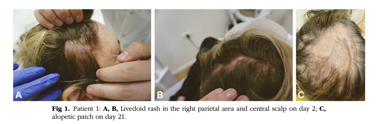
PATIENT 1 from the paper Landau M et al. Non-scarring alopecia after temporal lifting technique with dermal fillers. JAAD Case Rep. 2023 May 12;37:30-34. Images used with creative commons license.
PATIENT 2
The second case the authors report was a 44-year-old female Asian patient again injected using a temporal lifting technique. A bolus of 0.5 ml of HA-based filler was injected to each side. Two days after the procedure, during routine follow-up phone call with the patient, she described mild headache and tenderness of the left temple. A photo sent in by the patient showed no visible abnormality. Four days after the procedure, an examination performed in the office showed reticulated erythematous patches on the left forehead extending, temple, and parietal scalp were seen. Three thousand units of hyaluronidase were injected using a cannula following the original delivery path of HA. A total of 43,500 units of hyaluronidase were injected followed by additional 25,500 units on the following day. After a second round of hyaluronidase injections, she reported a significant reduction in pain.
On day 7, CT Angiography was performed and showed absence of visualization of the left frontal branch of the superior temporal artery. On day 9, despite complete normalization of the skin appearance after all the hyaluronidase injections, the patient noted new hair shedding. A patch of hair loss 5 cmx15 cm developed after 2 weeks. Four months later hair regrowth started.
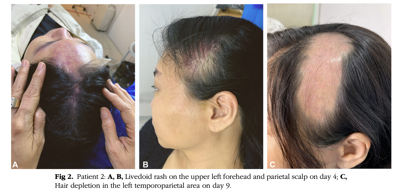
PATIENT 2 from the paper Landau M et al. Non-scarring alopecia after temporal lifting technique with dermal fillers. JAAD Case Rep. 2023 May 12;37:30-34. Images used with creative commons license.
6 Patients in Total Reported by the Authors.
In total, the authors share 4 other patients who developed hair loss after this temporal lifting technique bringing the total to 6.
In general, it appears that patients who go on to develop this side effect generally have pain and burning and bruising and redness. The redness may be quite delayed and skin can look quite normal for an extended period. Headache may be present. In one it was thought to be zoster (shingles infection). Pain can occur 3-4 hours after procedure or the next day.
Immediate treatment was with injection of hyaluronidase (sometimes a lot), as well as pain killers. Prednisone was given in one case.
Hair loss in this series started at day 9-21 but it was around 14 days for most. Patients were treated with various hair growth promoting agents like PRP, minoxidil, oral minxoidil and some had no treatment.
Long term follow up was limited but 2 seemed to recover reasonably well based on data available. But in general, most had follow up only a short duration. One patient followed for 12 months still had a small area of hair loss even though most other areas grew back.
PRIOR STUDIES ALSO SUPPORT THE CONCLUSIONS
The authors mention that these types of complications have been reported in a handful of other patients in the literature. They have occurred with HA fillers as well as with fat and calcium hydroxyapatite.
The mechanism of hair loss appears to be the compromised blood flood in the superficial temporary artery. The STA can be compromised by the increased external pressure induced by the filler’s bolus and the postprocedural edema. Even the filler itself might occasionally be injected by error into an artery compromising blood flow.
The authors remind has that in some studies, filler material was not found in the arterial lumen of the STA or its branches by duplex ultrasound examination in one case and was not seen on biopsy inside the arteries in two other cases examined histologically. In two other cases however, the authors point out that biopsies from areas of hair loss did actually show filler material in the arterial lumen. Thus in addition to mechanisms like compromised blood flow from pressure or edema, intraarterial injection is likely to be a third pathogenetic mechanism
CONCLUSION
All in all, this is a valuable cases series of 6 cases that reminds us that a non-scarring alopecia can develop after a temporal lifting technique.
REFERENCE
Landau M et al. Nonscarring alopecia after temporal lifting technique with dermal fillers. JAAD Case Rep. 2023 May 12;37:30-34
This article was written by Dr. Jeff Donovan, a Canadian and US board certified dermatologist specializing exclusively in hair loss.