h1.qusth1 { display: none !important; }

QUESTION OF THE WEEK

Dr. Donovan's Articles

QUESTION OF HAIR BLOGS


What is "trichotillomania" ?

TTM.jpg

Trichotillomania refers to hair loss due to an individual pulling out his or her hair. It was about 120 years ago that dermatologist Dr. François Henri Hallopeau introduced the term "trichotillomania" to describe a patient who pulled out his hair. 

Trichotillomania occurs in both adults and children.  My patients are often surprised when I tell them that trichotillomania is fairly common. In fact, about 3-5 % of women and 1-3 % of men will pull out hair at some point in their lives.  Many patients deny pulling out their hair at first meeting, so the diagnosis can be challenging and experience is needed to know when a patient visiting your office is likely to have trichotillomania.

What hairs are pulled out?

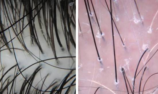
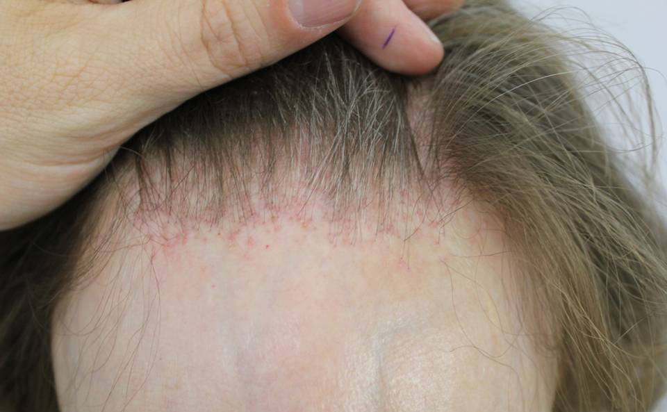
Individuals with trichotillomania may pull out any hair on the body.  Scalp hairs are the most common hairs to be pulled followed by the eyelashes, eyebrows and pubic hair. 

What do patients experience?

Patients with trichotillomania experience a sense of "tension" or "unease" before hair pulling occurs or while they are trying to resist the urge to pull the hair.  After the hair is pulled, individuals feel a great sense of relief or satisfaction.

How does a physician come to the diagnosis of trichotillomania?

According to the American Psychiatric Association's "Diagnostic and Statistical Manual of Mental Disorders" (DSM-IV), there are five key features that need to be met before a patient is said to have trichotillomania.

First, the patient pulls out their hair to the point at which it causes noticeable hair loss. Second, the patient normally has some sense of tension or unease before they pull out the hair or while they are trying to resist pulling out the hair. Third, there is some pleasure or sense of relief after the hair is pulled out.  Fourth, the hair pulling has to lead to some impairment in how the patient functions in social situations or at work or at school.  Finally, if the hair pulling is better explained by some other mental disorder, then the diagnosis of trichotillomania is not given.

These are helpful guidelines, but it's possible for a patient to have trichotillomania without all five of these features present.  There is some contraversery amongst doctors as to whether these five criteria are too restrictive.

Can trichtillomania occur in children?

Yes, In addition to adults, trichtillomania can occur in children and adolesecents.  In fact, it's probably more common in chidlren than adults. In very young children (under 6) it tends to be more of a habit phenomenon.  These younger children sometimes drop the habit of pulling their hair as time goes on.  However, trichotillomania occuring in young pre-teens and teenagers tends to be more concerning.  This hair pulling is often associated with underlying depression or anxiety and may last a long time.  It may affects school and home life. These children often need psychological or psychiatric help.

How much hair can be lost?

Trichotillmania can lead to small amounts of hair loss or rarely complete loss of all hair on the scalp.

Is there are cure?

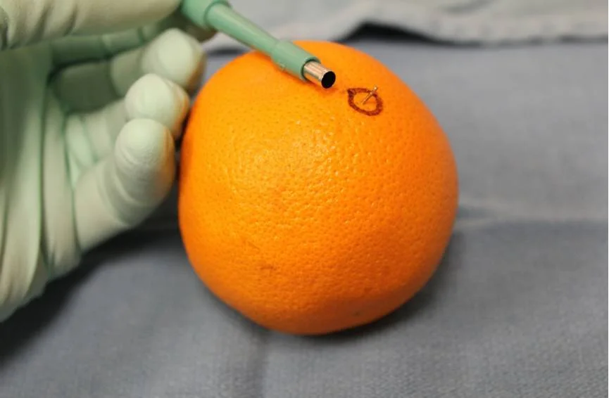
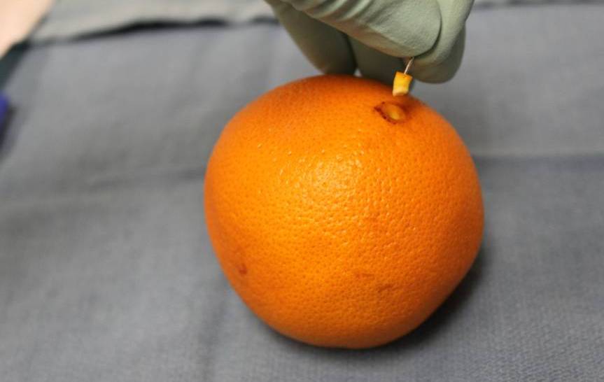
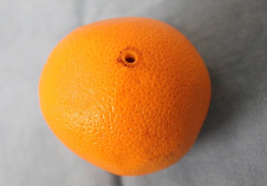
At present there is no cure.  Medications and psychotherapy provided by a psychiatrist or psychologist can be helpful in some cases. Wigs, hair pieces and other forms a scalp camouflage may be used by patients with trichotillomania. Hair Transplantation can also be performed in patients who are stable and have stopped pulling. (see article on Hair Transplantation for Trichotillomania).

References

1) American Psychiatric Association. (2000). Diagnostic and statistical manual of mental disorders. Fourth Edition. Text Revision Washington, DC: American Psychiatric Association.

2) Woods et al. Understanding and treating trichotillomania: What we known and what we don't. Pschiatr Clin N Am 2006; 29L 487-501


This article was written by Dr. Jeff Donovan, a Canadian and US board certified dermatologist specializing exclusively in hair loss.
Share This
No Comments

What's New in Hair Research?

Hair Follicles Help Guide Nerves

When the skin is wounded, many structures in the skin get damaged. Nerves, for example, get damaged and need to regenerate.  The ability of nerves to regenerate is very important to enable us to get sensation or 'feeling' back in the skin.

Recently, a fascinating research study was published which looked at a simple question – Do hair follicles have a role to help nerves to regenerate?

To answer this question, we need to back up a bit. It’s important first to understand that the hair follicle is not just a strand of keratin that somehow roots itself deep down under the skin. Rather, the hair follicle is an extremely complex structure that is richly innervated with nerves. Nerves wrap themselves around hair follicles. In the past, several researchers showed that hair follicles do, in fact, have an extremely important role in the way nerves grow and connect in the skin.

In a new study, researchers used a tissue engineered skin model to show that newly growing hair follicles helped newly growing nerves (neuritis) move about in the skin.  What was interesting was that newly growing nerves didn’t just connect up in a haphazard way, but rather seemed to be located around growing hair follicles and even within them.   The researchers concluded that growing hair follicles essentially ‘attract’ nerves and help to guide the way they develop.

nerves.jpg

Figure 1: Hair follicles play a key role to help nerve migration

To use an analogy, I like to think of hair follicles as traffic police working in an extremely chaotic traffic jam.  Without the traffic police, cars don’t move well. Without hair follicles, newly developing nerves in the skin don’t move well either. When traffic police are present, the cars move around the roads more efficiently.

Reference

Gagnon V et al. Hair follicles guide nerve migration in vitro and in vivo in tissue-engineered skin. J Invest Dermatol 2011; 131: 1375-8

Botcharev VA et al. Hair cycle-dependent plasticity of skin and hair follicle innervation in normal murine skin. J Comp Neur 386; 379-95

Zhang Y et al. Activation of beta-catenin signalling programs embryonic epidermis to hair follicle fate. Development 2008; 135: 2161-72.


This article was written by Dr. Jeff Donovan, a Canadian and US board certified dermatologist specializing exclusively in hair loss.
Share This
No Comments

Improving Eyebrow Growth: Does Bimatoprost (LATISSE®) Help?


Loss of eyebrows is common. A previous article reviewed a range of treatment strategies for eyebrow loss. These include topical medicines, like minoxidil and bimatoprost, as well as hair transplantation.

Bimatoprost is an interesting medication.  It is used for the treatment of glaucoma, an eye disease which leads to elevated eye pressures. Recently, it has found a new use – in the treatment of hair loss. The product is available by prescription under the name LUMIGAN® (used to treat glaucoma)  and LATISSE® (used to stimulate eyelash growth).  Both LUMIGAN® and LATISSE® contain the ingredient bimatoprost.

Chemically, bimatoprost is classified as prostaglandin analogue. Bimatoprost binds to prostaglandin receptors in the hair follicle and stimulates hair growth.  Several hair specialists including myself, have occasionally used this medication for patients with eyebrow loss. This is known as an “off-label” use as the drug is not formally approved for this use. Bimatoprost is formally approved for eyelash regrowth.

Doctors from Miami recently published an interesting report of two patients who achieved an improvement in their eyebrow density using bimatoprost solution.

Elias MJ et al. Bimatoprost ophthalmic solution 0.03 % for eyebrow growth. Dermatol Surg 2011; 37: 1057-59

Both patients used one 2.5 mL container each month and applied the medication nightly to each eyebrow. One patient, a 52 year old man had results after 16 weeks and the second patient, a 46 year old woman had results after 12 weeks. These two patients did not experience any side effects.

This is an exciting report and calls for more studies to be done to evaluate the use of bimatoprost (LATISSE®) in the treatment of eyebrow loss.

Reference

Elias MJ et al. Bimatoprost ophthalmic solution 0.03 % for eyebrow growth. Dermatol Surg 2011; 37: 1057-59.


This article was written by Dr. Jeff Donovan, a Canadian and US board certified dermatologist specializing exclusively in hair loss.
Share This
1 Comment

Hair Loss in the Infant: Does Sleeping Position Matter?

sleep on back babies sids.jpg

2 to 3 months after birth, some babies start losing hair at the back of the scalps.  The medical term for this hair loss pattern is "Neonatal Occipital Alopecia." 

For years, it was thought that this occurs because of pressure on the back of the scalp from babies sleeping on their backs.  It’s now understood that this hair loss occurs as a normal physiological phenomenom in some babies and has nothing to do with pressure. The condition always improves on its own and some parents don’t pay much attention to it at all.

But why do some babies develop this temporary hair loss condition and others do not?

Researchers in Korea set out to answer this question by examining 338 newborns over a time period of 1.5 years. They found that 20 % of babies developed hair loss in the back of the scalp.  Moreover, babies born to younger moms less than 35 years, moms who did not have C-sections and babies born after 37 weeks were most likely to develop this condition.

Conclusion: It is important for parents and grandparents to understand that hair loss at the back of the scalp occurring in a 2-4 month old baby has nothing to do with sleeping positions. It occurs as a normal phenomenon.  Babies should sleep on their backs rather than their stomachs, according to recommendations from the American Academy of Pediatrics.  The recommendation was put forth in order to reduce the incidence of Sudden Infant Death Syndrome (SIDS).

References of Interest

Kim MS et al. Prevalence and factors associated with neonatal occipital alopecia: a retrospective study.  Ann Dermatol 2011; 23: 288-92.

American Academy of Pediatrics AAP Task Forice on Infant Positioning and SIDS: Positioning and SIDS. Pediatrics 1992; 89: 1120-26

Gibson E et al. Infant sleep position following new AAP guidelines. American Academy of Pediatrics. Pediatrics 1995; 96: 69-72


This article was written by Dr. Jeff Donovan, a Canadian and US board certified dermatologist specializing exclusively in hair loss.
Share This
No Comments

Telogen Effluvium: Remembering Life's Events

Telogen effluvium is a common hair loss condition.  Patients note increased hair shedding - and hair starts collecting everywhere. There is hair around the house, hair on the floor, hair in the brush, hair in the sink, hair in the shower drain, hair in the car, hair at the office.  Spouses start to notice that there is more hair around than there used to be.  Even young children start picking up their parents hair and commenting that too much of mommy or daddy's hair is coming out. This is telogen effluvium.

Hair shedding can be caused by a wide range of 'triggers.'  These include physiologic stress, endocrine problems (including thyroid problems), nutritional problems, iron deficiency, and medications.  Other scalp diseases, including alopecia areata, and scarring alopecias can also cause hair shedding but these are different from telogen effluvium.

Sometimes it takes a bit of detective work to figure out what might be the trigger of a patient's hair shedding.  In many cases we find the cause, but in some cases we don't and just wait for the shedding to stop. 

There are many things I enjoy about being a hair doctor - but one aspect in particular I enjoy is pinpointing exactly in time when someone's hair loss 'trigger' might have occurred.  This usually occurs in a couple of classic scenarios.

Let me explain.

Suppose I see a patient who is worried about their hair shedding.   They tell me that the shedding used to be really bad a few months ago but is actually starting to get back to normal. After asking dozens of questions I proceed to examine the scalp.   I look for signs of various hair diseases, and there does not appear to be any.  Then as a final step, I lift 50-100 hairs straight up and - voila - I see a remarkable number of 3 cm hairs.  Normally, I would expect to see hairs of all different lengths - some 1 cm hairs, some 2 cm hairs, some 3 cm hairs, some 4 cm hairs and so on. But the patient in our example has many many 3 cm hairs. In fact - way too many 3 cm hairs!

What does this unusual number of 3 cm hairs tell me?

Well, it tells me the patient had some major trigger of hair shedding take place about 6 months ago. 

After a trigger like a surgery, or a crash diet, the hairs of some individuals can be rapidly shifted into a resting period called the "telogen phase". These hairs spend a mandatory period of three months in the telogen phase and then all get shed from the scalp at a similar time.   Because hairs normally grow back at a rate of about 1 cm per month - if I see alot of 3 cm hairs, I know they've been growing back for about three months and I know the trigger must have occured about 3 months before that - for a total of six months.

TE 2 color.jpg

So when I see lots of 3 cm hairs, I know there was some major trigger about 6 months ago (3+3=6).When I see lots of 5 cm hairs, I know there was some major trigger about 8 months ago (5+3=8). When I see lots of 7 cm hairs,  I know there was some major trigger of hair shedding about 10 months ago (7+3=10). It's as simple as that.

 So for me, telogen effluvium can sometimes boil down to helping patients remember life's events.  If the patient's shedding has stopped and they are growing back their hair, I can often pinpoint when the hair loss occured.  If the shedding is ongoing and has not yet stopped, a bit of detective work especially blood tests, will be necessary.

TE 1.jpg

So for the patient with 3 cm hairs, there is one simply question that can clinch the diagnosis... What happened in your life 6 months ago?  It sometimes takes patients a little bit of time and sometimes a calendar even gets pulled out from a bag, but the trigger often comes from remembering life's events:

Oh, now that you mention it, I was in the hospital, sick as a dog!

or ... That's when I started this new drug

or ... That's when my dad passed away

or ... That's when I have a bad flu and was off work for weeks

or ... That's when I had my surgery

The list goes on and on. Telogen effluvium can sometimes be challenging to diagnose. Every patient needs some basic blood work to make sure that there is no thyroid abnormality and no significant iron deficiency that caused the hair shedding. But if the hair shedding has already slowed down and is starting to grow back, one can often pinpoint exactly in time when the shedding started and then narrow down the exact cause.


This article was written by Dr. Jeff Donovan, a Canadian and US board certified dermatologist specializing exclusively in hair loss.
Share This
17 Comments

How is a Scalp Biopsy Performed?

biopsy pearls.jpg

There are over 100 reasons for an individual to have hair loss. Sometimes the reason for a patient's hair loss is evident within seconds of meeting the patient. At other times, the diagnosis requires a small scalp biopsy.  A scalp biopsy is a short procedure, performed under local anesthesia, that allows a few hairs (and the skin surrounding those hairs) to be removed for future examination under the microscope.

I routinely teach medical students, residents and fellows how to do proper scalp biopsies so that a good sample can be obtained.  It is absolutely essential to get a good sample - one that is deep enough, big enough and not damaged. Here, I outline the proper technique for obtaining a biopsy.  I often use an orange to demonstrate the procedure rather than showing the scalp. The orange background allows the key learning points to be very easily seen. Here, I'll use the orange as well to demonstrate the basic technique.

12 Steps to Performing a Scalp Biopsy

photo1.jpg

An orange is used to demonstrate the proper technique of performing a scalp biopsySTEP 1: Spend time finding an area of the scalp that best respesents the hair loss condition you think the patient has.

If the patient has an unusual pattern of hair loss, or I think that only some areas of the scalp are affected by the condition, I spend many minutes searching for the "perfect spot" to biopsy.  If I think the patient has a scarring alopecia such as lichen planopilaris, I look for an area with perifollicular erythema or scale. If I think the patient may have an unusual or atypical form of alopecia areata, I look for empty tracts or vellus like hairs.

photo2.jpg

STEP 2: Draw a circle around the area.

A blue Acculine Marking Pen can be used to outline the area. I like these pens because the marker does not wash off easily. Therefore, the circle will stay until it is wiped away at the end of the procedure.

photo3.jpg

STEP 3: Cut the hairs that are to be taken in the biopsy.

I routinely cut the hairs that I am going to sample. I clip the hairs to a distance of about 2-3 mm above the scalp. This way I can accurately see the angle that the hairs emerge from the scalp. This will be important in Step 6 (below).

photo4.jpg

STEP 4: Clean the skin.

The skin can be cleaned with many of the commercially available cleansing solutions. I typically use chlorhexidine.

photo5.jpg

STEP 5: Anesthetize the skin

The skin surrounding the hairs is frozen with a numbing solution such as 1% lidocaine with 1:200,000 epinephrine. After freezing the skin, I typically wait 10-15 minutes if possible. This allows the epinephrine to take full effect and drastically helps to reduce bleeding during the small procedure.

photo6.jpg

STEP 6: Use a 4 mm sized punch biopsy tool to puncture the skin.

The minimum size for a punch biopsy for scalp specimens is 4 mm.  These recommendations follow those of the North American Hair Research Society.

punch photo.jpg

The biopsy tool is placed directly over top of the hair - at the precise angle that the hairs are coming out of the scalp. The biopsy tool is then rotated back and forth quickly until the metal blade is completely submerged to the level of the subcutaneous fat. Then the biopsy tool is removed.

photo8.jpg

STEP 7: Delicately pull up on the specimen using the anesthetic needle.

Once the biopsy site has been made, it's important to treat the biopsy specimen very delicately. I use a 30 G needle (the same one used to freeze the skin) to "pull up" on the specimen.

photo9.jpg

STEP 8: Cut the biopsy specimen at the base.

Once the biopsy specimen has been pulled up, I cut it at the base in a horizontal manner using scissors. It is very important that this be done at the subcutaneous junction.

photo10.jpg

STEP 9: Remove the specimen.

After the specimen has been cut at the base, it can be easily removed from the scalp. It can be placed directly into 10% buffered formalin for transport to the histology lab.

empty.jpg

STEP 10: Suture the small opening.

The small opening can be closed with 3-0 or 4-0 nylon. Some physicians choose to leave it open without a suture or pack it with kaltostat.

photo13.jpg

STEP 11: Place petroleum jelly over the suture site to promote moist wound healing.

The biopsy site can be left uncovered. A bandage or dressing is not needed. A small amount of petroleum jelly can be placed over the incision site to keep it moist. Some physicians choose to use antibiotic ointment after the procedure and this is acceptable too. I typically advise patients to wash the area after 24 hours and to continue to apply petroleum jelly for an additional 5 days. It is well accepted that moist wounds heal better.

STEP 12: Remove the stitches in 10-14 days.

The small stitch is cut with scissors after 10-14 days.

All in all, performing a scalp biopsy is easy, and can be performed with little discomfort to the patient. The following represents a basic set up of a surgical tray:

tray set up.jpg

1. chlorhexidine solution 2. punch biopsy tool 3. needle driver 4. pick ups 5. scissors 6. q tips for applying petroleum jelly 7. suture 8. sterile drape 9. gauze


This article was written by Dr. Jeff Donovan, a Canadian and US board certified dermatologist specializing exclusively in hair loss.
Share This
18 Comments

Perifollicular Scale: A Clue to the Diagnosis of Lichen Planopilaris

normal vs lpp perifollicular scale.jpg

The photo on the left is from a patient with a normal healthy scalp. The hair follicles are all similar size and spacing between them is similar. There is no redness, and no flaking in the scalp.

The patient on the right has redness in the scalp. The hair density is decreased.  Another striking feature is also present - the white “scale” around many of the hair follicles. The medical term for this scale hugging each of the hair follicles is known as ‘perifollicular scale.’  Perifollicular scale is a potentially important clue to the diagnosis of a certain scarring hair loss conditions, especially a condition known as lichen planopilaris (LPP).

When I see perifollicular scale in a patient with a red itchy scalp, I suspect that the patient has a scarring hair loss condition and usually do a scalp biopsy to confirm the diagnosis. Lichen planopilaris is a cause of permanent hair loss.  When the condition first starts, patients may have itching, burning or pain in the scalp and possibly increased hair shedding too. It is important to diagnose this condition in the early stages in order to try to prevent further hair loss.


This article was written by Dr. Jeff Donovan, a Canadian and US board certified dermatologist specializing exclusively in hair loss.
Share This
2 Comments

Is it safe to recieve a ‘flu shot’ when I'm taking immunosuppressant medications for my hair condition?


Patients with hair diseases that cause extensive hair loss, including advanced alopecia areata, or certain types of scarring alopecias (i.e. lichen planopilaris or discoid lupus) require oral medications to treat their hair loss. Hair loss in these patients can not be managed simply with topical lotions and creams and medications such as hydroxychloroquine, cyclosporine, mycophenolate mofetil and sulfasalazine are frequently prescribed.

At this time of year, many patients taking these immunosuppressants ask a similar question... is it safe to get a flu shot when I'm taking an immunosuppressant drug?

To fully answer this question, let’s review the different types of vaccinations. There are two main types of vaccinations that people can receive – “inactivated” vaccines (like the flu shot, hepatitis B vaccine, and HPV vaccine) and “live” vaccines (like measles, mumps and rubella vaccines, yellow fever vaccines and nasal spray flu vaccines).  In general, "inactivated" vaccines are safe for patients taking immunosuppressive medications. However, I advise all patients to review the specific details with their physician. For example, children under 6 months of age, those with egg allergy and those with a previous ‘bad’ reaction to the flu shot should not get the vaccine. In addition, "live" vaccines should not be given to patients taking immunosuppressive medications – and are best given before patients go on these drugs.

The main messages I tell my patients are:

Patients who take certain types of immunosuppressive medications may be at increased risk for certain infections and vaccines can help prevent many of these infections.

Inactive vaccines, like the flut shot, are generally safe and helpful in patients taking immunosuppressive medications 

Live vaccines should not be given to patients taking immunosuppressive medications


This article was written by Dr. Jeff Donovan, a Canadian and US board certified dermatologist specializing exclusively in hair loss.
Share This
No Comments

Spiders, Rain and Hair Growth


Yesterday, by accident, I killed spider.  And then it rained.  In fact, it poured. To some, this information will come as no surprise.  After all, it’s common knowledge to many people that killing a spider will cause it to rain.  But is it really true? Does killing a spider cause it to rain?  To the best of my knowledge there has never been a scientific study examining the relationship between spider killing and rainstorm patterns. 

As a hair specialist, I hear myths about hair loss every day and consider it an important part of my practice to spend time with patients to help separate fact from fiction.  When I’m asked if a specific product will help with hair growth, I generally respond with one of the following 3 answers:

Yes, there is good scientific evidence that this is likely to be helpful ...

or

I think this will probably help your hair, but there is only a small amount of scientific evidence for this claim  ....

or

There is no scientific evidence at present that this does anything to help your hair


Why do we need "scientific evidence"? 

If a patient says to me that a product helped them grows hair, then doesn’t it help grow hair?  Unfortunately, the answer is no. It's not that I don't believe what a patient might tell me. It's simply that there are too many reasons why a patient’s hair might have grown  - even if he or she were to put water on top. Certain types of hair loss conditions improve - even if you do nothing.

Let’s revisit the spider story again. There is no scientific evidence that killing spiders causes it to rain. To prove whether killing spiders causes it to rain, we would need to design a "scientific study" using 1000’s of spiders.   To design a really good scientific study, we would need to design a study with at least two big groups of spiders. In one group the spiders would be stepped on and in the other group they would not be stepped on. And then we’d check to see if it was more likely to rain on the days that spiders were killed. But is that enough of proof? The answer is still no! We’d need to make sure that all the spiders were the same types of spiders and that they were the same age of spiders.  (After all, maybe stepping on young 'garden' spiders causes it to rain but stepping on older 'house' spiders has no effect.) Furthermore, we’d need to make sure that the killing was done at the same time of year.  (Maybe stepping on a spider in the month of September causes it to rain but stepping on spiders in the month of May has no effect).  We would also need to clearly define where it is that we’ll be watching for the rain drops to occur.  (Perhaps killing a spider in Paris, France causes it to rain in Sydney, Australia).

You can see that if would cost a lot of money and take a lot of time to design a study to prove if stepping on spiders causes it to rain.  And so, the study has never been done.  Do we really 100 % know if killing a spider has anything to do with rainfall patterns in the world ? The answer is no. And that’s why I try to avoid stepping on them and continue to spread the myth to help other people avoid killing spiders too. But deep down, do I really think that killing spiders causes it to rain? The answer is, of course, no. 

Thousands of Hair Studies Needed

There are thousands of products on the market for hair and hair loss.  Some products have no scientific evidence that they help hair loss or help with hair growth.  They might help hair growth, but we simply don't know. Certainly, some products do help. To test if a product helps, it would cost money and it would take time.  When a patient tells me that a product helped with hair loss, I tuck the fact into the back of my mind but I usually don’t get very excited. When two different patients tell me that the same product helped them, I still tuck the information into the fact of my mind but usually don’t get very excited. When 10-15 people tell me about a specific product, I think about designing a small study or researching the evidence that this could be true. I remain aware of the possibility that it may be turn out that the product still offers no help for hair loss.

Sound Science Needed for Everything Consumers Put Trust In

This past week I learned that Reebok was made to pay 25 milliion dollars in customer refunds to settle charges of deceptive advertising of a specific type of shoe.  Previous advertising by the Reebok company had claimed the the shoe helped to strengthen and tone muscles.  The company had advertized that this specific shoe could lead to 28 percent more strength and tone in the buttocks, 11 percent more strength in the hamstring muscles and 11 percent more strength in the calf muscles than an ordinary walking shoe. The US Federal Trade Commision stated that it wanted national advertisers to realize that they need to excercise responsibility to ensure claims are supported by "sound science."


The Future of Hair as a Science

Some incredibly exciting hair research discoveries are taking place all over the world. Incredible people are doing incredible work. This means that new treatments for hair loss are waiting us in the years ahead. We need to always make sure that detailed and rigorous scientific research methods are used to help clearly define which treatments help individuals with hair loss and which do not.


This article was written by Dr. Jeff Donovan, a Canadian and US board certified dermatologist specializing exclusively in hair loss.
Share This
1 Comment

Hair Transplantation in Frontal Fibrosing Alopecia

Hair transplants for FFA

Frontal  Fibrosing Alopecia is a cause of permanent scarring hair loss fibrosing alopecia is a  rare scarring hair loss condition that affects the frontal scalp. The condition occurs mostly in women although men can very rarely be affected. Frontal fibrosing alopecia causes permanent hair loss. Treatments for the condition include topical steroids and non-steroid medicines, steroid injections, as well as oral medications such as tetracyclines, hydroxychloroquine, mycophenolate mofetil and cyclosporine.


Patients with frontal fibrosing alopecia often ask me  - Is it possible to have a hair transplant?

To answer this question,  I remind myself of a interesting research study from Drs. Nusbaum and Nusbaum in Miami, Florida which was published in the journal Dermatologic Surgery.

Nusbaum BP and Nusbaum AG. Frontal fibrosing alopecia in a man: results of follicular unit test grafting. Dermatol Surg 2010; 36: 959-62.

In the journal, the doctors report a 44 year old man with frontal fibrosing alopecia who underwent a hair transplant.  Prior to the hair transplant he was treated with topical steroids and steroid injections. He also used the oral medicine hydroxychloroquine for 6 months.  His disease appeared to be ‘quiet.’

With the disease quiet, the man stopped the medications and underwent a very small ‘test’ hair transplant with 82 follicular units placed into the scalp.  Three months after the transplant, the hair started to grow and after 15 months after the transplant the hair had grown in normally. This would have been tremendously exciting for both the patient and surgeon.  Four years later, however, only 6 hairs remained – the remaining hairs were destroyed by the scarring alopecia. 


Comment: This report is very important. It reminds us of just how complex the disease frontal fibrosing alopecia can be. The disease can appear quiet for extended periods and then can reactivate.  It is challenging for doctors to monitor because patients often have no symptoms. 

We still can’t predict with certainty if a hair transplant will be successful in a patient with frontal fibrosing alopecia. Sometimes it can be - but sometimes it won't. The above study reminds us that hairs can grow in nicely, only to be lost in future years.  Further studies are needed to determine if patients who receive treatment with topical and oral medicines are less likely to lose their hair.


This article was written by Dr. Jeff Donovan, a Canadian and US board certified dermatologist specializing exclusively in hair loss.
Share This
No Comments

Dr. Seuss and Coping Strategies in Children

dr seuss.jpg

Some people are surprised to learn that children can be affected by hair loss. Conditions such as alopecia areata, tinea capitis, trichotillomania, traction aloepcia and telogen effluvium can affect children. Most of the very young patients in my practice have alopecia areata, an autoimmune condition which can lead to hair loss in circular patches, or total hair loss (alopecia totalis) or total body hair loss (alopecia universalis).

dr seuss books.jpg

Children, like adults, develop strategies to cope with their hair loss.  These coping strategies change as the child ages.  Many children with hair loss find going back to school especially stressful. It’s a time when the coping strategies they have developed are put to the test.

Parents usually tell me if their child is having problems coping with hair loss.  When I am concerned about how a child is coping I sometimes ask the child an indirect question. In the months of August and September I frequently ask:

“What would you say to another child who had hair loss and was worried about starting back up at school?”

When most children hear this question, there is a pause. Most children smile or laugh and then look at their parents. Some start their sentence only to pause for an extended period. One child had clearly thought about this in great detail and had developed some useful coping skills. The child answered:

I would tell them ... to be who you are and say what you feel, because those who mind don't matter, and those who matter don't mind.  

I knew right away that these could not be the original thoughts of the child. Not the words of the parents, the grandparents, the teacher or a friend.  I soon learned from the child that these were the words of Dr. Seuss.  Today marks the 20th anniversary of the loss of Theodor Seuss Geisel (March 2, 1904 – September 24, 1991), better known as Dr. Seuss. The words in his 46 children’s books impacted millions of children, including at least one child who used these words as a coping strategy for hair loss. Every now and then I find myself quoting Dr. Seuss when talking to children and their parents about hair loss.


This article was written by Dr. Jeff Donovan, a Canadian and US board certified dermatologist specializing exclusively in hair loss.
Share This
No Comments

Is destruction of the oil glands a key step in the development of scarring alopecia?

a scarring alopecia.jpg

Scarring Scarring Alopecia: Oil gland destruction may be a key early stepalopecias are a group of hair loss conditions that lead to permanent  hair loss. An example of a scarring alopecia is shown in the photos. Although we know how to recognize these conditions, and we understand how to treat them, we understand very little about their cause.

For years it has be thought that abnormalities developing in the sebaceous glands or "oil glands" of the hair follicle can lead to scarring alopecias.  Dr Stephen Lyle and colleages at the University of Massachusetts Medical School set out to determine if the oil glands are affected in patients with various types of scarring alopecias.

Al-Zaid T et al. Sebaceous gland loss and inflammation in scarring alopecia: A potential role in pathogenesis. Journal of the American Academy of Dermatology 2011; 65: 597-603

The researchers showed that scarring alopecias frequently showed loss of the sebaceous gland. For example, a reduction in sebaceous glands was seen in:

84 % of biopsy specimens from patients with the scarring alopecia lichen planopilaris

77% of biopsy specimens from patients with central centrifugal cicatricial alopecia

100 % of biopsy specimens from patients with follicultis decalvans

In many cases, the sebecaous glands were not only reduced but inflamed as well. For example, inflammation occurring in the sebaceous glands was seen in:

55 % of biopsy specimens from patients with lichen planopilaris

25 % of biopsy specimens from patients with central centrifugal cicatricial alopecia

50 % of biopsy specimens from patients with folliculitis decalvans

Conclusion: These data give further support to the current thinking that inflammation affects the sebaceous gland may be one of the "earliest steps" in the development of scaring alopecia.


This article was written by Dr. Jeff Donovan, a Canadian and US board certified dermatologist specializing exclusively in hair loss.
Share This
2 Comments

The Seasonality of Hair Shedding

fall in toronto.jpg

The Seasonality of Hair Shedding

As the Autumn in Toronto transitions from summer to early autumn approaches, I'm reminded of a remarkable feature of human hair growth - the increased tendancy for humans to shed hair in the early Fall.

Several research studies have shown that humans living in northern regions tend to shed more during the late summer and early autumn months.   Most of the time this goes undetected, but some individuals do notice this feature. A second phase of increase shedding in human beings may occur in Spring as well.

What causes hair shedding?

Of course, anyone coming into the office with concerns about hair shedding requires a thorough evaluation to determine the causes of increased hair shedding. These many include:

Physiological stress (i.e. surgery, labour and delivery, systemic diseases of the body, infections)

Endocrine problems (i.e. thyroid abnormalities)

Nutritional deficiencies (i.e. low iron, dieting)

Medications (i.e. anti-depressants, ACE inhibitors, heparin, beta blockers, lithium)

In addition to a thorough history and scalp examination, a patient with concerns about hair shedding requires blood work for complete blood count, thyroid studies and iron studies. Other studies may be needed as well.  All in all, there is a periodicity to how humans normally shed hair. Although loss of 50-100 hairs each and every day is considered normal, slightly increased rates can be observed in the Fall.

References of Interest

1) Courtois M et al. Periodicity in the growth and shedding of hair. Br J Dermatol 1996; 134;47-54.

2) Kunz M et al. Seasonality of hair shedding in healthy women complaining of hair loss. Dermatology 2009; 219: 105-10.

3) Randall CA and Ebling EJG. Seasonal changes in human hair growth.  Br J Dermatol 1991; 124: 146-51.


This article was written by Dr. Jeff Donovan, a Canadian and US board certified dermatologist specializing exclusively in hair loss.
Share This
No Comments

Flutamide May Help Women With Androgenetic Alopecia Who Don’t Respond to Spironolactone


By age 50, about 30 % of women will develop female pattern hair loss, also known as androgenetic alopecia. Treatments for this condition include topical minoxidil, hormone blocking oral medications such as spironolactone, flutamide and cyproterone actetate as well as hair transplantation. Spironolactone is considered among the first-line oral medications to treat female pattern hair loss.  

But what is the next step when spironolactone doesn’t seem to be helping?

Australian hair loss specialist Dr. Rodney Sinclair and his colleague Dr. Anosha Yazabadi suggested that the oral medication flutamide could be a helpful next step. They report the case of a 35 year old woman with androgenetic alopecia whose hair loss did not improve despite 5 years of spironolactone treatment at a daily dose of 200 mg and a 6 month course of 5 % minoxidil. A decision was made to stop the spironolactone and start flutamide at 250 mg per day and continue minoxidil. After 6 months of use, the patient’s hair loss ceased and her hair density improved.

Yazdabadi A adn Sinclair R. Treatment of female pattern hair loss with the androgen receptor antagonist flutamide. Australasian Journal of Dermatology 2011; 52: 132-34.

Comment: Flutamide may be a helpful medication in women with androgenetic alopecia whose hair loss does not improve with minoxidil and spironolactone and who are not candidates for hair transplantation. This oral medication blocks androgen hormones in several ways, including inhibiting update and the binding of androgens to the androgen receptor. Overall, it is a more potent androgen blocking medication than spironolactone. 

Consultation with a physician is necessarily to fully discuss the side effects of flutamide. This medication can rarely cause inflammation in the liver and so blood tests to monitor the liver are needed while using this drug. Furthermore, premenopausal women must not get pregnant while on flutamide and therefore contraceptive methods need to be carefully discussed with each patient.


This article was written by Dr. Jeff Donovan, a Canadian and US board certified dermatologist specializing exclusively in hair loss.
Share This
No Comments

Scalp Redness: More than the Summer Sun?

normal vs scalp erythema.jpg

Many Normal scalp (left) vs scalp redness (right) in patient with scarring alopeciascalp conditions can be associated with redness in the scalp. The medical term for redness is “erythema.” 

Scalp erythema always catches my attention and I prompts me to spend time examining the scalp for "worrisome" scalp conditions.  Although patients returning from summer holidays and long periods in the summer sun often attribute scalp redness simply to sun exposure, this is not always the case. Many conditions need to be considered. Here, I briefly review a dozen causes of scalp redness.

1) Irritation

Many products that required application to the scalp or hair can cause irritation. These include cosmetic products, hair sprays, mousse, gel,  and hair dyes. Some hair loss treatments can also be associated with irritation, including minxodil and other topical products containing irritants such propylene glycol.

2) Allergy

Products such as shampoos and hair dyes and even some cosmetic products can cause allergic reactions in the scalp. Although some patients with allergy have itching in the scalp, many patients do not.  These patients may have only a rash on the neck, ears or back where the product came into contact with the skin.

3) Seborrheic dermatitis

Seborrheic dermatitis is an inflammatory scalp condition which affects about 5 % of adults. Men are more commonly affected than women.  The condition occurs on body sites where the skin is oily such as the scalp, eyebrows, eyelids, sides of nose and chest.  Patients with seborrheic dermatitis of the scalp develop red, itchy and flaky skin. The scales can be yellow, white or grey colored and are often described as being "greasy." This differs from scalp psoriasis where the scales are often silver and powdery (see below).

4) Psoriasis

Psoriasis is complex immune-based disease which can affect the skin, nails and joints.  Psoriasis of the scalp occurs in about one-half of patients with skin psoriasis.  Patients have scalp redness, scaling, and flaking.  Patients may also have troublesome itching. Although the redness and flaking often cause embarrassment, scalp psoriasis does not usually cause hair loss.  The diagnosis of scalp psoriasis is typically quite straight forward as most patients also have evidence of psoriasis somewhere else on the body.

5) Infection

Bacterial, viral and fungal infections may cause redness in the scalp.  Determining the specific cause may come from a careful history and scalp examination and sometimes submission of a swab or piece of scalp tissue to the microbiology laboratory.  

Bacteria, such as staphylococci, may cause scalp infections. Bacteria may also contribute to infection of the hair follicle, which is a condition called "bacterial folliculitis." A variety of viral infections cause scalp redness. Chicken pox and shingles are two such examples. Scalp ringworm or “tinea capitis” refers to infection of the scalp by certain types of fungi. Scalp redness and scaling may be seen in these cases.

6) Alopecia areata

Alopecia areata is an autoimmune condition affecting about 2 % of the population. The scalp in patients with alopecia areata is usually normal in color but may be pink or peach colored in some cases. A variety of clinical types of alopecia areata are seen.

7) Scarring alopecias

Scarring or "cicatricial" alopecias are hair loss conditions which lead to permanent loss of hair. These conditions may be associated with scalp redness. The right panel of the accompanying photo shows scalp redness in a patient with early stages of a scarring alopecia known as lichen planopilaris.

8) Scalp Injury and Trauma

Patients with scalp injuries, either due to previous accidents or surgeries, may have persistent scalp redness. Burns from chemicals, fire or radiation can cause scalp redness. Burns from ultraviolet radiation, such as might occur on a sunny day, can cause scalp rendess. Patients receiving radiation for head and neck cancers and brain tumors can also develop scalp redness. 

9) Sun damage

Patients with extensive sun damage, from years of sun expose, may frequently have a red scalp.

10) Cancers

A variety of skin pre cancers and cancers, including non melanoma skin cancers, can cause redness in localized areas of the scalp. A biopsy may be obtained to reach the diagnosis.

11) "Red Scalp Syndrome"

'Red scalp syndrome" is a condition which occurs in patients who have persistent redness of the scalp that is not explainable by any other condition. The condition was first described in 1987 by Drs Thestrup-Pederson and Hjorth.  Patients with the Red Scalp Syndrome may have itching and burning but typically do not have scaling or flaking.

12) Other inflammatory diseases

A wide variety of other inflammatory conditions, including  lupus, dermatomyositis and rosacea can be associated with redness  in the scalp.

Conclusion

Many individuals with scalp redness attribute the redness to too much sun exposure. All in all, there are many causes of scalp redness and each needs to be carefully considered.  Fortunately, the cause of the redness can often be diagnosed from a thorough examination of the scalp. In complex or challenging situations, a scalp biopsy might be performed to confirm the diagnosis.


This article was written by Dr. Jeff Donovan, a Canadian and US board certified dermatologist specializing exclusively in hair loss.
Share This
4 Comments

AANS: Alopecic and Aseptic Nodules of the Scalp

Alopecic and Aseptic Nodules of the Scalp (AANS) Rare Hair Loss Condition Described in 15 Patients:

A very rare hair loss condition known as “pseudocyst of the scalp” has been known to hair loss specialists for some 20 years. In 2009, French researchers Drs. Abdennader and Reygagne proposed the condition be renamed “Alopecic and Aseptic Nodules of the Scalp (AANS)” to better describe this condition. Alopecia is the medical term for hair loss and aseptic is the medical term meaning free of microorganisms such as bacteria.

This year the same French research group published a larger study of patients with AANS. 

Abdennader et al. Alopecic and aspeptic nodules of the scalp (pseudocyst of the scalp): a prospective clinicopathological study of 15 cases. Dermatology 2011; 222: 31-35.

15 patients presented to their university hair clinic in Paris with one or more small coin-sized tender, areas of hair loss mainly at the top and back of the scalp. Most of the patients were men (14 of 15) and most were Caucasian (11 of 15 Caucasian and 4 of 15 Black).  13 of the 15 patients had symptoms of itching, pain or discomfort. The coin shaped areas of hair loss were slightly red, tender and swollen.  There was little to no hair overlying the bald areas.  In 4 patients, the swollen areas were found to contain fluid but consistently found to be free of bacteria. The hair loss was not permanent and most patients re-grew hair within a few months of treatment with the oral medication Doxycycyline.

More research is needed to understand the cause of this rare condition and how best to treat it. Other treatments such as injection with corticosteroids as well as various surgical treatment methods have been shown to be helpful for patients with AANS. Finally, it should be mentioned that despite the many bizarre terms that exist for the hundreds of hair loss conditions that exist, the seemingly lengthy term “aloepecic and aseptic nodules of the scalp” is a particularly good one. It nicely describes this condition. 


This article was written by Dr. Jeff Donovan, a Canadian and US board certified dermatologist specializing exclusively in hair loss.
Share This
No Comments

Cicatricial (Scarring) Alopecia and the "Trichologic Emergency"

The word cicatricial is derived from the Latin term ‘cicatrix’ meaning scar.  Cicatricial alopecias are a group of hair loss conditions that lead to permanent scarring in the scalp.  The cause of most of these conditions is not known. There are many different types of cicatricial alopecias that I see commonly in my practice, including lichen planopilaris, frontal fibrosing alopecia, central centrifugal cicatricial alopecia, discoid lupus, pseudopelade, folliculitis decalvans and dissecting cellulitis. There are dozens of other types of scarring alopecias as well.  Regardless of the type of scarring alopecia, all are characterized by one common entity – a disease process that leads to irreversible destruction of the hair follicle.  For patients, this means that these diseases lead to permanent hair loss – the patient will never regrow hair in areas where hair has been lost.

It was renowned dermatologist Dr. Jerry Shapiro who coined the term “trichologic emergency.”  This is an important term because it reminds us that we need to act quickly in diagnosing and treating patients with cicatricial alopecia.    Once a patient loses hair, it will not grow back.

photomicrograph scarring alopecia.jpg

The photo on the right shows the scalp of a patient with a scarring alopecia known as pseudopelade. The dotted border shows an area of permanent scarring. I know this patient has a scarring alopecia because the follicular openings (i.e. holes in the scalp where the hairs come out) are missing. This area appears very smooth, similar to a ice skating rink. There were once hairs underneath this area. However, these hairs have now been permanently loss and will not regrow in this area. A biopsy was done later and also confirmed the diagnosis of scarring alopecia.

But the diagnosis is only the first step in helping patients with scarring alopecia. The next step is to stop the process.  This border of hair follicle destruction will continue to move outward unless the patient is started on treatment. The small arrows show the direction of the scarring process. In fact, some of the healthy hairs at the outer border of the process are already starting to be affected by the disease.  You can see one hair follicle with the yellow arrow is starting to look very irregular with many twists and turns.  Within a few months this hair follicle will likely be permanently destroyed and fall out of the scalp. Treatment is needed to help slow or halt the process of hair follicle destruction.


This article was written by Dr. Jeff Donovan, a Canadian and US board certified dermatologist specializing exclusively in hair loss.
Share This
No Comments

Eight Genes Identified in Alopecia Areata


A few times each week, I pause from a busy clinic to explain to patients with alopecia areata an incredible study published in 2010.  I begin by telling my patients that what I’m about to tell them is among the most important studies in hair diseases published in the last few years.

Dr. Angela Christiano and her colleagues at New York’s Columbia University set out to investigate which genes are involved in the development of alopecia areata. The researchers addressed this question by comparing the DNA of about 1000 patients who had alopecia areata to about 3000 patients who did not have the disease. 

Petukhova et al. Genome-wide association study in alopecia areata implicates both innate and adaptive immunity. Nature 2010 Jul 1;466:113-7.


What did the study show?


1)      This study identified eight genes which are strongly implicated in alopecia areata. Three of the genes are expressed in the hair follicle and five of the genes are involved in the immune system. 

2)      The genes implicated in alopecia areata were found to be similar to the genes implicated in two other autoimmune diseases – namely type 1 diabetes and rheumatoid arthritis. 

3)      Patients who carried more of the implicated genes in their DNA were most likely to progress to alopecia universalis (complete loss of all scalp and body hair).


Why is the study important?

 1. New research questions to can be addressed. This information may now allow researchers to design future studies to address a multitude of important questions, including:

Why do some patients develop only a small patch of hair loss whereas others develop extensive hair loss?

Why do some patients develop alopeia areata at young ages whereas other develop the condition later in life?

Why do some patients respond well to certain treatments whereas others do not? 

2. New treatments for alopecia areata may be possible in the future. The findings of this study may allow researchers to design better treatments. There are many drugs already in various phases of research for type 1 diabetes and rheumatoid arthritis. Seeing that alopecia areata is more closely related to these two conditions than previously thought,  it might be possible to use diabetes and arthritis drugs to treat alopecia areata. 

3. New genetic tests might eventually become available for patients. Given that Dr. Christiano was able to show which subgroup of patients were likely to progress to severe disease (alopecia universalis), she is now creating a genetic test that could eventually be used in the clinic can predict which patients are most likley to progress to lose all of their hair.


This study is very important. For years it was thought that alopecia areata was closely related to diseases like psoriasis or vitiligo. Dr. Christiano's work points us in a different direction.


This article was written by Dr. Jeff Donovan, a Canadian and US board certified dermatologist specializing exclusively in hair loss.
Share This
No Comments

The Normal Scalp

one two three hairs.jpg

When doctors first learn to examine the scalp, it’s not uncommon for many to be surprised about its complexities.   One of the joys of teaching is helping others understand not only what is "abnormal" - but also what is "normal."  Recognizing what is abnormal in the scalp can only occur with a thorough understanding of what constitutes a normal scalp examination.

One of the first surprises is the realization that hair fibers don’t emerge from the scalp one-by-one, but rather in groups. These groups of hairs are called “follicular units.” Some follicles have three or four hairs coming out, some have two and some have just a single hair. The photo at the right shows these follicular units labelled 1 (single hair follicular unit), 2 (two hair follicular unit) and 3 (three hair follicular unit). Sometimes hair fibers emerge from the scalp in groups of 8, 10 or even 15 hairs. A previous article discussed the diseases that can be associated with such a phenomenon.

PHOTO: Follicular units: Hair follicles emerge in groups of 1-5 hair


This article was written by Dr. Jeff Donovan, a Canadian and US board certified dermatologist specializing exclusively in hair loss.
Share This
No Comments

Hair Loss After Pregnancy


Hair loss is common after pregnancy and can be extremely distressing. It typically occurs between 3 months and 6 months and can last a further 6 months.  The medical term is “post-partum telogen effluvium.”  Current research suggests that a drop in hormones, especially estrogen, after delivery results in hairs being shed. 


Why does hair loss occur after pregnancy?

To understand why hair loss occurs after delivery, it's important to understand how hair grows normally and the changes that occur during pregnancy:


Before pregnancy

About 85-90 % of hair are in the active "growing" phase. These growing hairs lengthen in size by 1 cm each month.

About 10-15 % of hairs on the scalp in the inactive "resting" phase. These hairs are preparing to be shed.

For most women, this means that there are about 100,000 hairs on the scalp at any time and between 50 to 100 hairs are lost or "shed" every day.


During pregnancy

Due to rising estrogen levels, fewer and fewer hairs get "shed" from the scalp with each passing day. More hairs accumulate in the active growing phase.

This means that the total number of hairs on the scalp actually increases during pregnancy. Hair counts may rise from 100,000 to 110,000 hairs. The result is thicker and more dense hair. 


After delivery

A decrease in hormones, especially estrogen and progesterone, causes the balance of growing and shedding hairs to again be disrupted in an effort to return back to pre-pregnancy patterns.

More and more hairs are shifted from the growing phase into the shedding phase. The result is increased hair shedding – usually all over the scalp.

This phenomenon typically occurs around 3-4 months after delivery

Full hair re-growth should occur by 12 months. A small proportion of women will note that hair density remains less than before pregnancy.


What tests are needed?

Extensive testing is not required in most patients. The resetting of the hair shedding patterns is a completely normal phenomenon, and there is no treatment or cure for post-partum hair shedding. I sometimes order blood tests to make sure that iron and thyroid levels are normal but only if there is some indication this may be a problem. All in all, I advise women that hair density should be regained by the time of celebrating their son or daughter’s first birthday.  Very rarely, hair shedding can extend to 15 months. If hair shedding does not stop, further investigation into other causes of hair loss should be undertaken.  Hair loss during pregnancy is abnormal, and I recommend women with hair loss in pregnancy seek medical advice.


Practical Advice for Women with Hair Shedding

1. Wash and shampoo as often needed.  More hair will come out on the days that the hair is shampooed but this will not affect the long term density of hair.  The use of a volumizing or thickening shampoo may help the hair look fuller and feel thicker.

2. Use a conditioner formulated for fine hair. I recommend that women with shedding avoid heavy conditioners as these tend to weigh down the hair. A conditioner formulated specifically for "fine hair" tends not to weigh the hair down as much.  The conditioner should be applied only to the ends of the hair.   If it is applied to the scalp and the entire hair it tends to weigh the hair down.

3. Avoid hair styles that puts stress on the hair.  This includes tight braids, pigtails, cornrows, or a tight pony tail. These hair styling practices can lead to more hair being pulled out.

4. Avoid excessive combing of hair when it is wet.  This can lead to more hair breakage. The use of a large tooth comb can be helpful.

5. Eat as healthy as possible.

6. Talk openly about hair loss concerns. With so much focus on the new baby, there is often little attention given to the concerns of the new mom. It is normal to be worried about hair loss. Talking with others, especially other mothers who experienced hair loss, can be helpful.

7. Wear a wig or hairpiece for a short time if it helps cope with hair loss. Very rarely, a new mom with extensive hair shedding will ask whether wigs or hair pieces are safe or whether they weigh down the hair and prevent it from breathing. Wearing a wig or hairpiece is completely safe. This can be a helpful camouflaging option for women whose scalp can be seen.

8. Consider cutting the hair shorter. This will give more lift to the hair and weigh it down less. This can help camouflage hair loss to some degree. However, cutting hair won’t make the shedding stop faster or hair grow back quicker.  Shorter hair can also be much easier to manage.


This article was written by Dr. Jeff Donovan, a Canadian and US board certified dermatologist specializing exclusively in hair loss.
Share This
5 Comments


Share This
-->