QUESTION OF THE WEEK

Dr. Donovan's Articles

QUESTION OF HAIR BLOGS

Filtering by Category: All 2022 Questions


Exosome Therapy for Hair Loss

What are your thoughts on exosome therapy?


I’ve selected this question below for this week’s question of the week. It allows us to review some concepts in new therapies, including exosomes.


Question

I have been suggested by my dermatologist exosome therapy, one injection per month for three months for MPB. I understand this is a novel therapy with limited studies.

I wanted to know your opinion, if you have had experience or know of results and/or side effects.

Thank you so much,


Answer

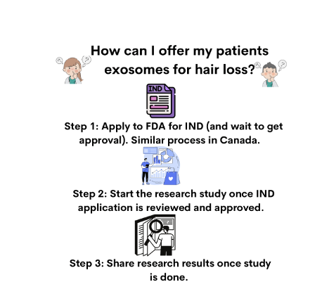
Thanks for the great question. I’m not sure what country you live in but here in North America (Canada and the United States), exosome therapy is not permitted - except under very special circumstances whereby a doctor has applied for and received a special IND application from the FDA (or equivalent in Canada).

I have yet to use it in my practice. In North America, if I wanted to use exosomes, I’d only be permitted to use it in the the context of a research study and I’d need to do all the paperwork to have my study approved. I’ll get back to that in a moment but you can already see how new and experimental exosome therapy truly is (as of the date of your question).

Exosome therapy is a potentially interesting therapy. To date, there have been very few published studies that actually show exosomes are all that helpful for treating hair loss. There was one study, however, by Chang-Hun et al back in 2019 in the Journal of the American Academy of Dermatology. That study had just 20 patients and followed the for just 12 weeks. However, the authors of that study found a 16 % increase in hair density and an 11% increase in thickness with use of exosome therapy. Not a lot - but some improvement. There were no serious side effects. That’s a nice change in hair density - but certainly not dramatic. With only 12 weeks of follow up, it’s unclear really what this means.

There have been a variety of in-vitro studies showing that exosomes can increase proliferation of dermal papillae (DP) cells, hair matrix cells, and outer root sheath cells as well as promote hair follicle stem cell proliferation and differentiation. I follow the world of exosomes closely!


Let’s dive deeper now.

What are exosomes?

Exosomes were only recently discovered - about 40 years ago. They are now being studied in various parts of medicine. Exosomes are extracellular vesicles that are produced by some type of cell. They are essentially tiny communication vesicles about 20-50 nm in size. It was first thought that exosomes were just cellular waste that got booted out of cells. Fortunately, it eventually came to be recognized that exosomes were important carriers of signaling molecules and were used for communication between cells. They contain many things including protein, nucleic acids, growth factors, lipids. It’s the growth factors in particular that are believed to be helpful in hair loss.

There are a wide range of cells that may be used to produce exosomes. However, donated human amniotic mesenchymal stem cells are one common source. Exosomes from adipose-derived stem cells are another source. There’s more yet - including exosomes from bone marrow derived mesenchymal stem cells.

Creation of exosomes. FROM: Graça Raposo and Willem Stoorvogel. Extracellular vesicles: exosomes, microvesicles, and friends. J Cell Biol . 2013 Feb 18;200(4):373-83. USED WITH CREATIVE COMMONS LICENSE. All credit to original source


The FDA Regulates Exosomes in the USA; Health Canada does the Same in Canada

The US Food and Drug Administration (FDA) has authority to regulate regenerative medicine products, including stem cell products and exosome products. The FDA has not given the green light to exosome therapy. It can only be used in a research setting by clinics that have a defined research study in place. The same is true in Canada. In Canada, exosomes fall under the jurisdiction of the Food and Drugs Act.

A US or Canadian physician who uses exosome therapy without having all the documents in place to show that they are conducting a research study could be subject to fines, and a variety of other penalties. It’s a serious issue and so exosome therapy is more common in other countries outside North America. In North America, a clinic needs to have applied for and received approval for an ‘investigational new drug” (IND) application before doing anything with exosomes.

I don’t do exosome therapy in my office. I’ve certainly had lots of conversations and phone calls with various regulatory bodies about these therapies. The message is always the same: A doctor can’t use these therapies unless they have been approved to do a clinical trial. Obviously in these kind of trials, the patient would sign a lot of forms indicating they know they are part of a clinical trial and the patient would be provided with proof that the clinic is part of such a trial. In other words, it would be pretty clear if a clinic in North America has an IND to study exosomes.

At the present time, there are no FDA or Health Canada approved exosome products for treating hair loss of any kind. Clinics can’t offer them (unless the patient receiving them is a research study patient).


The US FDA and Health Canada are very very worried about these types of therapies getting to the public without first doing the proper study. That’s why they want to study them ! In 2019, the FDA published a document for the public to warn of their concerns.

FDA’s Public Safety Message on Exosome Therapy

FDA’s Safety Alert for Exosome Therapy

CDC Warning About Exosomes

Health Canada Warnings on Autologous Therapies


Investigational New Drug Applications (IND Applications)

An “IND” is a submission to the FDA to request permission to study a drug. The goal of the entire IND process to help collect information that a given drug is indeed safe to use in humans. An ‘investigator IND’ is a special type of IND submission whereby the IND is submitted by a doctor who then initiates and conducts the clinical study. In order for a doctor in North America to conduct any kind of study with exosomes, they need an investigator IND.

Exosomes are considered “drugs” by many regulatory organizations including the FDA and Health Canada because they are used to treat some sort of medical issue. Here, in our discussion today, that medical issue we are speaking about here is hair loss. That’s the first point that makes an IND needed for anyone wanting to use or study exosomes for hair loss. The second issue is that the drug is used in human beings. Studies on human beings using drugs that have not been approved in the past need an IND. There are no exceptions for exosomes: all users need an IND application. Exosomes are not an approved ‘drug’.

A clinic or researcher with an approved IND application has permission to ship across the country and then to use exosomes in a formal study. It does not give physicians permission to use exosomes in any way they wish - only in a pre-approved research study.


Summary

In summary, exosomes are a potential new therapy. The data to date on effectiveness is promising (although not super exciting). The concept sure is exciting. We don’t really know much about exosome therapy but more information is going to emerge in the next few years. Stay tuned. It seems safe in small studies but these studies are extremely small. We have no idea how often treatments will be needed and no idea if 5 treatments is safe but 55 treatments is unsafe. We have no idea if 1 year of treatment poses different risk than 10 years of continuous treatment. Exosomes are completely new.

Exosome therapy is not FDA approved and not Health Canada approved. Here in North America, clinics can’t offer them without first formally registering and being approved to conduct a clinical trial with the US of Canadian government. A patient in North America can’t receive these therapies yet without being part of a clinical trial. In other words, exosome therapy not just another therapy option like PRP. It’s completely different. It’s not possible to walk into a clinic and choose exosome therapy like one would choose PRP or a hair transplant. Exosomes can only be offered to North American patients in the context of a clinical trial. That’s important for North American readers of this article to be aware of.


Thanks again for the great question.


REFERENCE

Chang-Hun et al. Exosome for hair regeneration: From bench to bedside. J Am Acad Dermatol. VOLUME 81, ISSUE 4, SUPPLEMENT 1, OCTOBER 01, 2019

Graça Raposo and Willem Stoorvogel. Extracellular vesicles: exosomes, microvesicles, and friends. J Cell Biol . 2013 Feb 18;200(4):373-83.


Share This
No Comments

Does inflammation in the scalp help minoxidil work better or worse?

How does inflammation affect how minoxidil helps over time?


I’ve selected this question below for this week’s question of the week. It allows us to review some concepts in treatment of androgenetic alopecia with minoxidil and the relationship to inflammation.

Question

I have androgenetic alopecia and my scalp is red at time as I have seborrheic dermatitis or some say psoriasis. I have heard that inflammation in the scalp may mean minoxidil works less well and other say it will work better.

What is the correct answer?

Answer

This is such a great question. The full answer as to whether inflammation causes minoxidil to work better or worse really comes down to what is causing the inflammation. There are 100 causes of inflammation in the scalp!!!! Some causes might make it work better and some might cause it to work less well.

As we tackle this subject, it’s important to keep in mind that few studies have actually been done.

Inflammation and fibrosis are known to affect responses to minoxidil. In 1993, Dr Whiting showed that patients with significant perifollicular inflammation and fibrosis have poorer responses to topical minoxidil. We don’t routinely evaluate inflammation in the scalp with males and females with androgenetic alopecia in deciding whether minoxidil will work or not. However, this 1993 study reminds us that it’s relevant.

There are other situations whereby inflammation probably makes the minoxidil work better. We sometimes add retinoic acid to minoxidil for example to make it more irritating and therefore get into the scalp better. Here is an example where we think inflammation helps minoxidil work better rather than worse. It’s not entirely clear if inflammatory states like seborrheic dermatitis, psoriasis, lupus, contact dermatitis are associated with better responses to minoxidil or not. The companies that make minoxidil warn users not to use if they have these sorts of inflammatory conditions. That is due to concern it might get into the scalp better.

All in all, there may be some inflammatory states where minoxidil works better and gets into the scalp more efficiently. This may also be associated with a greater chance of side effects like hair on the face or body, palpitations, headaches, etc. Be sure to discuss your specific situation with your dermatologist or hair specialist. If may not be a strict contraindication to use minoxidil if there is inflammation on the scalp but you will likely need a bit closer monitoring to ensure you are not developing worsening inflammation and not getting side effects from the treatment.

Share This
No Comments

Is PRP an option for Alopecia Areata Treatment?

Is PRP an option for treating alopecia areata?


I’ve selected this question below for this week’s question of the week. It allows us to review some concepts in treatment for alopecia areata.


Question

I have had patches of alopecia areata for the past few months. I’d like to use something natural rather than medications and things. I am afraid to use steroid injections or topical steroids given their side effects. Is PRP an option for alopecia areata?


Answer

Thanks for your question. I would encourage you to take your time and ask lots of questions. Your views on the safety of steroid injections and topical steroids may or may not be accurate. In general, the first line treatments for ‘patchy’ alopecia areata are steroid injections, topical steroids and topical minoxidil. For small patches, the safety is quite good with these three treatments. Side effects are uncommon but of course mild temporary ones can occur.

Here are typical treatments to consider for alopecia areata in the patchy stage. You can see that PRP is a second line option rather than a first. That means that we don’t typically turn to it first as a treatment but it may be an option IF first line options don’t work or a patient does not wish to use the first line options.


Alopecia is unpredictable. I would need to know a lot more information about your story to advise what I would recommend in your specific situation. For some patients, treatment is short term and recurrences uncommon. For others treatment is lifelong. Many patients with alopecia areata regrow their hair spontaneously even without treatment within 1 year. Steroid injections is far more effective and efficient as are all the first line options. That’s why they are called first line options. Many clinics do a wonderful job scaring patients out of the first line options so that patients can start whatever the clinic offers. ( A clinic that offers laser therapy or PRP or sells other remedies may say “Oh you wouldn’t want to do injections that’s so dangerous!).

In summary, PRP is an option - yes! But it’s not usually the first step for most.

Share This
No Comments

I have FPHL and have used everything imaginable. What else is there?

What are the other options for female pattern hair loss?


I’ve selected this question below for this week’s question of the week. It allows us to review some concepts in treatment of female androgenetic alopecia.

Question

I am 41 and have been diagnosed with FPHL and have used Rogaine, laser, PRP and spironolactone. Nothing works! What else is there? Have I exhausted all the options?

Answer

Thanks for the question. Let me being by saying that you’ll want to make sure that you have the right diagnosis. That’s always the first key step. My question when I see patients with a story like this is:

1) Is androgenetic alopecia the correct diagnosis ?

2) Are there other diagnoses here in addition to androgenetic alopecia ?

If there is any uncertainty, a biopsy may be needed. If you and your doctors are indeed confident it’s AGA then there are alternatives but what to use really depends on a person’s age, medical history, plans for pregnancy, emotional and psychiatric health, cardiovascular health and liver and kidney health. I’ll also assume that you have given each of these 6 months because that’s how long it takes to figure out if it’s working or not. I see patients every day who use Rogaine or spironolactone for 1-2 months and conclude it’s not working and stop. It takes a long time to evaluate effectiveness.

There are options for oral minoxidil, oral finasteride, oral dutasteride, topical finasteride, bicalutamide and hair transplantation. Be sure to give each and every treatment you try 9 months before you evaluate if it worked or not. I’ve included a list of first line, second line and third line treatments for premenopausal women. These may be a starting point for further discussions with your doctors. Some of these may not be options in young women on childbearing potential so you’ll want to discuss these in great detail. Often in a situation like you’ve described oral minoxidil or topical antiandrogens would be a next step with consideration given to a scalp biopsy to rule out any mimicking conditions.

Share This
No Comments

Switching from Oral to Topical Minoxidil

How easy is it to switch from Oral Minoxidil to Topical Minoxidil?


I’ve selected this question below for this week’s question of the week. It allows us to review some concepts in switching from oral to topical minoxidil.


Question

I am using 2.5 mg of oral minoxidil with success. Unfortunately, there is a shortage of oral minoxidil in my area so I’m going to need to switch to topical minoxidil until we get the supply back. Will I experience shedding when I start topical minoxidil? I hear that many people do!

Thank you.


Answer


Thanks for the question. You are likely to experience shedding but for a different reason than you ask about and perhaps a different reason than you think.


You are not likely to get shedding simply because you are “starting” topical minoxidil. No. However, you are likely to experience shedding because you are not able to supply your scalp with the equivalent amount of minoxidil by using the topical compared to the oral minoxidil. 5 % foam is NOT equivalent to 2.5 mg so you hair and scalp will likely say “where is my minoxidil?” “why am I suddenly being deprived?”

If you have been using oral minoxidil for a very short time, it may not be a big issue. But if you have been using oral minoxidil at 2.5 mg for some time it could be an issue. Be sure to discuss fully with your dermatologist.
The key point here is that 2.5 mg of oral minoxidil is not equivalent to topical minoxidil so you are now likely underdosing. There are options to have a compounding pharmacy in your area make up minoxidil pills for you. Not all pharmacies have this experience but many do. That is a far better option for most than switching to topical. But be sure to discuss with your health care providers.

Thank you again for the question!

Share This
No Comments

Hair Loss: What's causing my hair loss?

What’s causing my patch of hair loss?


I’ve selected this question below for this week’s question of the week. It allows us to review some concepts regarding clinical and trichoscopic examination of acute hair loss.


Question

Hi Dr. Donovan.

I’m a 31 year old female. While giving birth I almost died, went in to septic shock and lost a massive amount of blood. 2.5 months later I lost a lot of hair, I had thinning all over but more obvious around my ears, sides of my head and on the nape of my neck. My dermatologist (and biopsy) said it was TE and gave me steroid shots and my hair is growing back normally with no thinning.

I developed seborrheic dermatitis, my head is a little itchy and I’m on ketoconazole shampoo. 5 months after birth, I had to have major surgery on my kidney, the surgery itself lasted 8 hours. On the 2nd or 3rd day at the hospital I noticed a painful bump on my parietal lobe. On the 19th day after surgery, I washed my hair and then noticed the hair loss on the same area as the swelling. Ive attached photos of the first time noticing it.

My dermatologist injected it with steroids and it isn’t growing back. It’s been 3.5 months since it fell out. There are tiny hairs in the area so my dermatologist is sure it isn’t scarred.

But there are also exclamation looking hairs so we are not sure. The area is reddish. I have no hair loss anywhere else. It hasn’t gotten bigger and I don’t have any patches elsewhere. The picture labeled February 7 is the day I noticed it.

Answer

Thanks for submitting this question. I hope that you are feeling well. At first overview, it certainly would appear that the diagnosis is alopecia areata with some overlapping findings of seborrheic dermatitis. In addition, it appears that you first had a telogen effluvium (of alopecia areata again) that settled after delivery. I still favour alopecia areata as the diagnosis in the current photos but there are a few things in your story and some of your images that cause me to pause and ask “is it possible there is anything else going on here?”

The reason I’ve chosen this question is that it allows us to review some of these features today.

There are several scalp conditions that can cause localized hair loss in this manner with possible ‘exclamation mark like hairs”. The top 4 include:

  1. alopecia areata ** most likely **

  2. dissecting cellulitis

  3. pseudocysts (alopecic and aseptic nodules of the scalp

  4. infections (syphilitic alopecia)

Other diagnoses to consider here but do not have good evidence include:

  1. tinea capitis

  2. pressure alopecia

  3. infiltrative conditions.

  4. trichotillomania

Let’s take a look first at some of the images supplied in this question and then we’ll go into these possibilities a little further and come to some conclusions.

Submitted Image 1

This image shows patchy hair loss. There are broken hairs. Inflammation is mild. The top diagnosis at this magnification would be alopecia areata. The differential diagnosis from this image might include trichotillomania, tinea capitis, and pressure induced alopecia. Alopecia areata would be the top diagnosis. Exclamation mark hairs are not clearly seen in this image but are seen in other images. There is no evidence for a scarring alopecia. Density may be reduced in the more anterior portion of the scalp (top of the photo) suggesting ongoing TE or another hair loss diagnosis happening in this area.

Submitted Image 2

This image shows a well cicumscribed area with minimal inflammation. There are vellus hairs and broken hairs. Some hairs have hair shaft changes suggestive of a pseudo-monilthrix like change (Pohl Pinkus constrictions). Exclamation mark hairs are not clearly seen in this image but are seen in other images. There is no good evidence for a scarring alopecia. Alopecia areata remains a favoured diagnosis.

Submitted Image 3

Numerous exclamation mark hairs are seen in this image. Elbow hairs are seen. Yellow dots are seen. There is an inflammatory type change with whitish scale. There is a mild pigmentation alteration which is somewhat non specific. The exclamation mark hairs make other diagnoses quite unlikely as exclamation mark hairs of this kind do not occur in pressure alopecia nor in inflammatory connective issues issues. The appearance of the scalp in this image differs quite a bit from the appearance seen in other images.

Submitted Image 4

This image shows several exclamation mark hairs with regrowing vellus hairs. There is mild yellow scale which may be in keeping with seborrheic dermatitis (of psoriasis) or an artefact of the photo itself. There is no evidence for a scarring alopecia.


Further Discussion

Thanks again for submitting this case. I favour alopecia areata but of course it’s nice to have more information and see the entire scalp eyebrows eyelashes, and nails. The most accurate way to diagnosis hair loss is to collect all the information about the patient and then examine all the scalp.

The features that support alopecia areata are the exclamation mark hairs, vellus hairs, regrowing hairs and localized nature of the hair loss.


What other conditions cause exclamation mark hairs?

As we think about this question, it’s helpful to think about all that conditions that cause exclamation mark hairs. After all, one of the key features in the submitted images are the exclamation mark hairs.

Exclamation mark hairs are seen in alopecia areata, trichotillomania, thallium poisoning, dissecting cellulitis. Syphilitic alopecia has been rarely described to have a type of tapered hair closely resembling a true exclamation mark hair. This is very rare.

Trichotillomania

There does not appear to be good evidence here for trichotillomania. The story does not fit. It’s one of the famous causes of exclamation mark like hairs. Certainly extensive broken hairs can be a feature but other findings like black dots, V hairs, coiled hairs, hook hairs, hair powder just don’t appear to be a feature of this patient’s hair loss. I don’t think we’re dealing with trichotillomania.

Thallium poisoning

Of course, thallium poisoning is rare.

Dissecting Cellulitis and Alopecic and Aseptic Nodules of the Scalp

The description of the ‘painful bump’ is a bit unusual in the submitted question. It’s not typical of alopecia areata. It may be a ‘red herring’ and unrelated to the case here or it may truly be a valuable clue. Also, it would be helpful to know more about what is meant by a painful bump and how big of a bump is the individual referring to.

As we think about painful bumps, we need to think about small bumps and things like a folliculitis. As we get into larger and larger bumps we need to consider more significant inflammatory conditions of the scalp. Dissecting cellulitis can cause a larger dome shaped bump when it occurs and is famous for mimicking alopecia. Another closely related entity is “alopecic and aspetic nodules of the scalp” (AANS). AANS can resemble alopecia areata. The condition was first called “pseudocyst” but the name AANS was proposed in 2009 by Abdennader and Reygagne when it became clear that not all of these lesions show a pseudocyst morphology under the microscope.

The back of the scalp is a common area for AANS. Often patients present with just a single painful bump. Some authors feel that AANS is closely related to dissecting cellulitis.

Exclamation mark hairs have not been described in AANS but have been described in dissecting cellulitis.

Dome shaped area of hair loss on the vertex scalp, consistent with a diagnosis of alopecic and aseptic nodules of the scalp. Image from Anna Isabel Lázaro-Simó et al. Alopecic and Aseptic Nodules of the Scalp with Trichoscopic and Ultrasonographic Findings. Indian J Dermatol. Sep-Oct 2017;62(5):515-518. Image used with creative commons license.


Trichoscopic image from alopecic and aseptic nodules of the scalp (AANS), also known as pseudocysts. There are black dots, yellow dots, vellus hairs and broken hairs. Image from Anna Isabel Lázaro-Simó et al. Alopecic and Aseptic Nodules of the Scalp with Trichoscopic and Ultrasonographic Findings. Indian J Dermatol. Sep-Oct 2017;62(5):515-518. Used with creative commons license.

Trichoscopy of alopecic and aseptic nodules of the scalp. Image from Khalil I. Al-Hamdi and Anwar Qais Saadoon. Alopecic and Aseptic Nodules of the Scalp with a Chronic Relapsing Course. Int J Trichology. 2019 Nov-Dec; 11(6): 244–246. Used with creative commons license.

There are three stages of appearance to AANS lesions as described by Al-Hamdi and colleagues. It’s important to understand this - especially in this case.

Stage 1: Firm nodule. A firm and often tender nodule is present and the nodule lasts 1-3 weeks. There may be lymphadenopathy. If the nodule is punctured, it does not usually express any fluid. But if it does, the fluid is sterile and does not grow bacteria

Stage 2: Fluctuant Nodule with Hair Loss. In this stage, the nodule becomes less tender and hair loss is clearly seen. If the lesion is punctured in this stage a yellow fluid is expressed. This stage lasts 3-7 days.

Stage 3: Patchy Hair Loss Stage. In this stage, the nodule is no longer present as it has flattened either spontaneously or by puncture. This stage may last 2-3 month at which point hair growth normally occurs. It’s common in this stage for the patchy hair loss to be given a diagnosis of alopecia areata.

Was the bump described by the patient in this case actually stage 1 or stage 2 of AANS? Clearly, more information is needed. I would say it’s still quite unlikely.

Infections (Syphilitic Alopecia)

In a case like the one presented, one must never lose sight of alopecia areata as the most likely diagnosis. Most things fit well and it could be simply that this patch is more refractory and needs further steroid injections. However, we do need to consider rare mimickers (like AANS) - and another rare mimicker of syphilitic alopecia.

I don’t think that there is much in this case that makes a diagnosis of syphilitic alopecia high on the list. However, it can be a cause of patchy hair loss - especially with tapered exclamation mark like hairs, scale and redness like we see in the photos sent in by the patient.

Atypical trichoscopy of a patient with syphilitic alopecia in a 32 year old male. Exclamation mark like hairs are seen. Tapered bended hairs, erythematous background, diffuse scaling and perifollicular hyperkeratosis were present. Testing revealed a positive Venereal Disease Research Laboratory (VDRL) at a titer of 1:256 and a reactive Treponema pallidum particle hemoagglutination assay. Image from Linda Tognetti et al. Syphilitic alopecia: uncommon trichoscopic findings. Dermatol Pract Concept. 2017 Jul; 7(3): 55–59.


Other Diagnoses to Consider


There are several other diagnoses to consider here but they do not really have good evidence. These include:

  1. tinea capitis

  2. pressure alopecia

  3. infiltrative conditions.


Tinea capitis

Tinea capitis can be a mimicker and the appearance can be altered by steroid injections. I don’t know the patient’s history well enough to know if there are predisposing factors that might make tinea capitis more likely. (In fact, I don’t have enough information in this patient’s history including information about the kidney surgery at 5 months post partum). It’s always possible that an inflammatory tinea developed and was flattened by steroid injections and persists in some manner. Of course, that’s unlikely and there do not really appear to be any trichoscopic features of tinea. There are no corkscrew hairs, comma hairs, bent hairs, i hairs, morse code hairs and no zig zag hairs. I don’t think this is tinea.


Pressure alopecia

In anyone with patchy hair loss after surgery, we need to consider pressure alopecia. It’s thought that hypoxia and altered blood flow predisposes to hair loss. Studies of patients with pressure alopecia have not suggested that exclamation mark hairs are part of the pressure alopecia diagnosis. Therefore, a diagnosis of pressure alopecia would not be likely in this case. According to Neema et al, trichoscopic findings of pressure alopecia include comedone- like black dots, black dots and area of scarring. In 2016, Francine Papaiordanou et al proposed that black dots, broken and dystrophic hairs were main features of pressure alopecia. In 2020, Tortelly et al proposed that black dots and vellus hairs were key features.

It’s not impossible that pressure from surgery facilitated the development of alopecia areata. in fact, R L Zuehlke et al in 1981 suggested that pressure may be a risk for alopecia areata too. So it’s going to be important to review if this area on the scalp shown in the photos had pressure during surgery. I don’t think it’s likely that what we’re seeing is related to pressure alopecia.

Trichoscopy of pressure alopecia showing black dots, broken and dystrophic hairs. In Image from Papaiordanou F et al. Trichoscopy of Noncicatricial Pressure-induced Alopecia Resembling Alopecia Areata. Int J Trichology. Apr-Jun 2016;8(2):89-90. Used with creative commons license.

Infiltrative conditions.

The term “infiltrative conditions” refers to a massively long list of cells that can enter into an area of the scalp (infiltrate) and cause localized hair loss. A variety of inflammatory and neoplastic cells can trigger patchy hair loss so one needs to always keep these in mind. They don’t usually cause exclamation mark hairs. Alopecia neoplastica refers to hair loss from metastatic cancer and can mimic alopecia areata in some cases.

This would not be expected in this case but this is added to the list and discussed here for completeness as we review patchy hair loss and considerations in the setting of refractory patchy hair loss. It does seem that hair is growing back in your case which makes infiltrative type causes quite unlikely. I don’t have a good sense of the time course of the photos you’ve submitted so that too would need to be carefully reviewed.

Alopecia neoplastica due to breast cancer. Image from Efthymia Skafida et al. Secondary Alopecia Neoplastica Mimicking Alopecia Areata following Breast Cancer.Case Rep Oncol. 2020 Jun 11;13(2):627-632. Image used with creative commons license.

Alopecia neoplastica due to breast cancer. Image from Efthymia Skafida et al. Secondary Alopecia Neoplastica Mimicking Alopecia Areata following Breast Cancer.Case Rep Oncol. 2020 Jun 11;13(2):627-632. Image used with creative commons license.

Conclusion and Summary

Thank you for this question. There are a few important points here in this case as we conclude. The first is that a full history and full examination are needed. This area is the area that you have photographed but one needs to always examine the entire scalp. A full history is needed of health and medical conditions over the past 31 years. The reason for the kidney surgery is completely unknown and would need to be included in the full story. I need to know everything about patients to confirm diagnoses with absolute certainty.

That said, the photos and clinical case still fit with undertreated alopecia areata as a top diagnosis. The exclamation mark hairs here and vellus hairs and regrowing hairs support this diagnosis. There are mimickers of course and these need to be considered.

For my own patients with similar stories I would first take a full history and do a full examination of the scalp, eyebrows, eyelashes and body hair. Then I might inject with 2.5 mg per mL triamcinolone acetonide (steroid) with 2 to 3 mL injected into the area. I would not be too concerned if hair does not immediately grow back as it might take 2-3 sessions one month apart. I would not do more frequent than this. There is an option to add topical minoxidil to the plan but you’d want to review side effects with your supervising doctor.

If the area was slow to regrow I might add periodic use of clobetasol and minoxidil at home while doing these steroid injections.

Your photos would suggest you are already growing back significant hair.

If the area did not respond, I might do a biopsy. I don’t see this as necessary right now. The area needs to be properly treated and then if it does not respond to proper treatment, one can move on to step 2.

I don’t see AANS as a likely diagnosis (alopecic and aseptic nodules of the scalp), or pressure alopecia as being likely. We don’t see exclamation mark hairs in most cases of pressure alopecia. It would be helpful to know just how lumpy or raised this area was when you noticed it as this might lead one to at least consider AANS. The reality is that even if it is AANS and it’s some unusual pattern of exclamation mark hairs, it should respond to steroid injections at this point. I don’t think it’s likely we’re dealing with AANS.

Finally, anyone with this story should have blood tests for CBC, TSH, ferritin, vitamin D, creatinine. An antidandruff shampoo should continue to be used. Ketoconazole is reasonable. As mentioned, a biopsy will be needed if the area is not responding to appropriate doses of steroid injections (+/- minoxidil or clobetasol).

A full scalp examination is needed to determine if there are any other issues too. The area to the front may be thinner than prior years and that needs to be evaluated. It could be part of a resolving telogen effluvium or another diagnosis. A full examination of the scalp is needed in this case (as well as full examination of eyebrows, eyelashes and body hair as mentioned)

Thanks again

REFERENCE

Anna Isabel Lázaro-Simó et al. Alopecic and Aseptic Nodules of the Scalp with Trichoscopic and Ultrasonographic Findings. Indian J Dermatol. Sep-Oct 2017;62(5):515-518.

Khalil I. Al-Hamdi and Anwar Qais Saadoon. Alopecic and Aseptic Nodules of the Scalp with a Chronic Relapsing Course. Int J Trichology. 2019 Nov-Dec; 11(6): 244–246.

Abdennader S, Reygagne P. Alopecic and aseptic nodules of the scalp. Dermatology. 2009;218:86.

Linda Tognetti et al. Syphilitic alopecia: uncommon trichoscopic findings. Dermatol Pract Concept. 2017 Jul; 7(3): 55–59.

Neema S et al. Trichoscopy of Pressure-Induced Alopecia and Alopecia Areata: A Comparative Study. Int J Trichology. Jan-Feb 2022;14(1):17-20.

Papaiordanou F et al. Trichoscopy of Noncicatricial Pressure-induced Alopecia Resembling Alopecia Areata. Int J Trichology. Apr-Jun 2016;8(2):89-90.

Tortelly et al,Pressure-Induced Alopecia: Presence of Thin Hairs as a Trichoscopic Clue for the Diagnosis Skin Appendage Disord. 2020 Jan; 6(1): 48–51.

R L Zuehlke et al. Pressure-potential alopecia areata. Am J Orthod . 1981 Apr;79(4):437-8.

Efthymia Skafida et al. Secondary Alopecia Neoplastica Mimicking Alopecia Areata following Breast Cancer.Case Rep Oncol. 2020 Jun 11;13(2):627-632.


Share This
No Comments

Microblading for FFA Patients Using Isotretinoin

Is eyebrow microblading safe for FFA patients using isotretinoin?


I’ve selected this question below for this week’s question of the week. It allows us to review some concepts regarding isotretinoin and procedures for patients with FFA.


Question

I am a 54 year old female with frontal fibrosing alopecia. Similar to many women, I have lost a significant amount of eyebrows. My scalp FFA has improved a great deal with use of dutasteride, isotretinoin, pimecrolimus and steroid injections. My eyebrow hair loss has halted but unfortunatley it has not grow back.

I am considering microblading for my eyebrows. The lady I see won’t do the microblading if I’m on isotretinoin and says I need to be off 6 months or more. Obviously, I am terrified to stop as it’s helped my scalp and facial bumps a very significant amount.

Should I stop the isotretinoin?

Thank you for your question section of the website so that we can ask questions of this kind.

Answer

Thanks for the great great question.

Isotretinoin is a vitamin A derivative and can affect many parts of the skin including the epidermis, sebaceous gland, and even collagen formation. The diverse affects of isotretinoin on wound healing have generated a long debate about whether patients who use isotretinoin are more likely to have poor wound healing or worse yet - develop a keloid or hypertrophic scar - if they undergo surgery or a procedure on the skin.

In fact, the US FDA advises against patients having laser resurfacing procedures within 6 months of use of isotretinoin.

The general recommendations to avoid surgical procedures have been called into question by various experts around the world in the last few years.

Tattooing in the Eyebrow Region in FFA

It appears that tattooing in the eyebrow area is likely to be safe in patients with FFA who use low doses of isotretinoin (under 10 mg daily). One should be aware that large scale studies have not yet been conducted in FFA but certainly we have had many stable patients in our clinic on low doses of isotretinoin undergo various tattooing procedures without issue.

For those who question the safety, a thorough review of two articles would be advised:

Spring et al, 2017

In 2017 a panel of experts (Spring et al 2017) put forth recommendations on the safety of isotretinoin in various dermatological procedures. Thirty-two relevant publications reported outcomes of 1485 procedures. Overall, the authors concluded that “there was insufficient evidence to support delaying manual dermabrasion, superficial chemical peels, cutaneous surgery, laser hair removal, and fractional ablative and nonablative laser procedures for patients currently receiving or having recently completed isotretinoin therapy. Based on the available literature, mechanical dermabrasion and fully ablative laser are not recommended in the setting of systemic isotretinoin treatment” (Source: Spring et al 2017).

Mysore et al, 2017

In 2017, the Association of Cutaneous Surgeons of India also put forth a recommendation “that microneedling and microdermabrasion treatment can safely be performed in patients administered with isotretinoin”. The level of evidence was 2+ (backed up by well conducted case control studies or cohort studies) and the grade of the recommendation was given a C rating.

The formal view of the Association of Cutaneous Surgeons of India was that “there is insufficient evidence to support the current protocol of avoiding and delaying treatments in the patient group under consideration and recommends that the current practice should be discontinued. The task force concludes that performing procedures such as laser hair removal, fractional lasers for aging and acne scarring, lasers for pigmented skin lesions, fractional radio-frequency microneedling, superficial and medium-depth peels, microdermabrasion, dermaroller, biopsies, radio-frequency ablation, and superficial excisions is safe in patients with concurrent or recent isotretinoin administration.

Conclusion and Comment

My general feeling is that tattooing of eyebrow is likely to be pretty safe for patients with FFA who use low doses of isotretinoin. We have not had problems with patients who use 10 mg daily or less AND whose FFA is fairly stable. I am not a big fan of eyebrow tattooing in patients with super active FFA simply because I have never guided patients through tattooing unless the FFA is somewhat calm. Likely tattooing is even active FFA is safe but of course no data is widely available. We also don’t have patients on higher doses of isotretinoin.

If one is willing to accept a really low risk of problems, then eyebrow tattooing is wonderful - assuming an experienced and competent practitioner is doing the tattooing. Close follow up with the dermatologist is advised so that immediate action can be taken in the event there are problems (ie. administration of topical steroids, steroid injections, topical calcineurin inhibitors). It is fortunately very rare to have problems other than the side effects that anyone experiences with tattooing. The improvement in self confidence and feeling of well being after successful tattooing procedures is great.

If a patient with FFA is not comfortable with any risk whatsoever, then eyebrow tattooing might not be the right procedure for them. At minimum, such a patient should be off isotretinoin for 6 months if they are not comfortable with risk - but again I must emphasize that this is without evidence and one risks worsening their scalp FFA if they stop isotretinoin and the isotretinoin was helping them.

Eyebrow microblading has its own inherent risks (even without isotretinoin!!) so all risks need to be reviewed with the practitioners performing the procedure. Persistent redness, tattoo reactions, bumps, poor uptake of pigment, rapid fading of pigment, allergic reactions are all part of the side effects for anyone with or without FFA. One should never undergo tattooing without first reviewing what sorts of rare side effects occur in anyone.

Finally, one must be aware that pigments don’t always taken up quite as well in FFA. The skin is different in some patients and takes of pigments differently - so multiple procedures may be needed. One must never ever assume that the microblading outcome they will have will necessarily be the same as anyone else who does not have FFA. The skin is different and it’s possible for variations in outcomes.

Fortunately, most patients have really nice results and the vast majority of patients are really pleased with the decision to have microblading and other pigmentation procedures. One should carefully review the risks and benefits with the dermatologist overseeing care as well as the tattoo expert performing the procedure. A full understanding of risks and benefits is needed.

REFERENCE

Mysore V et al. Standard Guidelines of Care: Performing Procedures in Patients on or Recently Administered with Isotretinoin J Cutan Aesthet Surg. 2017 Oct-Dec; 10(4): 186–194.

Spring L et al. Isotretinoin and Timing of Procedural Interventions: A Systematic Review With Consensus Recommendations. JAMA Dermatol . 2017 Aug 1;153(8):802-809.

Share This
No Comments

What level of ferritin should I aim for ?

What level of ferritin should I aim for to keep my hair growing?


I’ve selected this question below for this week’s question of the week. It allows us to review some concepts in iron deficiency and the threshold level of ferritin to aim for.


Question

I have hair loss and my ferritin is 33. I’m a 37 year old woman and in excellent health. I am not vegetarian. I eat well and exercise every day. I’m wondering what level I should aim for? I’ve heard 40 is a good target but some say 70. What level is reasonable?


Answer

One of the biggest myths in the field of hair loss is that there is a magic number. Not so. I wish there was, but there is not.

Certainly it makes sense to aim for a ferritin level of 40-50 if one has hair loss.

However, the reality is many many people have zero iron related issues provided the ferritin is above 30. Sure, a very small proportion might. Now as ferritin levels dip down into the 20s, there will be some that have iron issues that are related to the hair. But lots and lots and lots of females have ferritin 24-32 and have zero hair loss issues at all. So it’s not a clear cut number!

The following table provides some insights into how I think about ferritin levels for most women. One can seen that as the ferritin dips down below 70, it becomes more and more likely that taking iron could help - especially if the levels are less than 26. With ferritin levels in the teens - it’s clear iron is needed!

In summary, there are hundreds of patients today with ferritin levels 32-40 who will be told “you just need to bring up your iron to 50 or 70 and you’ll be fine.” Most of the time increasing ferritin (when levels are in the 30s is simply not going to have any effect on the hair. Is it worth trying? Well that requires input from a health care provided. As ferritin gets lower and lower below 30 it becomes more and more likely that iron supplementation will help.

I would like to point out that I never just look at the ferritin. I look at B12 (which can make ferritin levels look artificially good if B12 is low) and I look at hemoglobin, MCV, MCH and RDW and transferrin saturation if available. Most importantly I look at prior labs to see how things used to be. If one wants a magic cut off number, I’d say 40 but it’s really not so simple and lots of people eat iron pills and get constipated just to reach a number that has absolutely no benefit.

If a patient has had ferritin of 34 for 20 years and now developed hair loss last year with a ferritin of 35, are we do tell the patient “Oh you lost hair because of low iron! Bring your ferritin up to 40 to 50 and you’ll be fine.” No, this is nonsense but a common scenario.

I hope this helps with understanding the complexities of iron.

Share This
No Comments

How long do I need treatment for my FFA?

How long do I need treatment for my FFA?


I’ve selected this question below for this week’s question of the week. It allows us to review some concepts in treating FFA.


Question

I was diagnosed with frontal fibrosing alopecia last month. I’ve started on hydroxychloroquine and steroid injections. How long am I going to need to do this treatment?

Answer

Thanks for your question. Some patients with FFA need treatment for 1-2 years and some patients with FFA need treatment for 10-20 years. Some never can stop treatment without losing hair. It’s difficult to say for any given person how long they will need treatment but over time you’ll come to know the answer for yourself and your specific case.

The first step in treatment is to stop the disease so that it does not keep getting worse and worse. Once your doctor stops the disease, he or she will want to continue medications a bit longer and see if it stays quiet for many years or starting acting up again and causing more hair loss. If the conditions stays quiet, it may be possible to starting slowly ‘tapering” medications or reducing the dose bit by bit. If the disease stays quiet and you don’t start losing hair again when the dose is tapered then it means that things are truly quiet.

Step 1 therefore is to stop the disease

Step 2 is to keep the disease stopped

Step 3 is to slowly taper some medications (if possible)

There is a view out there in the internet that scarring alopecia simply burn out after a year or two. This is not correct. Some patients of course do have a form that goes inactive rather quickly. But not all do. Some patients need to try many different medications before they find a combination that finally stops the disease. some get the treatment right on the first try.

Share This
No Comments

Should I get another biopsy?

Should I get another biopsy?


I’ve selected this question below for this week’s question of the week. It allows us to review some concepts in interpreting biopsy results.


Question

I’m a 34 year old female with previously beautiful hair. My biopsy came back showing “non-scarring alopecia with features consistent with androgenetic alopecia.” I’m wondering if I should get another biopsy? Could it be wrong?


Answer

Thanks for the question.

Without knowing more about your medical history, and seeing your scalp (or photos), it’s difficult for me to say.

There are a number of possibilities here:

a) Your story and examination confirm you have androgenetic alopecia and doing a biopsy was not needed but simply served to confirm the diagnosis. Doing another biopsy would not be advised.

b) Your story and examination confirm you have androgenetic alopecia but there is a suggestion in the story or examination that a second diagnosis is also present. In this case, doing another biopsy could be a good idea if one is not 100% confident about this second diagnosis. If one is 100% confident in the second diagnosis, then another biopsy is not needed.

c) Your story and examination do not suggest androgenetic alopecia and there is a suggestion in the story or examination that a different diagnosis is present. In this case, doing another biopsy could be a good idea if one is not 100% confident about this diagnosis. If one is 100% confident in the diagnosis, then another biopsy is not needed.


I hope this helps. I don’t know your diagnosis because I haven’t seen your scalp and I don’t know your story. Certainly, if your practitioner is highly experienced in diagnosing women’s hair loss and says “This can’t be androgenetic alopecia” then I’m more likely to feel something is strange here and more likely to feel that a re-evlauation of things is needed. Early androgenetic alopecia is tricky to diagnose so many women have AGA even though it might not look like it.

If the practitioner is less experienced in diagnosing women’s hair loss, I’m less bothered by him or her saying “This can’t be AGA.” it’s tricky to diagnose some of the early forms of AGA. The reality is that if the terminal to vellus hair ratio on biopsy is dipping down below 4:1 and sebaceous glands are present in the biopsy there’s a good chance we’re dealing with AGA. The key question is whether this is the only diagnosis or the correct diagnosis.

Finally, I’d like to point out that we don’t diagnose hair loss just with biopsy results alone. One needs to take the biopsy finding and see if it ties in with the history of hair loss and the clinical examination findings. I would never every commit to saying a person has a certain diagnosis without the chance to see their hair and know their story. Even with the biopsy sitting in front of me, I need to know the story and see the scalp.


Share This
No Comments

The Risk of Lichen Planopilaris in Patients with Oral Lichen Planus

What is my risk of lichen planopilaris if I have oral lichen planus?


I’ve selected this question below for this week’s question of the week. It allows us to review some concepts in oral lichen planus.


Question

I have oral lichen planus and I’m wondering what my risk of LPP is. Am I likely to develop LPP in the future? I’m feeling terrified about the whole thing actually.

Answer

Thanks for the question.

The short answer is that you probably won’t develop lichen planopilaris. The risk is not zero that you’ll develop LPP - but it’s really really low. Actually, it’s really really really low. I hope you get the point.

Oral lichen planus is a condition that develops in the 30s and 40s. Women are slightly more affected than males. F:M ratio of 1.4 to 1. Oral lichen planus affects about 1-2 % of the population. There are many different forms of oral lichan planus inlcuding variants such as the reticular, papular, plaque-like, erosive, atrophic, and bullous variants. The inner sides of the cheeks (buccal mucosa), tongue and gums are the most commonly affected.

In contrast, lichen planopilaris affects about 0.03% fo the population or roughly 1 in every 3000 to 5000 people. Most people with oral lichen planus don’t have lichen planopilaris at the time they are diagnosed with oral lichen planus and most never go on to develop it! So if you forced me to bet, I would bet you will not develop lichen planopilaris. The odds are dramatically stacked in my favour.

Most people with oral lichen planus never develop LPP. On the contrary, the risk of a person diagnosed with lichen planopilaris developing oral lichen planus is probably 1-2 %. This number is probably similar to the general population risk of 1-2 % or possibly a very slight amount higher. Good studies have not been done to really convincingly know if patients with LPP have a higher risk of oral lichen planus than the general population or not. It’s possible they do but large studies that generate good data just haven’t been done.

Share This
No Comments

How long does it take for shedding to stop once you've corrected the trigger?

How long does it take for hair shedding to stop ?


I’ve selected this question below for this week’s question of the week. It allows us to review some concepts in telogen effluvium.


Question

I think I have telogen effluvium from a drug I took. I have now stopped the drug and so it’s going out of my system. How long does it take shedding to stop once a person has found the right trigger and stopped it? I’ve heard it takes 9-12 months. Is that true?


Answer

Thanks for the question.

Shedding stops far sooner that this if truly you’ve found the right trigger. In fact, shedding should start really slowing down in 1-2 months and be quite back to normal rates of shedding by month 6 at the latest (but probably month 3-5 for most patients). There are many many patients that note that shedding seems to “shut off like a tap” when a person has really found the right trigger.

It’s important not to confuse two things:

1) The timeline for the hair shedding to slow.

2) The timeline for the hair density and thickness to come back.

These two timelines are not the same!

It takes a matter of months for the shedding to slow but it takes about 6-9 months from the time of stopping the trigger for hair density to really be growing in nicely. In other words, there will be many months where a patient will say “Ok, my shedding stopped but my hair is still so thin.” This is followed by a period where the patient notes that not only is shedding remaining low but hair thickness and volume is coming back.

I have outlined some of this timing below:

Summary

To summarize. it will take up to 6 months for shedding to return to normal once the trigger is fixed. For many patients, the shedding stops much sooner than this. It’s usually just a matter of a few months before shedding is back to the normal expected rates and for some it’s a matter of weeks rather than months. If shedding is not stopping after 6 months, one really should ask themselves?

a) do I really have the correct diagnosis?

b) do I have the correct diagnosis - but have I missed another diagnosis that is also present?

Share This
No Comments


Share This
-->