QUESTION OF THE WEEK

Dr. Donovan's Articles

QUESTION OF HAIR BLOGS

Filtering by Category: Blood tests


Hair Loss: What's causing my hair loss?

What’s causing my patch of hair loss?


I’ve selected this question below for this week’s question of the week. It allows us to review some concepts regarding clinical and trichoscopic examination of acute hair loss.


Question

Hi Dr. Donovan.

I’m a 31 year old female. While giving birth I almost died, went in to septic shock and lost a massive amount of blood. 2.5 months later I lost a lot of hair, I had thinning all over but more obvious around my ears, sides of my head and on the nape of my neck. My dermatologist (and biopsy) said it was TE and gave me steroid shots and my hair is growing back normally with no thinning.

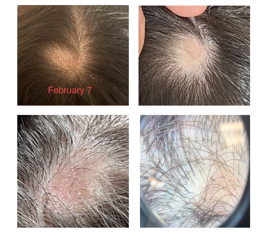
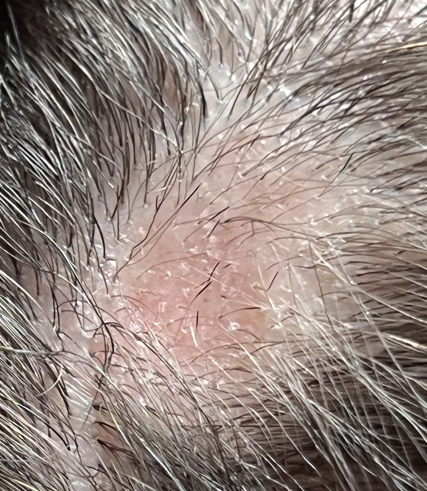
I developed seborrheic dermatitis, my head is a little itchy and I’m on ketoconazole shampoo. 5 months after birth, I had to have major surgery on my kidney, the surgery itself lasted 8 hours. On the 2nd or 3rd day at the hospital I noticed a painful bump on my parietal lobe. On the 19th day after surgery, I washed my hair and then noticed the hair loss on the same area as the swelling. Ive attached photos of the first time noticing it.

My dermatologist injected it with steroids and it isn’t growing back. It’s been 3.5 months since it fell out. There are tiny hairs in the area so my dermatologist is sure it isn’t scarred.

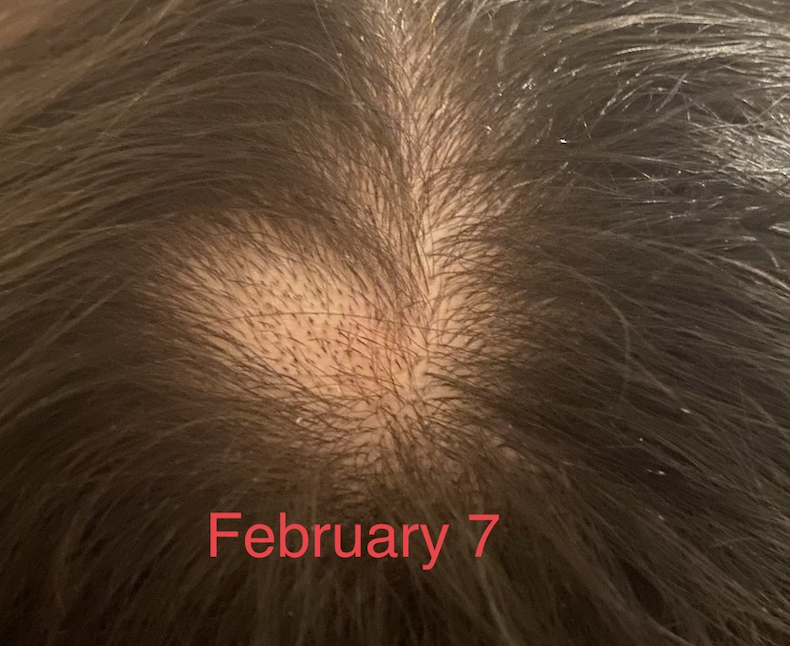
But there are also exclamation looking hairs so we are not sure. The area is reddish. I have no hair loss anywhere else. It hasn’t gotten bigger and I don’t have any patches elsewhere. The picture labeled February 7 is the day I noticed it.

Answer

Thanks for submitting this question. I hope that you are feeling well. At first overview, it certainly would appear that the diagnosis is alopecia areata with some overlapping findings of seborrheic dermatitis. In addition, it appears that you first had a telogen effluvium (of alopecia areata again) that settled after delivery. I still favour alopecia areata as the diagnosis in the current photos but there are a few things in your story and some of your images that cause me to pause and ask “is it possible there is anything else going on here?”

The reason I’ve chosen this question is that it allows us to review some of these features today.

There are several scalp conditions that can cause localized hair loss in this manner with possible ‘exclamation mark like hairs”. The top 4 include:

  1. alopecia areata ** most likely **

  2. dissecting cellulitis

  3. pseudocysts (alopecic and aseptic nodules of the scalp

  4. infections (syphilitic alopecia)

Other diagnoses to consider here but do not have good evidence include:

  1. tinea capitis

  2. pressure alopecia

  3. infiltrative conditions.

  4. trichotillomania

Let’s take a look first at some of the images supplied in this question and then we’ll go into these possibilities a little further and come to some conclusions.

Submitted Image 1

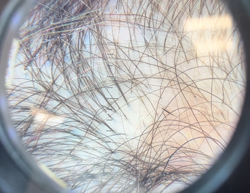
This image shows patchy hair loss. There are broken hairs. Inflammation is mild. The top diagnosis at this magnification would be alopecia areata. The differential diagnosis from this image might include trichotillomania, tinea capitis, and pressure induced alopecia. Alopecia areata would be the top diagnosis. Exclamation mark hairs are not clearly seen in this image but are seen in other images. There is no evidence for a scarring alopecia. Density may be reduced in the more anterior portion of the scalp (top of the photo) suggesting ongoing TE or another hair loss diagnosis happening in this area.

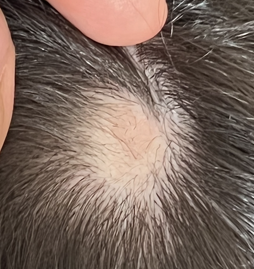
Submitted Image 2

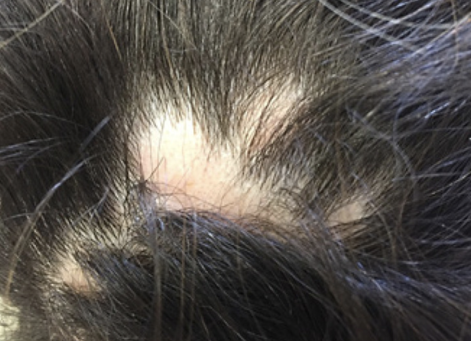
This image shows a well cicumscribed area with minimal inflammation. There are vellus hairs and broken hairs. Some hairs have hair shaft changes suggestive of a pseudo-monilthrix like change (Pohl Pinkus constrictions). Exclamation mark hairs are not clearly seen in this image but are seen in other images. There is no good evidence for a scarring alopecia. Alopecia areata remains a favoured diagnosis.

Submitted Image 3

Numerous exclamation mark hairs are seen in this image. Elbow hairs are seen. Yellow dots are seen. There is an inflammatory type change with whitish scale. There is a mild pigmentation alteration which is somewhat non specific. The exclamation mark hairs make other diagnoses quite unlikely as exclamation mark hairs of this kind do not occur in pressure alopecia nor in inflammatory connective issues issues. The appearance of the scalp in this image differs quite a bit from the appearance seen in other images.

Submitted Image 4

This image shows several exclamation mark hairs with regrowing vellus hairs. There is mild yellow scale which may be in keeping with seborrheic dermatitis (of psoriasis) or an artefact of the photo itself. There is no evidence for a scarring alopecia.


Further Discussion

Thanks again for submitting this case. I favour alopecia areata but of course it’s nice to have more information and see the entire scalp eyebrows eyelashes, and nails. The most accurate way to diagnosis hair loss is to collect all the information about the patient and then examine all the scalp.

The features that support alopecia areata are the exclamation mark hairs, vellus hairs, regrowing hairs and localized nature of the hair loss.


What other conditions cause exclamation mark hairs?

As we think about this question, it’s helpful to think about all that conditions that cause exclamation mark hairs. After all, one of the key features in the submitted images are the exclamation mark hairs.

Exclamation mark hairs are seen in alopecia areata, trichotillomania, thallium poisoning, dissecting cellulitis. Syphilitic alopecia has been rarely described to have a type of tapered hair closely resembling a true exclamation mark hair. This is very rare.

Trichotillomania

There does not appear to be good evidence here for trichotillomania. The story does not fit. It’s one of the famous causes of exclamation mark like hairs. Certainly extensive broken hairs can be a feature but other findings like black dots, V hairs, coiled hairs, hook hairs, hair powder just don’t appear to be a feature of this patient’s hair loss. I don’t think we’re dealing with trichotillomania.

Thallium poisoning

Of course, thallium poisoning is rare.

Dissecting Cellulitis and Alopecic and Aseptic Nodules of the Scalp

The description of the ‘painful bump’ is a bit unusual in the submitted question. It’s not typical of alopecia areata. It may be a ‘red herring’ and unrelated to the case here or it may truly be a valuable clue. Also, it would be helpful to know more about what is meant by a painful bump and how big of a bump is the individual referring to.

As we think about painful bumps, we need to think about small bumps and things like a folliculitis. As we get into larger and larger bumps we need to consider more significant inflammatory conditions of the scalp. Dissecting cellulitis can cause a larger dome shaped bump when it occurs and is famous for mimicking alopecia. Another closely related entity is “alopecic and aspetic nodules of the scalp” (AANS). AANS can resemble alopecia areata. The condition was first called “pseudocyst” but the name AANS was proposed in 2009 by Abdennader and Reygagne when it became clear that not all of these lesions show a pseudocyst morphology under the microscope.

The back of the scalp is a common area for AANS. Often patients present with just a single painful bump. Some authors feel that AANS is closely related to dissecting cellulitis.

Exclamation mark hairs have not been described in AANS but have been described in dissecting cellulitis.

Dome shaped area of hair loss on the vertex scalp, consistent with a diagnosis of alopecic and aseptic nodules of the scalp. Image from Anna Isabel Lázaro-Simó et al. Alopecic and Aseptic Nodules of the Scalp with Trichoscopic and Ultrasonographic Findings. Indian J Dermatol. Sep-Oct 2017;62(5):515-518. Image used with creative commons license.


Trichoscopic image from alopecic and aseptic nodules of the scalp (AANS), also known as pseudocysts. There are black dots, yellow dots, vellus hairs and broken hairs. Image from Anna Isabel Lázaro-Simó et al. Alopecic and Aseptic Nodules of the Scalp with Trichoscopic and Ultrasonographic Findings. Indian J Dermatol. Sep-Oct 2017;62(5):515-518. Used with creative commons license.

Trichoscopy of alopecic and aseptic nodules of the scalp. Image from Khalil I. Al-Hamdi and Anwar Qais Saadoon. Alopecic and Aseptic Nodules of the Scalp with a Chronic Relapsing Course. Int J Trichology. 2019 Nov-Dec; 11(6): 244–246. Used with creative commons license.

There are three stages of appearance to AANS lesions as described by Al-Hamdi and colleagues. It’s important to understand this - especially in this case.

Stage 1: Firm nodule. A firm and often tender nodule is present and the nodule lasts 1-3 weeks. There may be lymphadenopathy. If the nodule is punctured, it does not usually express any fluid. But if it does, the fluid is sterile and does not grow bacteria

Stage 2: Fluctuant Nodule with Hair Loss. In this stage, the nodule becomes less tender and hair loss is clearly seen. If the lesion is punctured in this stage a yellow fluid is expressed. This stage lasts 3-7 days.

Stage 3: Patchy Hair Loss Stage. In this stage, the nodule is no longer present as it has flattened either spontaneously or by puncture. This stage may last 2-3 month at which point hair growth normally occurs. It’s common in this stage for the patchy hair loss to be given a diagnosis of alopecia areata.

Was the bump described by the patient in this case actually stage 1 or stage 2 of AANS? Clearly, more information is needed. I would say it’s still quite unlikely.

Infections (Syphilitic Alopecia)

In a case like the one presented, one must never lose sight of alopecia areata as the most likely diagnosis. Most things fit well and it could be simply that this patch is more refractory and needs further steroid injections. However, we do need to consider rare mimickers (like AANS) - and another rare mimicker of syphilitic alopecia.

I don’t think that there is much in this case that makes a diagnosis of syphilitic alopecia high on the list. However, it can be a cause of patchy hair loss - especially with tapered exclamation mark like hairs, scale and redness like we see in the photos sent in by the patient.

Atypical trichoscopy of a patient with syphilitic alopecia in a 32 year old male. Exclamation mark like hairs are seen. Tapered bended hairs, erythematous background, diffuse scaling and perifollicular hyperkeratosis were present. Testing revealed a positive Venereal Disease Research Laboratory (VDRL) at a titer of 1:256 and a reactive Treponema pallidum particle hemoagglutination assay. Image from Linda Tognetti et al. Syphilitic alopecia: uncommon trichoscopic findings. Dermatol Pract Concept. 2017 Jul; 7(3): 55–59.


Other Diagnoses to Consider


There are several other diagnoses to consider here but they do not really have good evidence. These include:

  1. tinea capitis

  2. pressure alopecia

  3. infiltrative conditions.


Tinea capitis

Tinea capitis can be a mimicker and the appearance can be altered by steroid injections. I don’t know the patient’s history well enough to know if there are predisposing factors that might make tinea capitis more likely. (In fact, I don’t have enough information in this patient’s history including information about the kidney surgery at 5 months post partum). It’s always possible that an inflammatory tinea developed and was flattened by steroid injections and persists in some manner. Of course, that’s unlikely and there do not really appear to be any trichoscopic features of tinea. There are no corkscrew hairs, comma hairs, bent hairs, i hairs, morse code hairs and no zig zag hairs. I don’t think this is tinea.


Pressure alopecia

In anyone with patchy hair loss after surgery, we need to consider pressure alopecia. It’s thought that hypoxia and altered blood flow predisposes to hair loss. Studies of patients with pressure alopecia have not suggested that exclamation mark hairs are part of the pressure alopecia diagnosis. Therefore, a diagnosis of pressure alopecia would not be likely in this case. According to Neema et al, trichoscopic findings of pressure alopecia include comedone- like black dots, black dots and area of scarring. In 2016, Francine Papaiordanou et al proposed that black dots, broken and dystrophic hairs were main features of pressure alopecia. In 2020, Tortelly et al proposed that black dots and vellus hairs were key features.

It’s not impossible that pressure from surgery facilitated the development of alopecia areata. in fact, R L Zuehlke et al in 1981 suggested that pressure may be a risk for alopecia areata too. So it’s going to be important to review if this area on the scalp shown in the photos had pressure during surgery. I don’t think it’s likely that what we’re seeing is related to pressure alopecia.

Trichoscopy of pressure alopecia showing black dots, broken and dystrophic hairs. In Image from Papaiordanou F et al. Trichoscopy of Noncicatricial Pressure-induced Alopecia Resembling Alopecia Areata. Int J Trichology. Apr-Jun 2016;8(2):89-90. Used with creative commons license.

Infiltrative conditions.

The term “infiltrative conditions” refers to a massively long list of cells that can enter into an area of the scalp (infiltrate) and cause localized hair loss. A variety of inflammatory and neoplastic cells can trigger patchy hair loss so one needs to always keep these in mind. They don’t usually cause exclamation mark hairs. Alopecia neoplastica refers to hair loss from metastatic cancer and can mimic alopecia areata in some cases.

This would not be expected in this case but this is added to the list and discussed here for completeness as we review patchy hair loss and considerations in the setting of refractory patchy hair loss. It does seem that hair is growing back in your case which makes infiltrative type causes quite unlikely. I don’t have a good sense of the time course of the photos you’ve submitted so that too would need to be carefully reviewed.

Alopecia neoplastica due to breast cancer. Image from Efthymia Skafida et al. Secondary Alopecia Neoplastica Mimicking Alopecia Areata following Breast Cancer.Case Rep Oncol. 2020 Jun 11;13(2):627-632. Image used with creative commons license.

Alopecia neoplastica due to breast cancer. Image from Efthymia Skafida et al. Secondary Alopecia Neoplastica Mimicking Alopecia Areata following Breast Cancer.Case Rep Oncol. 2020 Jun 11;13(2):627-632. Image used with creative commons license.

Conclusion and Summary

Thank you for this question. There are a few important points here in this case as we conclude. The first is that a full history and full examination are needed. This area is the area that you have photographed but one needs to always examine the entire scalp. A full history is needed of health and medical conditions over the past 31 years. The reason for the kidney surgery is completely unknown and would need to be included in the full story. I need to know everything about patients to confirm diagnoses with absolute certainty.

That said, the photos and clinical case still fit with undertreated alopecia areata as a top diagnosis. The exclamation mark hairs here and vellus hairs and regrowing hairs support this diagnosis. There are mimickers of course and these need to be considered.

For my own patients with similar stories I would first take a full history and do a full examination of the scalp, eyebrows, eyelashes and body hair. Then I might inject with 2.5 mg per mL triamcinolone acetonide (steroid) with 2 to 3 mL injected into the area. I would not be too concerned if hair does not immediately grow back as it might take 2-3 sessions one month apart. I would not do more frequent than this. There is an option to add topical minoxidil to the plan but you’d want to review side effects with your supervising doctor.

If the area was slow to regrow I might add periodic use of clobetasol and minoxidil at home while doing these steroid injections.

Your photos would suggest you are already growing back significant hair.

If the area did not respond, I might do a biopsy. I don’t see this as necessary right now. The area needs to be properly treated and then if it does not respond to proper treatment, one can move on to step 2.

I don’t see AANS as a likely diagnosis (alopecic and aseptic nodules of the scalp), or pressure alopecia as being likely. We don’t see exclamation mark hairs in most cases of pressure alopecia. It would be helpful to know just how lumpy or raised this area was when you noticed it as this might lead one to at least consider AANS. The reality is that even if it is AANS and it’s some unusual pattern of exclamation mark hairs, it should respond to steroid injections at this point. I don’t think it’s likely we’re dealing with AANS.

Finally, anyone with this story should have blood tests for CBC, TSH, ferritin, vitamin D, creatinine. An antidandruff shampoo should continue to be used. Ketoconazole is reasonable. As mentioned, a biopsy will be needed if the area is not responding to appropriate doses of steroid injections (+/- minoxidil or clobetasol).

A full scalp examination is needed to determine if there are any other issues too. The area to the front may be thinner than prior years and that needs to be evaluated. It could be part of a resolving telogen effluvium or another diagnosis. A full examination of the scalp is needed in this case (as well as full examination of eyebrows, eyelashes and body hair as mentioned)

Thanks again

REFERENCE

Anna Isabel Lázaro-Simó et al. Alopecic and Aseptic Nodules of the Scalp with Trichoscopic and Ultrasonographic Findings. Indian J Dermatol. Sep-Oct 2017;62(5):515-518.

Khalil I. Al-Hamdi and Anwar Qais Saadoon. Alopecic and Aseptic Nodules of the Scalp with a Chronic Relapsing Course. Int J Trichology. 2019 Nov-Dec; 11(6): 244–246.

Abdennader S, Reygagne P. Alopecic and aseptic nodules of the scalp. Dermatology. 2009;218:86.

Linda Tognetti et al. Syphilitic alopecia: uncommon trichoscopic findings. Dermatol Pract Concept. 2017 Jul; 7(3): 55–59.

Neema S et al. Trichoscopy of Pressure-Induced Alopecia and Alopecia Areata: A Comparative Study. Int J Trichology. Jan-Feb 2022;14(1):17-20.

Papaiordanou F et al. Trichoscopy of Noncicatricial Pressure-induced Alopecia Resembling Alopecia Areata. Int J Trichology. Apr-Jun 2016;8(2):89-90.

Tortelly et al,Pressure-Induced Alopecia: Presence of Thin Hairs as a Trichoscopic Clue for the Diagnosis Skin Appendage Disord. 2020 Jan; 6(1): 48–51.

R L Zuehlke et al. Pressure-potential alopecia areata. Am J Orthod . 1981 Apr;79(4):437-8.

Efthymia Skafida et al. Secondary Alopecia Neoplastica Mimicking Alopecia Areata following Breast Cancer.Case Rep Oncol. 2020 Jun 11;13(2):627-632.

Share This
No Comments

What level of ferritin should I aim for ?

What level of ferritin should I aim for to keep my hair growing?


I’ve selected this question below for this week’s question of the week. It allows us to review some concepts in iron deficiency and the threshold level of ferritin to aim for.


Question

I have hair loss and my ferritin is 33. I’m a 37 year old woman and in excellent health. I am not vegetarian. I eat well and exercise every day. I’m wondering what level I should aim for? I’ve heard 40 is a good target but some say 70. What level is reasonable?


Answer

One of the biggest myths in the field of hair loss is that there is a magic number. Not so. I wish there was, but there is not.

Certainly it makes sense to aim for a ferritin level of 40-50 if one has hair loss.

However, the reality is many many people have zero iron related issues provided the ferritin is above 30. Sure, a very small proportion might. Now as ferritin levels dip down into the 20s, there will be some that have iron issues that are related to the hair. But lots and lots and lots of females have ferritin 24-32 and have zero hair loss issues at all. So it’s not a clear cut number!

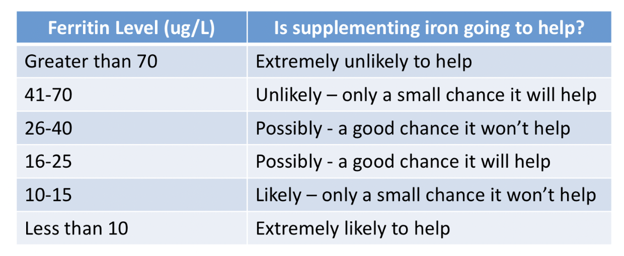
The following table provides some insights into how I think about ferritin levels for most women. One can seen that as the ferritin dips down below 70, it becomes more and more likely that taking iron could help - especially if the levels are less than 26. With ferritin levels in the teens - it’s clear iron is needed!

In summary, there are hundreds of patients today with ferritin levels 32-40 who will be told “you just need to bring up your iron to 50 or 70 and you’ll be fine.” Most of the time increasing ferritin (when levels are in the 30s is simply not going to have any effect on the hair. Is it worth trying? Well that requires input from a health care provided. As ferritin gets lower and lower below 30 it becomes more and more likely that iron supplementation will help.

I would like to point out that I never just look at the ferritin. I look at B12 (which can make ferritin levels look artificially good if B12 is low) and I look at hemoglobin, MCV, MCH and RDW and transferrin saturation if available. Most importantly I look at prior labs to see how things used to be. If one wants a magic cut off number, I’d say 40 but it’s really not so simple and lots of people eat iron pills and get constipated just to reach a number that has absolutely no benefit.

If a patient has had ferritin of 34 for 20 years and now developed hair loss last year with a ferritin of 35, are we do tell the patient “Oh you lost hair because of low iron! Bring your ferritin up to 40 to 50 and you’ll be fine.” No, this is nonsense but a common scenario.

I hope this helps with understanding the complexities of iron.

Share This
No Comments

What is More Accurate for Diagnosing Early Stages of Hair Loss : A Scalp Biopsy or Clinical (Trichoscopic) Examination?

Biopsy or Up Close (Trichoscopic) Examination: What’s better for diagnosing the early stages of hair loss?

I’ve selected this question below for this week’s question of the week. It allows us to the review some key concepts in diagnosing hair loss via clinical scalp examination and through a biopsy.

trichoscopy vs bx


QUESTION


What is more accurate - a scalp biopsy or a scalp exam with a dermatoscope? My biopsy results said telogen effluvium and androgenic alopecia with the diagnosis of androgenetic alopecia being favored.

As for me, I’m a 30 year old female. My scalp is itchy, likely from seborrheic dermatitis which was diagnosed by a dermatologist. I’ve suffered from alopecia areata in the past (1 small bald patch at a time and treated with cortisone injections) . I have a lot of food and environmental allergies that I’m treating naturally. My hair started shedding excessively at the end of February 2021 after a very traumatic event in December 2020. I’m not on any prescription medications but I do take supplements (iron, vitamin D and C, coQ10, quercetin, probiotic, l-lysine, caprylic acid, and a multivitamin for hair). The shedding has been diffuse and I have lost density. My family members insist that no one would know I’m having issues with my hair. In the past few weeks I have had days with minimal shedding. I have been treating the seborrheic dermatitis with medicated shampoos. I have been treating the hair loss naturally, through dietary changes, lowering stress levels with meditation, etc; I have not used any medications.

The dermatologist that performed the biopsy said it’s “age related” (I’m a 30 year old female) and therefore not even considered an early stage AGA. The second dermatologist I saw (for a second opinion) did a scalp exam with a dermatoscope and said there was “maybe one” miniaturized follicle at the biopsy site on my crown. Throughout the rest of the top of my scalp she said about 1 in 100 follicles are miniaturized. She gave me a diagnosis of just telogen effluvium. So far all of my test results (iron, ferritin, vitamin D, vitamin B12, thyroid panel, and hormone panel) have been normal. I’m very confused and not sure if and what treatment would be best for me. Thank you!


ANSWER

Thank you for the question. In order for me to advise you on what treatment would be best for you, we need a diagnosis.

So what is your diagnosis then?

Well, in order for me to give you a diagnosis, I would need to know a bit more about your story from birth until today, and see your scalp up close myself and review your blood tests. Those are the three key steps in order to make a diagnosis for anyone!. Because I don’t have any of these pieces of information in your case, I can’t actually say what your diagnosis is.

However, there are still some very important points to be aware of and that’s why I’ve selected your question for this week’s question. It’s such a good one with so many things for us to review.

So let’s get to it.

You have what I call early hair loss. You yourself know there is a change, but your friends and family think everything is just fine. Even one of the dermatologists thinks it’s simply a telogen effluvium. This is early hair loss.

As you have correctly outlined, this can often be due to androgenetic alopecia or telogen effluvium …. or both.

As I review all your information about what your biopsy showed and what your doctors actually said, I need to know how reliable each of these three pieces of information are. If dermatologist 2 is a world expert in hair loss and doesn’t think its AGA - does this carry more “influence” as I think about your case than if dermatologist 1 thinks it’s AGA but really has only seen a handful of hair loss patients in his or her career?

Yes it most certainly does.

Your question is really all about the reliability of these three pieces of information - the 2 doctors and the 1 biopsy.

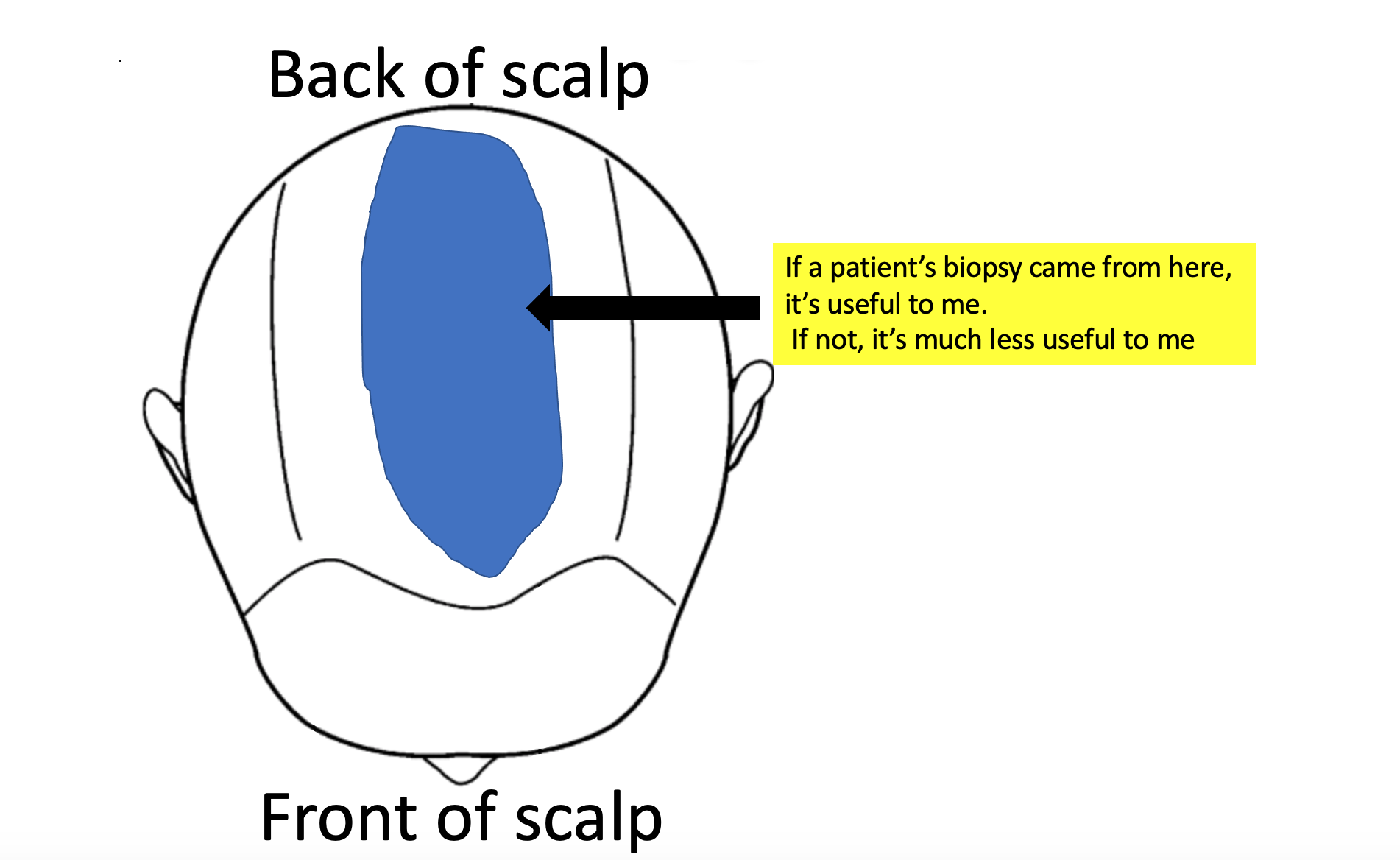
And what if the biopsy was taken from an area on the scalp that is really not so useful for making a diagnosis (like the temples) - am I to trust this result? Well, no.

So, let’s take a look at these four scenarios below in order for us to better understand when a biopsy is better than a clinician’s interpretation and when a clinician’s interpretation is to be trusted more than a biopsy report.

In general, the very early stages of hair loss can be challenging to decipher from one another. The more experience and expertise the clinician has in treating hair loss … the more reliable his or her view will be on the cause of hair loss. The less experience the clinician has, the less reliable his or her view is and the more a biopsy result is to be trusted. However, biopsies are not all the same. The only biopsy result that I really trust is one taken from the correct area of the scalp and interpreted properly by expert dermatopatholgist.


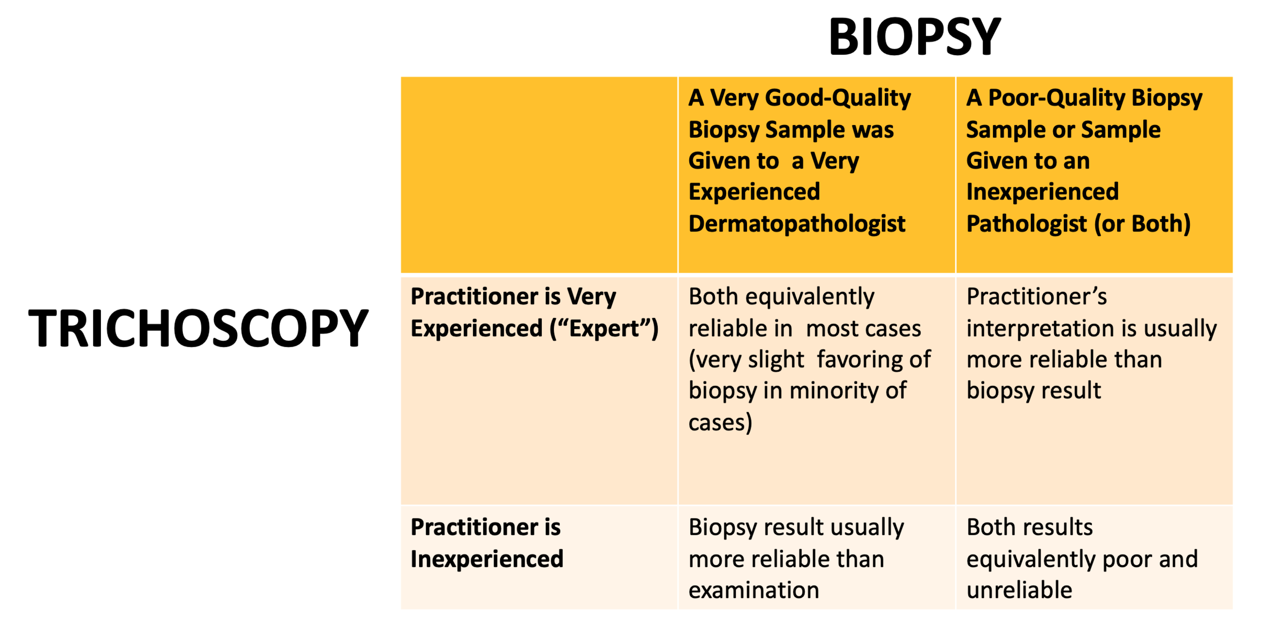
Let’s take a look at the following chart and then we’ll break it down some more.

biopsy vs clinical

SCENARIO 1. The practitioner evaluating the scalp is a VERY EXPERIENCED hair loss expert and a 4 mm punch biopsy was taken from a correct area of the scalp and interpretations were done by a VERY EXPERIENCED dermatopathologist.


In this case, both the dermatologist’s opinion and the dermatopathologist’s opinion are fairly reliable. In fact, in most cases, they are fairly equivalent. A highly experienced clinician can examine all areas of the scalp and can determine just how much variation in the caliber of hair follicles (ie “miniaturization”) is seen in the various regions including the front, middle, top and back. If the clinician appreciates that density is slightly different in one area compared to another it’s like their is some androgenetic alopecia going on - especially if the thinner area show a greater degree of miniaturization.

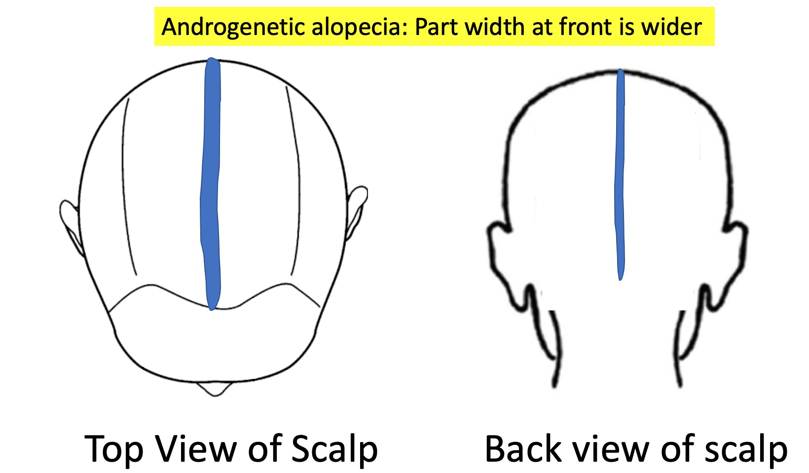
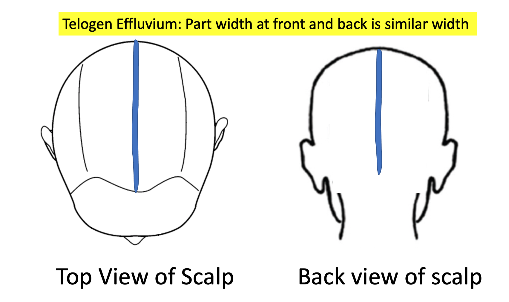
A clinician can also evaluate density in the frontal area and compare this to the back. If there is a subtle increase in “part width” in the frontal and mid scalp compared to the back, this gives a suggestion there could be some androgenetic alopecia going on.

aga
te


So an astute clinician can look at the scalp, look at the part width, look a the density in various regions of the scalp and look at what the trichsocopy shows and come up with a conclusion.

Clinical examinations of the early stages of hair loss are tricky to interpret. It takes expertise to appreciate subtle changes in hair follicle caliber. It’s not something that is learned overnight. It’s not a result that pops up on any sort of screen when one places a dermatoscope one the scalp. Of course, it one’s dermatoscope its connected to a computer and the caliber of follicles can actually be measured in various areas, this really increases the reliability of the interpretation for less experienced practitioners.

But if a practitioner is less experienced with hair and scalp issues, simply placing a dermatoscope on the scalp and concluding “I don’t see any miniaturization” does not give me a great amount of confidence in diagnosing early hair loss issues.

What about a biopsy? Biopsies in early hair loss can be wonderful! A biopsy taken from the area of androgenetic alopecia can also show a DECREASING terminal to vellus ratio from a normal low 7:1 or 8:1 down to 4:1 or less. In true telogen effluvium, the terminal to vellus ratio stays well above 6 or 7 to 1. An experienced dermatopathologist who interprets a biopsy from a patient with early hair loss and says ‘the T:V ratio is 3.5:1 and sebaceous glands appear enlarged and there is no real shift in catagen to telogen ratios and there is no peribular inflammation” is telling me this is likely androgenetic alopecia. I trust that report if I know the dermatopathologist is experienced.

To summarize, a very experienced practitioner can often make a diagnosis of androgenetic alopecia fairly reliably even without a scalp biopsy. However, if a scalp biopsy is done, the results should be similar trusted as the findings of a very experienced practitioner provided the biopsy is interpreted by an expert pathologist.


SCENARIO 2. The practitioner evaluating the scalp is an INEXPERIENCED practitioner and a 4 mm punch biopsy was taken from a correct area of the scalp and interpretations were done by a VERY EXPERIENCED dermatopathologist.


In this case, the biopsy report is MORE reliable than the view of the clinician. We need to remember here that early hair loss stages are really difficult to diagnose! There is no harm in saying that and I’ll be the first to point that out.

It can take anywhere from 6 months to 5 years from the time some types of hair loss first start before a patient themselves figure out that something is changing on their scalp. So, the early stages of hair loss are tricky to spot. The early stages of hair loss can sometimes look normal. The less experience the practitioner has …. the more the scalp will look normal to them ! That’s just a fact. Any practitioner who takes a quick 5-10 second glance at the scalp and says to their patient ‘your scalp looks fine to me… don't worry” is by definition an inexperienced practitioner. This is pretty much a rule. The early stages of hair loss are hard to spot sometimes and take a bit of poking and prodding in the scalp to see what all the 100,000 hairs are doing and a bit of sleuthing to gather information from the patient as to exactly what’s been happening over the past months.

If a very experienced clinician says ‘This scalp is normal” then it’s pretty unlikely there is any androgenetic alopecia. Not 100% guarantee of course….. but pretty unlikely. If an inexperienced clinician says ‘This scalp is normal” then it carries less meaning. Of course, it could be normal, but I’m a bit more skeptical. I am sent referrals every day of the year that say “ Normal scalp exam. Patient thinks they have hair loss. Please see in consultation.”

What do many of these patients end up having as a diagnosis ? Well, many have androgenetic alopecia !

Suppose I’m meeting up with a friend for dinner and I tell my friend that I have been getting some pretty bad headaches lately. If my friend tells me everything sounds fine, do I believe it? Well, if my friend is a neurologist I’m a bit more likely to trust this information than if my friend is an accountant. The quality of the information makes a difference.

So to summarize, if a clinician is less experienced with diagnosing hair loss but takes a biopsy from a correct area of the scalp (ie where the hair loss is most affected) and the biopsy lands in the hands of an expert dermatopatholgist …. then I would usually trust the dermatopathoglist report over the clinician’s interpretation of what’s causing the early hair loss.

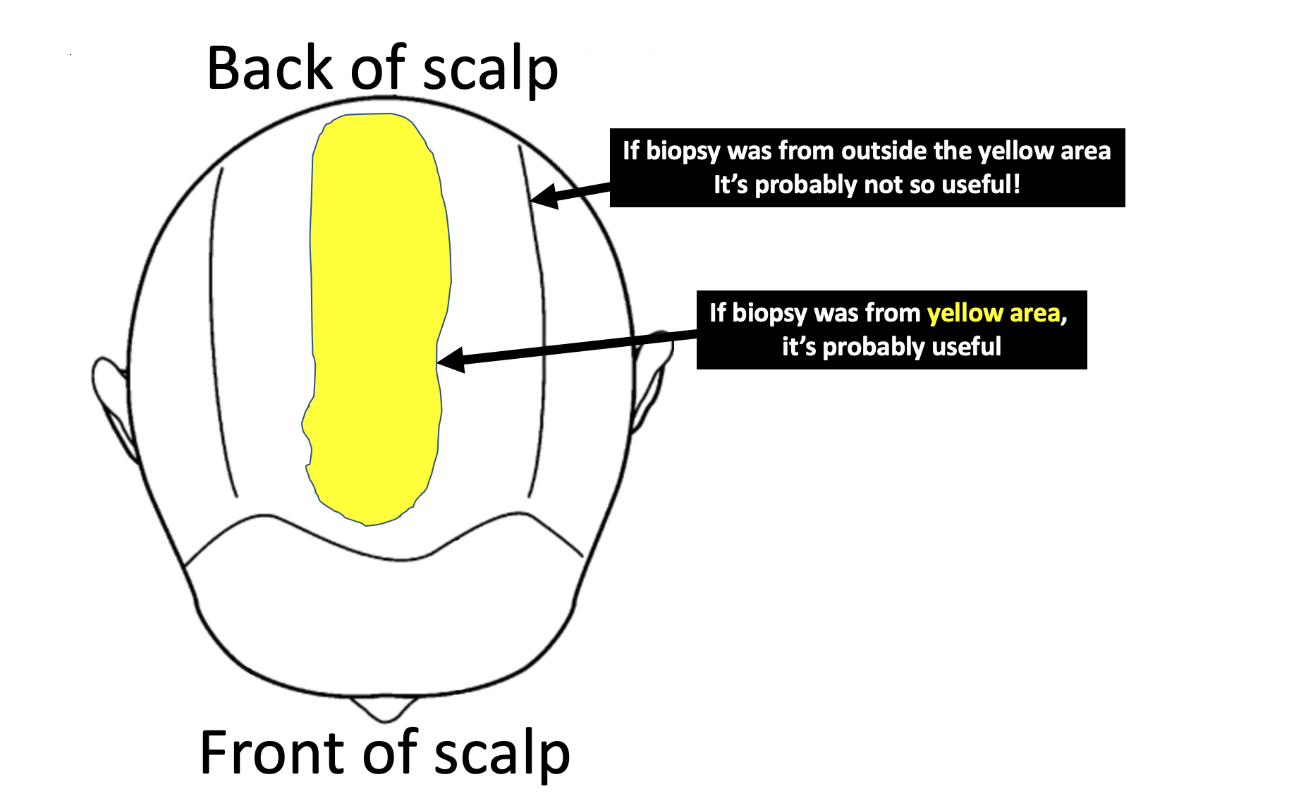
So what’s a good biopsy in your case? Well, in your case this likely means that biopsy was taken from somewhere in the yellow area shown below. I would prefer if the biopsy was 4 mm in size. I would also like if the biopsy was processed with horizontal sections as personally that increases my confidence in these early stages of hair loss. It’s only with horizontal sections that the pathologist can give a measurement of the terminal to vellus ratio. This can’t be done with vertical sections. If your T:V ratio is less than 4:1, we might begin to think there is some androgenetic alopecia present as a diagnosis.


biopsy

SCENARIO 3. The practitioner evaluating the scalp is an EXPERIENCED hair loss expert and a suboptimal biopsy was taken from an incorrect area of the scalp and/or interpretations were done by an INEXPERIENCED pathologist.

This would be an unusual situation whereby an experienced clinician took a biopsy from a wrong spot. But this situation could be an experienced clinician is trying to decide what diagnosis a patient has and the patient brings in a biopsy report they had at another clinic showing a certain result.


In this case, I trust the result from the clinician any day over the biopsy report. Every day, I see biopsy reports that are taken form the back of the scalp or the sides of the scalp or the temples. These are not the ideal areas to be taking biopsies from if we want determine whether or not the patient has androgenetic alopecia!!!

Sometimes, the doctor does not want to cause a scar…. and so takes it from the sides of the scalp so as to hide any scar. Sometimes, a patient asks the doctor to take it from the temples because that’s where they are most worried and where they see the changes every day of their life when they look in the mirror. These are not where we should be taking biopsies to confidently assess androgenetic alopecia !

If a biopsy returns showing “no evidence of androgenetic alopecia” but was taken from the sides fo the scalp does it mean the patient does not have androgenetic alopecia? No! Not at all,. That biopsy was not helping in making the proper diagnosis.

If a biopsy returns showing “no evidence of androgenetic alopecia” but was taken from the main area of hair loss in the central scalp zone, does it mean the patient does not have androgenetic alopecia? Probably that is the correct interpretation.

SCENARIO 4. The practitioner evaluating the scalp is an INEXPERIENCED practitioner and a suboptimal biopsy was taken from an incorrect area of the scalp and interpretations were done by an INEXPERIENCED pathologist.


A particularly challenging situation is when a less experienced practitioner is not sure what the diagnosis is but proceed to take the biopsy from an area of the scalp which is less than ideal. Typically this is a well meaning practitioner who wishes to take the biopsy from an area that will best be hidden in the future should the area form a small scar. So the biopsy is taken from the sides of back of thee scalp and typically returns showing no evidence of androgenetic alopecia. The only thing that can be interpreted in this situation is that the patient does not have androgenetic alopecia down the sides of their scalp. However, we can’t conclude anything at all about what might be happening in the middle of the scalp - the area where the patient is most concerned about the hair!

bx not to take

I often use the following analogy when I explain the concept to doctors that I teach.

Suppose you have a mold of some kind in your home. The house smells like mold! Terrible, right?

And so you call a mold specialist for help. Unfortunately, all the mold specialists in town are away at a convention so you decide to call a plumber. After all, mold grows in water and damp conditions, and you figure that a plumber knows a lot about water and damp conditions in homes.

The plumber answers the call and says he or she knows how to take mold samples because they learned how to do so in a course they took.

Voila!

You are happy with the answer and invite the plumber to your home to get some help.

The plumber finds a bit of water in the basement and takes some mold samples. It all comes back negative.

You are all relieved there is no mold!

The problem is that the smell continues.

When the mold specialist in town returns from the convention, you invite him or her now into your home. Within a few minutes the source of the mold is located in the attic of the house. Their is a leak in the roof and this is causing the roof to leak and the attic to grow mold !!!! Samples are taken and the mold is finally proven.

Did it matter where the samples were taken? You bet it did!

An experienced specialist is more likely to know where to take the sample .

Conclusion

Your question is really a great one. Thanks again for submitting. It’s difficult, if not impossible for patients to know if their biopsy was taken from the correct spot or whether their clinician really has a lot of experience or not. It’s tough to navigate the medical world sometimes.

The short answer to your question , however, is that a very experienced clinician can often diagnose hair loss with a similar degree of accuracy to a biopsy interpreted by an expert dermatopathlogist. If the skills of the clinician change or the skills of the dermatopathologist now change, this no longer holds true and you’ll need to see the chart about as to which is better.

It is quite likely with your story that at least one of your diagnoses was telogen effluvium that was triggered by the stress of December 2020. With your story, I think it’s really important that someone make sure that your seborrheic dermatitis is under good control and someone keep an eye on the possibility that a diffuse alopecia areata is not part of the reason for your shedding. I think that would be unlikely given that shedding has settled now and that the biopsy did not capture this.

With this one biopsy that you do have I can’t exclude that there is not some degree of androgenetic alopecia present. There certainly is a possibility with this information you’ve given. oOf course, I would need to see the scalp or a photo of your scalp myself to know for sure one way or another.

Please keep taking photos of your scalp to show your doctors. If you feel that your hair returns to full by September 2021 and you are really pleased with the way your hair looks and feels at that time, then it’s pretty unlikely there is any AGA. However, if your hair does not return to full by September, I would encourage you to further explore ways to confirm this diagnosis with certainty one way or another so that you might get connected with the correct treatment in the event you do have androgenic alopecia.

Thank you again for your question.

Share This
No Comments

Daily Shedding... with high DHEAS.... What should I be doing?

Why am I still shedding ?

I’ve selected this question below for this week’s question of the week. It allows us to discuss shedding issues in young women with high DHEAS.


QUESTION.

Dr. Donovan, I'm hoping you may be able to shed some light onto my hair shedding situation. I started experiencing increased hair shedding 6 months ago. Prior to the shedding, I'd started a birth control pill (low androgen) 2.5 months before, and Spironolactone 5.5 months before (for hirsutism). I experienced mild stress but it was nothing out of the ordinary. A month into the shedding, I began to experience tingling, crawling, and pain in my scalp. 1.5 months after the hair loss started, I went off of Spironolactone, and 2.5 months into the shedding, I went off birth control. My scalp became itchy as well.

I went to 3 dermatologists, all of whom diagnosed me with telogen effluvium. I recently had a biopsy done that stated telogen effluvium as well. I was also diagnosed with seborrheic dermatitis (which I've never had before in my life). I tried ketoconazole shampoo. I've had blood work done that stated my thyroid was normal, ferritin within normal range, (was 95 in October due to brief supplementation and then 26 in January), and I was deficient in vitamin D. My vitamin B12 level was too high, I'm not sure what that means.

I have symptoms of androgen excess (acne and excess hair growth on my face / body), dating back to my teen years (17), I'm 21 now, soon to be 22. The facial hair growth appeared in November of 2019 at age 20. I have elevated DHEAs (512), and had an ultrasound ruling out PCOS (no irregular periods, no polycystic ovaries).

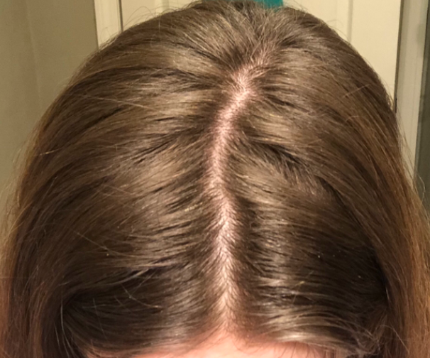
Photo of the patient’s scalp.

Photo of the patient’s scalp.


Since the hair shedding began, I experienced massive emotional stress as a result. I was diagnosed with anxiety and depression - which I'm sure I've had for years but got extremely bad once my hair started to fall out. I am thin (weighing only 104lbs) but lost 10 pounds presumably due to stress of the hair loss. I've had two episodes of what was probably telogen effluvium in the past - one was related to low iron (ferritin of 7) back when I was 15, and one episode when I was 18 most likely due to a bad case of the flu. Those episodes only lasted 3-6 months and I grew all of my hair back.

This time, I've been consistently shedding for 6 months with no sign of improvement. My scalp is very tender, sensitive, flaky and itchy still. On an average day, I used to shed maybe 5-10 hairs, now I am shedding roughly 50-150 hairs - which is not normal for me. I'm at a loss for what could be causing this and what I can do about it. Any advice would be greatly appreciated


ANSWER

Thanks for the question.

I’d like to discuss several important things in the question you ask and the information you have submitted.

Before we do go further, I’d like to point out that the ideal way to diagnose hair loss is using what I termed the ”Diagnostic S.E.T.” I refer to these as the diagnostic “set” because theses 3 aspects all go together. These 3 items include:

1) the patient’s story

2) the findings uncovered during the process of the scalp examination including trichoscopy

3) the results of relevant blood tests. 

The first letter of each of the three words 1) story, 2) examination and 3) tests spell out the word “S.E.T.” - again a helpful reminder of how the information obtained from reviewing each of these 3 aspects helps solidify a proper diagnosis.

There is lots more to your story that I need. I would want to know about other medications you have started and stopped. I would want to know about other symptoms like joint pains, headaches, fatigue, weight loss, eyebrow changes, eyelashes changes, body hair changes, nail changes, and rashes.

The 2 key questions here in your case are:

a) Is the diagnosis ONLY telogen effluvium … and …. if so what is the trigger?

b) Is this a telogen effluvium with the starting stages of androgenetic alopecia?


Let’s go further into your story.

POINT 1. Many people who take birth control pills shed for the first few months.

First, I think there’s little doubt that at least one of your diagnoses is telogen effluvium. We don’t actually need to debate that. The debate we will get into in a moment is whether anything else is going on.

You have several reasons why you could have a telogen effluvium, including starting the birth control pill and starting spironolactone. A large proportion of women shed when starting these treatments, especially birth control pills. The shedding starts 2-3 months after taking the first pill and the shedding lasts 3-6 months provided the pill is continued every day. A lot of women shed when stopping these pills too, especially the birth control pill. The shedding starts 2-3 months after stopping the pill and the shedding lasts 3-6 months provided the pill is not restarted.

So what would I expect to hear from a 21 year old woman who starts spironolactone and then starts a birth control pill? Shedding.

And what would I expect to hear from a 21 year old woman who starts spironolactone and then starts a birth control pill and then stops these pills? Shedding.

What is your story? Shedding.

So in some ways, it’s possible this is entirely consistent with your story.


POINT 2: All patients with hair loss, acne and hirsutism and androgen excess need a proper work up. A work up should be done off birth control.


You have DHEAS 512 (which is 13.9 umol/L in SI units). Any female age 21 with DHEAS 512 and acne and hirsutism and hair loss needs a thorough endocrine work up in my opinion. We need to rule out PCOS and late onset CAH.

With the limited information you have provided here, it would be false to say that you “don’t” have PCOS.

The correct way to say it is more likely “you have a low likelihood of having PCOS.”

Women with PCOS who are thin with low BMI often have regular periods and often have no cysts visualized on ultrasound examinations … but still have elevated androgens. This is a bit more advanced type of knowledge, but I think it’s important especially since you have hyperandrogenism. Anyone who claims there is zero chance you have PCOS is wrong. Anyone who thinks there is a very low chance you have PCOS is correct. I have seen many women with your story exactly who go from being thin to being heavier in their 20s and 30s and ‘develop’ PCOS. I’m not saying that is your case, but sometimes weight gain brings about insulin resistance that then promotes a fuller PCOS clinical presentation.

So what work up do you need? Well, I would advise a proper work up on day 3-5 of your cycle for my own patients that come to see me with a story like yours. The fact that you are off birth control again is a good time to do this. We can’t do these tests when women are on birth control.

The tests that I order on day 3-5 of the cycle for my patients with similar stories are: LH, FSH, estradiol, testosterone, free testosterone, SHBG, glucose, insulin, hemoglobin 1A1c, AM cortisol, prolactin, androstenedione and 17 hydroxyprogesterone, AST, ALT, and cholesterol. These should be done fasting and day 3-5 of the cycle. You have already had your DHEAS measured so there is not a lot of good reason to do this again unless someone suspected levels could be climbing. I would probably include it again for completeness.

What am I looking for in these tests?

a) a high 17 OHP level on day 3-5 that would lead us to a diagnosis of late onset congenital adrenal hyperplasia

b) a high testosterone, high fasting insulin, high LH that would point is towards a PCOS like state

c) A normal prolactin and AM cortisol that reassures us that no other issues are present

In your case, I would want more blood tests if I was your doctor. I would want to know if there is any evidence of insulin resistance that would push me towards PCOS. I would want to know if the other hormones were normal. I would want to know your free testosterone and SHBG. I would want to know your 17 OHP levels to rule out late onset congenital adrenal hyperplasia before moving on.

POINT 3: When it comes to ultrasound examinations, there is a lot that patients don’t realize.

When I hear that a patient had an ultrasound that showed no cysts, my first response is usually that I’m glad to hear that news. But there are a few points to keep in mind. First, it depends on whether the ultrasound examination was a transabdominal ultrasound or transvaginal. There is a big difference in the quality of the studies and what it all means. Transvaginal studies with modern ultrasound techniques are the most helpful. Many people don’t have these studies done. Transvaginal studies can pick up a lot of cysts that the transabdominal can not. Of course, it’s very unlikely this is even an issue with your story but it’s something that we need to keep in mind.

With your ultrasound, I would want to know where it was done (what center? what clinic?) and whether it was transabdominal or transvaginal? What was the volume of the ovaries noted in the report ? Were any ovary measurements more than 10 mL ?

POINT 4: In the early stages of hair loss, TE and AGA can look the same and have the same story. Rarely, they can have a similar biopsy too.

When I look at your photos, I immediately say to myself :

Could this person have AGA?

Could this person have TE?

Could this person have both conditions?

(Also …. whenever we use the word TE, we need to immediate shout out hey could this be diffuse alopecia areata …. but I don’t think that’s what this is. But I include this for completeness of this write up).

In the early stages TE and AGA look the same. Of course, an up close examination with use of trichoscopy is going to help in your situation. In fact, it’s critical this be done! If the back of the scalp is convincingly thicker than the front of the scalp, I am pushed more towards thinking this could be AGA (with your TE). If the back and front areas are similar density, we are more likely thinking about a sole diagnosis of isolate TE. If there is no evidence of “follicular miniaturization” or variation in the caliber of your hairs when your scalp is examined with trichoscopy, we are likely dealing with an isolated TE. If there is a convincing variation in the caliber of hairs, it could be an early AGA.

Biopsies can be tricky. There is so much more to doing a biopsy than just doing it and so much more to interpreting a biopsy than just reading the information that comes printed on report. My ability to accurately interpret a biopsy depends where on the scalp it was taken from!. It depends how it was processed (horizontal vs vertical section). It depends who read the biopsy (dermpath vs general path). If a biopsy was taken from the sides or the back or somewhere just to prevent the patient from having a visible scar, then the biopsy is often useless. Biopsies in your case need to come from the top.

Here is where I would need your biopsy to have been taken from for me to feel better about the situation:


sites of biopsy

Also, if someone is going to tell me all you have in your biopsy is a telogen effluvium, I’m going to hope that horizontal sections were used. It’s a huge stretch to diagnose a telogen effluvium confidently from vertical sections. In horizontal sections, the pathologist gets to see 20-40 hairs in order to give their best guess about what could be going on with the rest of the scalp. With vertical sectioned biopsies, they just get to see 3-6 hairs. I don’t want to leave my patient’s hair loss diagnosis to interpretations as to what is seen with 3-6 hairs.

POINT 5: TE and seborrheic dermatitis (and sometimes even AGA) give tingling and symptoms.

With the biopsy result you have in your possession, I’m much much less worried about the tingling, crawling and pain. Of course, I wish you did not have it. But I’m not suspecting anything inflammatory that would make me want to act with a much different course. This is assuming your biopsy came from an area that was tender) I’m always worried when a patient says they have scalp pain. But this worry evaporates to a large degree when the biopsy from that area shows non-scarring alopecia. If your biopsy was from a random area and not from a tender area, then it becomes more difficult to interpret what it means.

Pain and tingling in your case can come from seborrheic dermatitis. it can come from TE, It can come from depression. It can come from allergy or irritation from a current shampoo. it can come from irritation of allergy from other cosmetics.

We still need to keep an eye on this pain. I often encourage my patients to commit to treating their seborrheic dermatitis with a rotating schedule of shampoos. Zinc pyrithione one day. Ketoconazole the next regular shampooing day and selenium sulphide shampoo the next shampooing day. Shampooing must be done 2-3 times per week and left on 2 minutes. I advise my patients to not over do the time as this often just dries the hair and scalp out further. Also, putting a prescription topical steroid on the scalp like betamethasone valerate lotion 0.1 % aa few times per week right after showering is often helpful (of before bed). 10 drops to 15 drops of betamethasone valerate lotion two times per week for a few months is very safe and anyone who says otherwise has little understanding, knowledge or training in the area.

If the pain is still present in 3-5 months, this needs to be looked into further.


POINT 6: What to do next depends on the blood test results and your prior response to spironolactone.

What exactly to do next and how do help your shedding depends partly on your next set of blood test results. If you have elevated 17 OHP on your blood tests, you may want to see an endocrinologist. If you have high LH or evidence of insulin resistance you’ll want to see a really experienced endocrinologist for evaluation of PCOS. Not all women with PCOS are overweight and in fact, women with PCOS who are thinner or have low body mass index often have regular periods and no cysts on ultrasound.

If you tolerated spironolactone well (in the past) and tolerated birth control well, it may even be an option to return to these and stay on these. Did it control your acne? Did it stop hirsutism? Did you feel good on it? Anyone starting these medications has a chance to get shedding so I’m not necessarily worried by a story of shedding. You stopped too soon to really get any sense what the long term outcome was. If there is any evidence of AGA with an up close examination, this could be a good option again. If not, you might want to treat your acne and hirsutism differently - perhaps topically.. The other option is to wait longer in hopes the TE resolves. Continuing iron and vitamin D and shampooing your hair diligently with these anti dandruff shampoos is going to be important no matter what is going on up on the scalp.

Finally, with any TE, we need to always keep in mind that maybe we have not found the trigger. If your ferritin was low and your hemoglobin was low (less than 12.0), a work up could be important. I often test for example a celiac panel in patients with BOTH low HGB and low ferritin. I’m not worried about your high B12. I would want to know about other medications you have started and stopped. As mentioned above, I would want to know about other symptoms like joint pains, headaches, fatigue, weight loss, eyebrow changes, eyelashes changes, body hair changes, nail changes, and rashes. Sometimes we consider ordering autoimmune tests in women with shedding but only if the history points us to ordering these. Ordering these tests ‘just to cover all bases’ is usually not a good idea.

Summary

I can instantly tell by your question that you’ve read a lot and thought a lot about your issues and what all this information means. Congratulations for that. That is important. You need to figure out if late onset CAH is a possibility or not ….. and whether insulin resistance/PCOS its truly off the list or not. In my opinion, these blood tests on day 3-5 are important to you. I’m glad you are off the birth control pill now because it allows you to get these tests done.

If your biopsy was taken from the area I have noted above, then that’s probably very helpful provided it was analyzed in the lab with horizontal sections. . The key point now is figuring out if there is any possibility of an evolving androgenetic alopecia that just could not be picked up in the early stages with the work up you had. A biopsy with horizontal sections and a good trichoscopic examination by a specialist who understands hair loss will uncover these answers.

Regardless, photos should be taken every 3 months. Not every day and not every week. If there is any kind of evolving pattern of hair loss, a photos will also capture these changes over time.

If TE is truly what you have and there are no underlying concerns, doing minimal additional things could be the best plan. However, if your hyperandrogenism is part of a bigger endocrine issue (like PCOS or CAH), getting advice from an endocrinologist would be a good way to proceed. These blood tests will be a really important guide. Some women just have elevated DHEAS and some women with elevated DHEAS develop AGA but some don’t. I never recommend patients start treatment because of what the labs say - treatment is started because of what the skin or hair is doing.

Thanks again for sending in the question. I hope this helps you are your team of specialists.


Share This
No Comments


Share This
-->