QUESTION OF THE WEEK

Dr. Donovan's Articles

QUESTION OF HAIR BLOGS

Filtering by Category: Hair Collection Tests


How do we prevent progression from eyebrow FFA to scalp FFA?

How do I stop by scalp from developing FFA?


I’ve selected this question below for this week’s question of the week. It allows us to review some concepts in the management of frontal fibrosing alopecia (FFA).


Question

I am 47 years old and have eyebrow loss that my dermatologist feels is FFA. My mother has FFA and so we think my recent loss is also FFA. I have menopause at 41 which seems to fit well with the condition.

I want to know if there is anything I can do right now to stop the stop from becoming involved. My dermatologist does not see any signs whatsoever of FFA in the scalp.

My eyebrows are doing okay with Latisse and Rogaine as well as steroid injections every few months.

Answer

Thank you for this question.

The immune system has a plan for each person and we do not know what exactly that plan is for any given person and it's different for different people. If one wants to reduce the chances of scalp involvement then systemic medications may be needed.

Eyebrow loss can be treated with the options in the following chart. You are already on a solid plan with many of these. I often start with ONE OR MORE of minoxidil, bimatoproast, pimecrolimus and steroid injections and then see how the eyebrows respond. If we are successful then these are the options.

Reducing the chances of scalp involvement in the future may require one or more of the systemic agents (pills) shown in the list below. You may want to have a good discussion with your dermatologist about these various options as there are some reasons why some women can not use these medications at all. The key decisions in my mind would be for you and your dermatologist to figure out where dutasteride, finasteride, isotretinoin or hydroxychloroquine fit in. For many patients, it could be that getting on dutasteride is among the best steps and then waiting to see if any FFA develops.

Preventive Measures in FFA Have Not Been Studied

Your question is such a great one. The best way to prevent FFA of the scalp in someone with eyebrow FFA has not been well studied. For now, most physicians treat and address hair loss as it happens. This is probably not the best plan but most things we do in modern medicine are reactive rather than proactive.

Your point is a very good one and we do need to be thinking about the potential for future scalp hair loss. My feeling is that confirmed eyebrow FFA requires at least one systemic treatment and very very very close follow up. I would advise that a patient take photos every 4-6 months of the eyebrows, eyelashes, frontal hairline, crown, back of the scalp and sideburns.

If there is any evidence that FFA of the frontal hairline develops, then oral isotretinoin together with pimecrolimus cream and steroid injections can be started. Again, close follow up every 3-5 months will be needed to determine if this treatment plan is effective. If not, adding an antihistamine like cetirizine together with hydroxychloroquine would be the way I’d go in the present day (date of this post!)

Thanks again for the question

Share This
No Comments

What is More Accurate for Diagnosing Early Stages of Hair Loss : A Scalp Biopsy or Clinical (Trichoscopic) Examination?

Biopsy or Up Close (Trichoscopic) Examination: What’s better for diagnosing the early stages of hair loss?

I’ve selected this question below for this week’s question of the week. It allows us to the review some key concepts in diagnosing hair loss via clinical scalp examination and through a biopsy.

trichoscopy vs bx


QUESTION


What is more accurate - a scalp biopsy or a scalp exam with a dermatoscope? My biopsy results said telogen effluvium and androgenic alopecia with the diagnosis of androgenetic alopecia being favored.

As for me, I’m a 30 year old female. My scalp is itchy, likely from seborrheic dermatitis which was diagnosed by a dermatologist. I’ve suffered from alopecia areata in the past (1 small bald patch at a time and treated with cortisone injections) . I have a lot of food and environmental allergies that I’m treating naturally. My hair started shedding excessively at the end of February 2021 after a very traumatic event in December 2020. I’m not on any prescription medications but I do take supplements (iron, vitamin D and C, coQ10, quercetin, probiotic, l-lysine, caprylic acid, and a multivitamin for hair). The shedding has been diffuse and I have lost density. My family members insist that no one would know I’m having issues with my hair. In the past few weeks I have had days with minimal shedding. I have been treating the seborrheic dermatitis with medicated shampoos. I have been treating the hair loss naturally, through dietary changes, lowering stress levels with meditation, etc; I have not used any medications.

The dermatologist that performed the biopsy said it’s “age related” (I’m a 30 year old female) and therefore not even considered an early stage AGA. The second dermatologist I saw (for a second opinion) did a scalp exam with a dermatoscope and said there was “maybe one” miniaturized follicle at the biopsy site on my crown. Throughout the rest of the top of my scalp she said about 1 in 100 follicles are miniaturized. She gave me a diagnosis of just telogen effluvium. So far all of my test results (iron, ferritin, vitamin D, vitamin B12, thyroid panel, and hormone panel) have been normal. I’m very confused and not sure if and what treatment would be best for me. Thank you!


ANSWER

Thank you for the question. In order for me to advise you on what treatment would be best for you, we need a diagnosis.

So what is your diagnosis then?

Well, in order for me to give you a diagnosis, I would need to know a bit more about your story from birth until today, and see your scalp up close myself and review your blood tests. Those are the three key steps in order to make a diagnosis for anyone!. Because I don’t have any of these pieces of information in your case, I can’t actually say what your diagnosis is.

However, there are still some very important points to be aware of and that’s why I’ve selected your question for this week’s question. It’s such a good one with so many things for us to review.

So let’s get to it.

You have what I call early hair loss. You yourself know there is a change, but your friends and family think everything is just fine. Even one of the dermatologists thinks it’s simply a telogen effluvium. This is early hair loss.

As you have correctly outlined, this can often be due to androgenetic alopecia or telogen effluvium …. or both.

As I review all your information about what your biopsy showed and what your doctors actually said, I need to know how reliable each of these three pieces of information are. If dermatologist 2 is a world expert in hair loss and doesn’t think its AGA - does this carry more “influence” as I think about your case than if dermatologist 1 thinks it’s AGA but really has only seen a handful of hair loss patients in his or her career?

Yes it most certainly does.

Your question is really all about the reliability of these three pieces of information - the 2 doctors and the 1 biopsy.

And what if the biopsy was taken from an area on the scalp that is really not so useful for making a diagnosis (like the temples) - am I to trust this result? Well, no.

So, let’s take a look at these four scenarios below in order for us to better understand when a biopsy is better than a clinician’s interpretation and when a clinician’s interpretation is to be trusted more than a biopsy report.

In general, the very early stages of hair loss can be challenging to decipher from one another. The more experience and expertise the clinician has in treating hair loss … the more reliable his or her view will be on the cause of hair loss. The less experience the clinician has, the less reliable his or her view is and the more a biopsy result is to be trusted. However, biopsies are not all the same. The only biopsy result that I really trust is one taken from the correct area of the scalp and interpreted properly by expert dermatopatholgist.


Let’s take a look at the following chart and then we’ll break it down some more.

biopsy vs clinical

SCENARIO 1. The practitioner evaluating the scalp is a VERY EXPERIENCED hair loss expert and a 4 mm punch biopsy was taken from a correct area of the scalp and interpretations were done by a VERY EXPERIENCED dermatopathologist.


In this case, both the dermatologist’s opinion and the dermatopathologist’s opinion are fairly reliable. In fact, in most cases, they are fairly equivalent. A highly experienced clinician can examine all areas of the scalp and can determine just how much variation in the caliber of hair follicles (ie “miniaturization”) is seen in the various regions including the front, middle, top and back. If the clinician appreciates that density is slightly different in one area compared to another it’s like their is some androgenetic alopecia going on - especially if the thinner area show a greater degree of miniaturization.

A clinician can also evaluate density in the frontal area and compare this to the back. If there is a subtle increase in “part width” in the frontal and mid scalp compared to the back, this gives a suggestion there could be some androgenetic alopecia going on.

aga
te


So an astute clinician can look at the scalp, look at the part width, look a the density in various regions of the scalp and look at what the trichsocopy shows and come up with a conclusion.

Clinical examinations of the early stages of hair loss are tricky to interpret. It takes expertise to appreciate subtle changes in hair follicle caliber. It’s not something that is learned overnight. It’s not a result that pops up on any sort of screen when one places a dermatoscope one the scalp. Of course, it one’s dermatoscope its connected to a computer and the caliber of follicles can actually be measured in various areas, this really increases the reliability of the interpretation for less experienced practitioners.

But if a practitioner is less experienced with hair and scalp issues, simply placing a dermatoscope on the scalp and concluding “I don’t see any miniaturization” does not give me a great amount of confidence in diagnosing early hair loss issues.

What about a biopsy? Biopsies in early hair loss can be wonderful! A biopsy taken from the area of androgenetic alopecia can also show a DECREASING terminal to vellus ratio from a normal low 7:1 or 8:1 down to 4:1 or less. In true telogen effluvium, the terminal to vellus ratio stays well above 6 or 7 to 1. An experienced dermatopathologist who interprets a biopsy from a patient with early hair loss and says ‘the T:V ratio is 3.5:1 and sebaceous glands appear enlarged and there is no real shift in catagen to telogen ratios and there is no peribular inflammation” is telling me this is likely androgenetic alopecia. I trust that report if I know the dermatopathologist is experienced.

To summarize, a very experienced practitioner can often make a diagnosis of androgenetic alopecia fairly reliably even without a scalp biopsy. However, if a scalp biopsy is done, the results should be similar trusted as the findings of a very experienced practitioner provided the biopsy is interpreted by an expert pathologist.


SCENARIO 2. The practitioner evaluating the scalp is an INEXPERIENCED practitioner and a 4 mm punch biopsy was taken from a correct area of the scalp and interpretations were done by a VERY EXPERIENCED dermatopathologist.


In this case, the biopsy report is MORE reliable than the view of the clinician. We need to remember here that early hair loss stages are really difficult to diagnose! There is no harm in saying that and I’ll be the first to point that out.

It can take anywhere from 6 months to 5 years from the time some types of hair loss first start before a patient themselves figure out that something is changing on their scalp. So, the early stages of hair loss are tricky to spot. The early stages of hair loss can sometimes look normal. The less experience the practitioner has …. the more the scalp will look normal to them ! That’s just a fact. Any practitioner who takes a quick 5-10 second glance at the scalp and says to their patient ‘your scalp looks fine to me… don't worry” is by definition an inexperienced practitioner. This is pretty much a rule. The early stages of hair loss are hard to spot sometimes and take a bit of poking and prodding in the scalp to see what all the 100,000 hairs are doing and a bit of sleuthing to gather information from the patient as to exactly what’s been happening over the past months.

If a very experienced clinician says ‘This scalp is normal” then it’s pretty unlikely there is any androgenetic alopecia. Not 100% guarantee of course….. but pretty unlikely. If an inexperienced clinician says ‘This scalp is normal” then it carries less meaning. Of course, it could be normal, but I’m a bit more skeptical. I am sent referrals every day of the year that say “ Normal scalp exam. Patient thinks they have hair loss. Please see in consultation.”

What do many of these patients end up having as a diagnosis ? Well, many have androgenetic alopecia !

Suppose I’m meeting up with a friend for dinner and I tell my friend that I have been getting some pretty bad headaches lately. If my friend tells me everything sounds fine, do I believe it? Well, if my friend is a neurologist I’m a bit more likely to trust this information than if my friend is an accountant. The quality of the information makes a difference.

So to summarize, if a clinician is less experienced with diagnosing hair loss but takes a biopsy from a correct area of the scalp (ie where the hair loss is most affected) and the biopsy lands in the hands of an expert dermatopatholgist …. then I would usually trust the dermatopathoglist report over the clinician’s interpretation of what’s causing the early hair loss.

So what’s a good biopsy in your case? Well, in your case this likely means that biopsy was taken from somewhere in the yellow area shown below. I would prefer if the biopsy was 4 mm in size. I would also like if the biopsy was processed with horizontal sections as personally that increases my confidence in these early stages of hair loss. It’s only with horizontal sections that the pathologist can give a measurement of the terminal to vellus ratio. This can’t be done with vertical sections. If your T:V ratio is less than 4:1, we might begin to think there is some androgenetic alopecia present as a diagnosis.


biopsy

SCENARIO 3. The practitioner evaluating the scalp is an EXPERIENCED hair loss expert and a suboptimal biopsy was taken from an incorrect area of the scalp and/or interpretations were done by an INEXPERIENCED pathologist.

This would be an unusual situation whereby an experienced clinician took a biopsy from a wrong spot. But this situation could be an experienced clinician is trying to decide what diagnosis a patient has and the patient brings in a biopsy report they had at another clinic showing a certain result.


In this case, I trust the result from the clinician any day over the biopsy report. Every day, I see biopsy reports that are taken form the back of the scalp or the sides of the scalp or the temples. These are not the ideal areas to be taking biopsies from if we want determine whether or not the patient has androgenetic alopecia!!!

Sometimes, the doctor does not want to cause a scar…. and so takes it from the sides of the scalp so as to hide any scar. Sometimes, a patient asks the doctor to take it from the temples because that’s where they are most worried and where they see the changes every day of their life when they look in the mirror. These are not where we should be taking biopsies to confidently assess androgenetic alopecia !

If a biopsy returns showing “no evidence of androgenetic alopecia” but was taken from the sides fo the scalp does it mean the patient does not have androgenetic alopecia? No! Not at all,. That biopsy was not helping in making the proper diagnosis.

If a biopsy returns showing “no evidence of androgenetic alopecia” but was taken from the main area of hair loss in the central scalp zone, does it mean the patient does not have androgenetic alopecia? Probably that is the correct interpretation.

SCENARIO 4. The practitioner evaluating the scalp is an INEXPERIENCED practitioner and a suboptimal biopsy was taken from an incorrect area of the scalp and interpretations were done by an INEXPERIENCED pathologist.


A particularly challenging situation is when a less experienced practitioner is not sure what the diagnosis is but proceed to take the biopsy from an area of the scalp which is less than ideal. Typically this is a well meaning practitioner who wishes to take the biopsy from an area that will best be hidden in the future should the area form a small scar. So the biopsy is taken from the sides of back of thee scalp and typically returns showing no evidence of androgenetic alopecia. The only thing that can be interpreted in this situation is that the patient does not have androgenetic alopecia down the sides of their scalp. However, we can’t conclude anything at all about what might be happening in the middle of the scalp - the area where the patient is most concerned about the hair!

bx not to take

I often use the following analogy when I explain the concept to doctors that I teach.

Suppose you have a mold of some kind in your home. The house smells like mold! Terrible, right?

And so you call a mold specialist for help. Unfortunately, all the mold specialists in town are away at a convention so you decide to call a plumber. After all, mold grows in water and damp conditions, and you figure that a plumber knows a lot about water and damp conditions in homes.

The plumber answers the call and says he or she knows how to take mold samples because they learned how to do so in a course they took.

Voila!

You are happy with the answer and invite the plumber to your home to get some help.

The plumber finds a bit of water in the basement and takes some mold samples. It all comes back negative.

You are all relieved there is no mold!

The problem is that the smell continues.

When the mold specialist in town returns from the convention, you invite him or her now into your home. Within a few minutes the source of the mold is located in the attic of the house. Their is a leak in the roof and this is causing the roof to leak and the attic to grow mold !!!! Samples are taken and the mold is finally proven.

Did it matter where the samples were taken? You bet it did!

An experienced specialist is more likely to know where to take the sample .

Conclusion

Your question is really a great one. Thanks again for submitting. It’s difficult, if not impossible for patients to know if their biopsy was taken from the correct spot or whether their clinician really has a lot of experience or not. It’s tough to navigate the medical world sometimes.

The short answer to your question , however, is that a very experienced clinician can often diagnose hair loss with a similar degree of accuracy to a biopsy interpreted by an expert dermatopathlogist. If the skills of the clinician change or the skills of the dermatopathologist now change, this no longer holds true and you’ll need to see the chart about as to which is better.

It is quite likely with your story that at least one of your diagnoses was telogen effluvium that was triggered by the stress of December 2020. With your story, I think it’s really important that someone make sure that your seborrheic dermatitis is under good control and someone keep an eye on the possibility that a diffuse alopecia areata is not part of the reason for your shedding. I think that would be unlikely given that shedding has settled now and that the biopsy did not capture this.

With this one biopsy that you do have I can’t exclude that there is not some degree of androgenetic alopecia present. There certainly is a possibility with this information you’ve given. oOf course, I would need to see the scalp or a photo of your scalp myself to know for sure one way or another.

Please keep taking photos of your scalp to show your doctors. If you feel that your hair returns to full by September 2021 and you are really pleased with the way your hair looks and feels at that time, then it’s pretty unlikely there is any AGA. However, if your hair does not return to full by September, I would encourage you to further explore ways to confirm this diagnosis with certainty one way or another so that you might get connected with the correct treatment in the event you do have androgenic alopecia.

Thank you again for your question.

Share This
No Comments

Do I have AGA? Everything thinks I am Crazy!

Why am I losing hair ?

I’ve selected this question below for this week’s question of the week. It allows us to discuss the diagnosis of hair loss in the early stages and the use of the 5 day modified hair wash test.

QUESTION

Dear Doctor Donovan,

I'm a 27 year old female. I've seen a lot of dermatologists over the years but none of them could solve my problem and I'm desperate!! It's been three years that I'm losing hair. It started during a stressful period in 2018 with a significant amount of shedding (300-400 hair a day).

The shedding stopped for 4 months and then the shedding started again until now. The only thing I noticed during these years is that hair loss is focused on the side of the head and temples and hair always grows back, even if I have less. The dermatologists have all said the diagnosis is telogen effluvium or chronic telogen effluvium (CTE) because in their opinion I still have a lot of hair and no miniaturized hair (but I can see them!)

In 2019 I decided to starting using minoxidil 2% because I was losing my mind and I was scared to go bald. My mother uses it for AGA. I suffer from hypothyroidism, I have regular periods but low levels of ferritin. I'm also losing hair in different lengths (short, long, thick, thin).

I am afraid I have AGA. Nobody believes me and they think I'm crazy!.

Thank you very much for listening.

central


Screen Shot 2021-04-25 at 6.50.00 AM.png
regrowth
Screen Shot 2021-04-25 at 6.34.26 AM.png
before and after


ANSWER

Thanks for submitting this very interesting question.

I’d like to discuss several important things in the question you ask and the information you have submitted.

Before we do go further, I’d like to point out that the ideal way to diagnose hair loss is using what I termed the ”Diagnostic S.E.T.” I refer to these as the diagnostic “set” because theses 3 aspects all go together. These 3 items include:

1) the patient’s story

2) the findings uncovered during the process of the scalp examination including trichoscopy

3) the results of relevant blood tests. 

The first letter of each of the three words 1) story, 2) examination and 3) tests spell out the word “S.E.T.” - again a helpful reminder of how the information obtained from reviewing each of these 3 aspects helps solidify a proper diagnosis. I don’t have a full story and I only have a trichoscopy photos from one area and I don’t have the opportunity to review all tests …. so I am limited to some degree in my helpfulness. Nevertheless, I do think the discussion here will be helpful.

In the early stages of hair loss, nobody can tell you have hair loss. So, when people tell you they think your hair looks normal, they are being honest. You know you hair the best.


What are the Most Likely Diagnoses in this Case?

We are deciding here between three situations:

1) Do you have androgenetic alopecia (AGA) and telogen effluvium (TE) ?

or

2) do you have telogen effluvium (TE) only ?

or

3) Do you have both of these conditions?

I think there is little doubt in your story that telogen effluvium has been present at some time in the past. Whether it is still present NOW (today) is a bit more challenging to say as we will see in a while. I will explain in a moment how we can better distinguish between the two diagnoses.

It’s fairly unlikely that other diagnoses like alopecia areata incognito are present this long and similarly the photos don’t really lead me to believe we’ve dealing with other diagnoses. Of course, a full examination can confidently exclude that. But it’s unlikely.


What do I see in the photos?

The photos seem to show less density ‘now’ compared to 2018. This simply tells me you have some type of hair loss. It’s true that the part width is wider in these photos but without a sense of the part width in the back (to compare to part width in the front) it would be a mistake to conclude that this widened part width equates to AGA. The temples have some subtle changes but we see these minor changes in AGA and TE. The changes in the temples do not allow me to determine the diagnosis.

The trichoscopic images you have included here are great quality. However, I don ’t know where on the scalp they are from. It’s really important when fully interpreting trchoscopy to know where the images are from.

Trichoscopy IMAGE 1

The top image appears pretty normal. Is it from the back of the scalp from more posterior regions on the head? Hairs in the first image are grouped in groups of 3 and 4 hairs. There are really no single hairs. There is slight variation in caliber of hairs but this is just a few percent that show this. It is nowhere near the 10-20 % variation in caliber of hairs that’s needed to make a diagnosis of AGA. There are no upright regrowing hair seen. The scalp is pretty healthy other than than some minor redness.. There is a bit of scale but that’s what a scalp looks like.

trichoscopy 1

Trichoscopy IMAGE 2

The second image is different than the first which makes it important to know where exactly where it’s taken from. Whenever we interpret trichosocpy we need to know where it’s taken from or else we can’t say all that much. What stands out to me in image 2 is that it’s slightly different than image 1. This could be important but again I would need to know where on the scalp it is from. There are more single hairs seen in image 2 and the density is subtly less compared with image 1. If image 2 was from the front of the scalp and image 1 was from the back I’d be of the opinion that there was at least some good evidence for androgenetic alopecia. But I don’t know where these were taken from so I can’t say very much. However, the fact that the two images look slightly different makes me wonder about pattered hair loss - the most common being andrgoenetic hair loss.

trichoscopy 2


Key Questions I Would Want to Know and Why I Would Need this Information

There is lots more to your story that I need to know if order to help determine the diagnosis in a convincing way. We need to determine if there is evidence for androgenetic alopecia and we need to determine if there is evidence for a telogen effluvium. I would want to know the answers to the following questions::


PART 1: IS THERE EVIDENCE TO SUPPORT A DIAGNOSIS OF ANDROGENETIC ALOPECIA?

Q1. Where does it feel the thinnest?

It would be helpful to know from your perspective where the hair feels the thinnest. Does it simply feel thinner “all over” (diffusely) or does one area of the scalp feel more than another? Most patients with classic TE feel that the hair is thinner all over and can’t usually point to a single area that is thinner (EXCEPT maybe the temples). In AGA, the central scalp often feels a bit thinner although some women do have a diffuse pattern or loss.

Q2. Is the part width similar in the back of the scalp as it is in the front of the scalp?

Why do I need to know? Le't’s talk now about the physical examination of the scalp and the steps your doctors would take when examining the scalp. it’s critical to know if the part width in the frontal region is the same as in the back or whether it’s different. You’ve shown a very good photo of your central part in the FRONT which allows me to see that there is more hair loss than 2018. However, what I am not able to tell now is what the part in the BACK OF THE SCALP looks like. If it’s quite similar “width” to the frontal region, that’s not really very supportive of AGA and is more supportive of TE. However, it it’s clear that you have lost more hair in the frontal region that would be more consistent with androgenetic alopecia.

AGA
TE part width

Q3. What was the exact timing of starting minoxidil ?

Why do I need to know? There is evidence of regrowth on the scalp. As I see in the photos, there is considerable growth of 12-15 cm hairs which means that these hairs in the middle of the scalp started growing well about 1 to 1.5 years year ago. It would be helpful to know if this is where minoxidil was applied and if it was late 2019 rather than early 2019 that minoxidil was started.

Q4. Is that 12-15 cm regrowth seen just in the middle of the scalp or is that seen everywhere equally.

Why do I need to know? When your doctors examine the scalp, it’s going to be really important to determine if this growth of these hairs is truly everywhere or just I the central scalp where I imagine you would be applying minoxidil. If the hair regrowth is just central and these 12-15 cm hairs are not found so easily in the back of the scalp, then it’s more suggestive of an early androgenetic alopecia that is present that is responding well to minoxidil.

minox


Q5. Is the hair density today less than 1 year ago or about the same as one year ago?

One of the most important questions in this case is whether or not you are still losing hair density. Here I am not referring to shedding but rather density and how thick your hair feels overall. Is it less thick? …. or just as thick as it used to be. I understand that you are still shedding but that is a bit different from whether you are still losing density. If you are still losing density and you feel that you have less hair on your scalp today than 1 year ago, that would be slightly more suggestive of possible AGA than TE. If you are still having problems with shedding but don’t actually feel your density is getting less and less, then TE still remains a very likely diagnosis.

shedding

Q6. Do you have acne or excessive hair growth elsewhere?

As we evaluate hair loss, it important to get a sense if there are clinical signs of hyperandrogenism (high androgens). Having hyperandrogenism does not necessarily mean the patient has AGA but does increase the odds just that much more.

Acne and hirsutism are two of the more important signs to enquire about. If there is any clinical evidence of hyperandrogenism, it will be important to have blood tests for testosterone and DHEAS. Only 10 % of women with AGA have hyperandrogenism so the finding of normal hormone levels still makes AGA possible. In addition, many women with hyperandrogenism do not have AGA at all so this is just a fact that gets considered as part of the bigger picture.


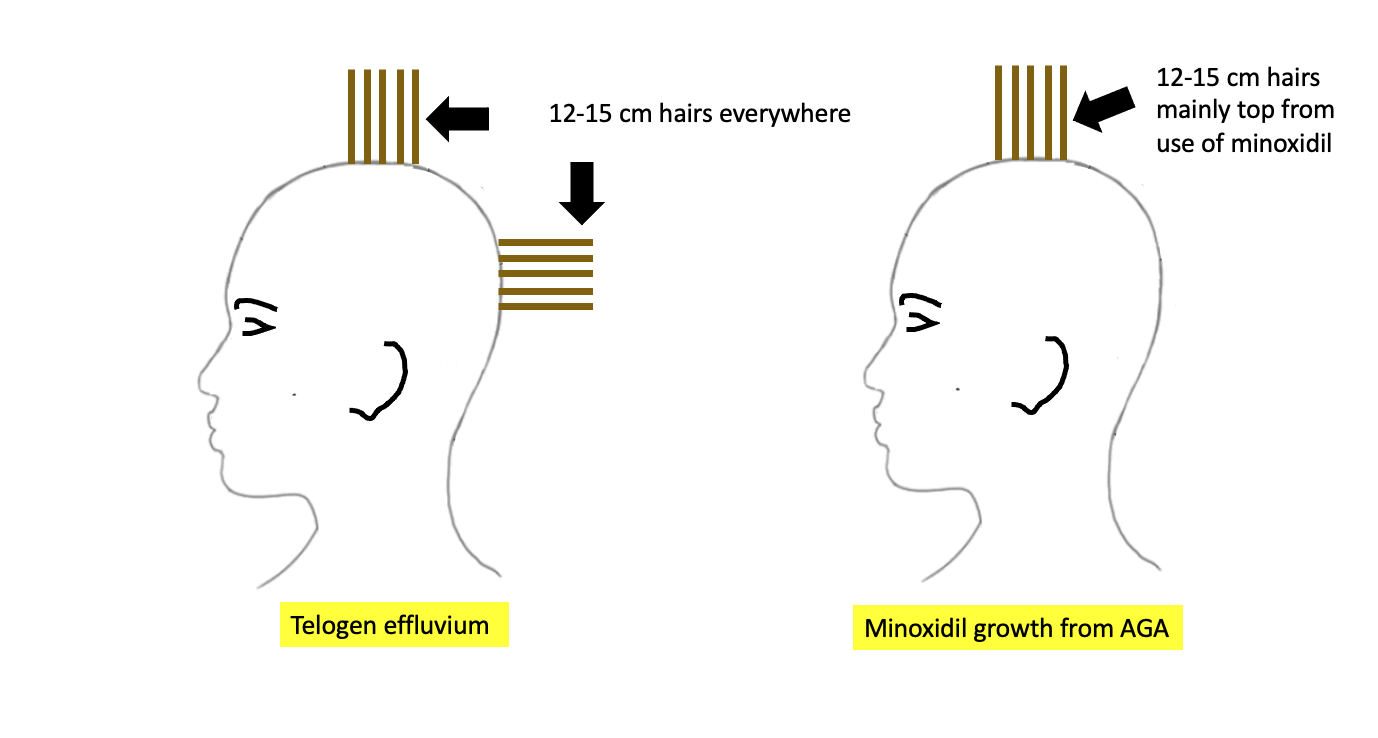
PART 2: IS THERE EVIDENCE TO SUPPORT A DIAGNOSIS OF ONGOING TELOGEN EFFLUVIUM?
It’s clear that a telogen effluvium was present in the past for you. The key question now is whether there is some kind of ongoing TE. The causes of telogen effluvium include stress, low iron, thyroid problems, medications, diets, systemic illness. So we need to understand more about these potential issues or ‘triggers’ for anyone who is shedding. In CTE, shedding can occur without a clear trigger.

Q7. Did you start and stop other medications or supplements?

Why do I need to know? Other medications and supplements can trigger shedding. Even taking some medications and herbal medications can give an ongoing shedding. I’m assuming in this scenario that minoxidil is the only product used. Clearly, if other products are used they need to be considered.

Q8. Have you had eyebrow changes, eyelashes changes, body hair changes, nail changes?

Why do I need to know? Eyebrow and eyelash changes and body hair changes don’t occur as part of AGA but can occur in TE. They can also occur in other immune based conditions. I am not suspecting any immune based issues but certainly it would be quite unexpected if you were experiencing a lot of body hair loss. If there was any eyebrow or eyelash loss, the key is to figure out if it affects both the right and left side symmetrically or whether one side is affected more. Eyebrows and eyelashes can reduce in density from telogen effluvium, alopecia areata, trichotillomania, seborrheic dermatitis as well as cicatricial alopecia (and other conditions). It would not be surprising if you have some minor eyebrow changes over the last 3 years but it should be quite symmetrical. If you do have eyebrow changes, some of the eyebrow changes could be due to the hypothyroidism.

Similarly, if there was an increase in body hair this too would be unexpected here and a possible sign of increased androgens. Increased hair on the thighs, abdomen and nipples can be a sign of hyperandrogenism and raise suspicions for PCOS (even if periods are regular). I would think that it issei's quite unlikely you would report this to be the case. This would be information that your doctors should confirm.


Q9. Have you had weight loss or weight gain? What is the current weight?

In the setting of a likely telogen effluvium, it’s important to know whether there has been weight loss that could be triggering a telogen effluvium. In addition, we need to make sure that your current weight is high enough (i.e. BMI is above 18) otherwise the result can be an ongoing TE from poor nutritional status.

In addition, weight loss and weight gain can be reflective of underlying medical conditions. So, if weight has fluctuated either way, it could be important.


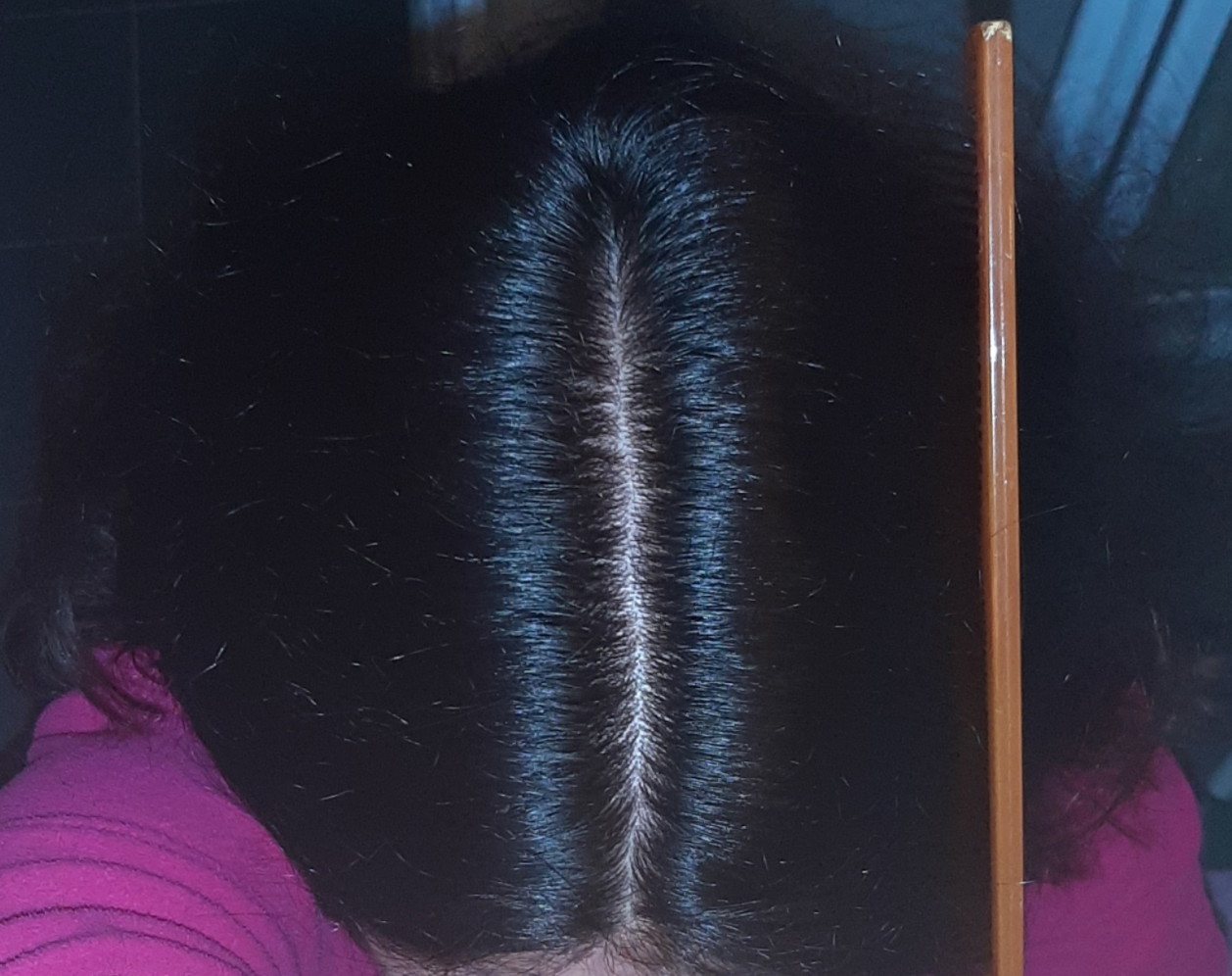
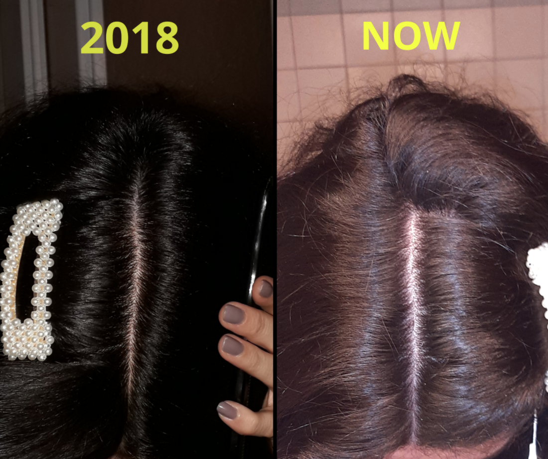
Q10. Do you have any scalp itching, burning or tenderness?

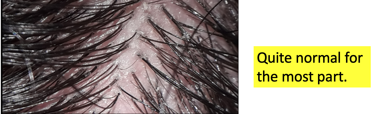
Scalp symptoms are important to know for every single patient. Your scalp looks quite healthy so I would be surprised to learn there is any itching or burning or tenderness. I’m usually not too worried about a slight among of itching from time to time, but if there was burning or tenderness in the scalp, that would be worrisome. I don’t think this is very likely to be relevant here for your case but we need to always keep in mind that there are conditions that mimic telogen effluvium.


Q11. Do you feel you are getting hair breakage?

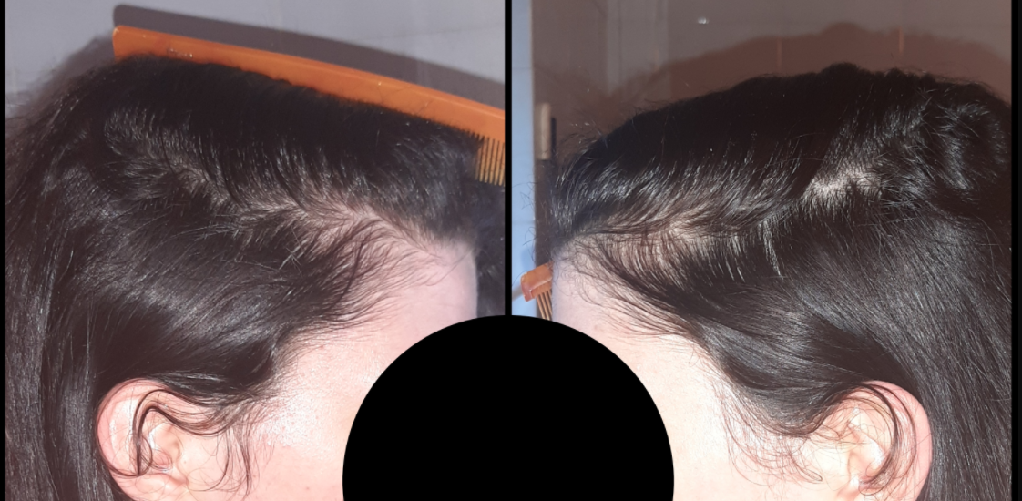
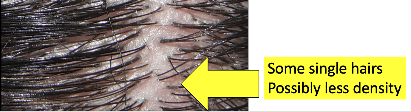
I am not appreciating much in the way of hair breakage, but it’s important to inquire about. Some patients confuse hair breakage with actual new growth so what you are seeing on the top of the scalp appears to be new growth. It’s not uncommon for patients to exclaim - “look at all my hair breakage!” In your case, this is not breakage from what I can see. However, if you felt there truly was increased breakage that would need further exploration. Breakage can come from heat and chemicals and can give the feeling of ongoing shedding and loss. A proper examination can completely exclude breakage, but I would be surprised if that’s even an issue here. your scalp and hair are healthy.

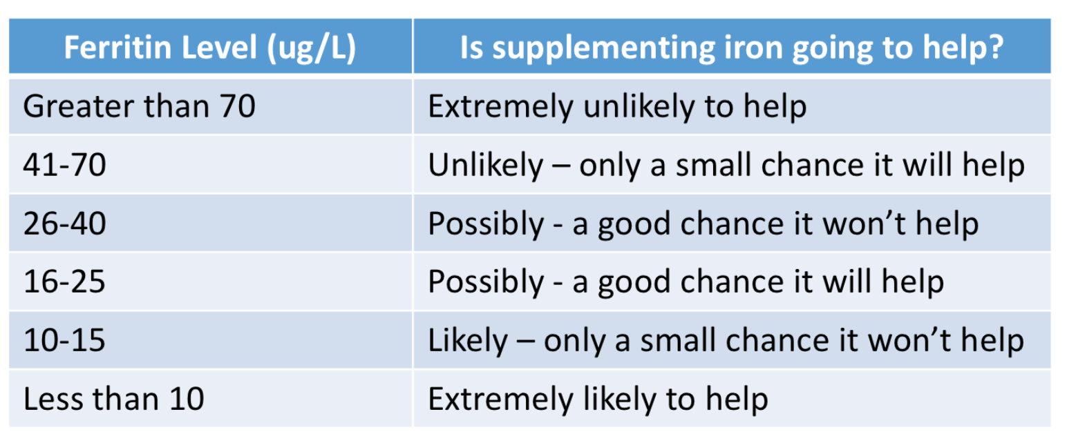
Q12. How low your ferritin dropped over this period ?

I understand that you have a history of low ferritin. It will be important to know what your ferritin level is right NOW. That is what will be relevant to ongoing shedding. A ferritin of 10 is likely associated with hair loss. A ferritin of 40 is probably not. Therefore, it will be important to know what the ferritin level is now and how it’s been changing over the last few years.

is it possible you’ve had a ferritin of 22 and it’s been responsible for your shedding for three years? Perhaps. The higher your ferritin level - the less likely it is that your iron is the culprit in your hair loss. But you have not given me the number so I don’t know the current level.

Certainly, if you have a ferritin less than 15, it’s probably a problem and probably one of the reasons why your hair is shedding. Taking iron supplements in these situations has a high likelihood of helping. However if your ferritin is currently 40, 50 or 60, it’s less likely that you currently have an iron problem.

Most women need ferritin levels in the 30-40 range for healthy hair. However, some women definitely need those levels to be higher than that and into the 50 - 70 range. If you have a history of low ferritin, it will be important for your doctors to review why you have low ferritin and how the levels have been changing over time and what they are now. Many women have low ferritin levels. However, when low ferritin is combined with low hemoglobin (which we call iron deficiency anemia) I’m much more concerned. Excessive bleeding from menstrual cycles, poor diet, celiac disease, gastrointestinal issues all need to be explored.

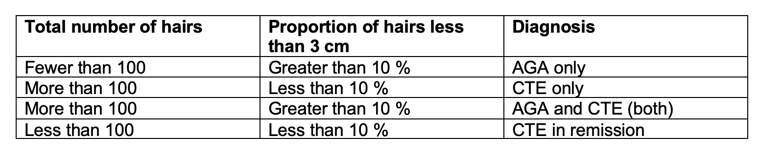
The following table gives an estimate of how likely it is that taking iron supplements will help your hair according to the ferritin levels.

ferritin levels


Q13. When did you start thyroid medications? Have your thyroid levels (TSH) changed during this time? What is the exact reason for the thyroid disease (Is it autoimmune Hashimoto’s?)

It would be helpful to understand when your thyroid disease was diagnosed and whether the TSH has been changing over this period. Some patients occasionally have fluctuation TSH levels which give shedding. Also, it would be important to know if you are on thyroid medications NOW and when this started and whether the dose has been changing over time. It is important to know the exact causes of the thyroid disease.

Q14. Were your periods always regular?

It’s always reassuring to know that your periods are regular at the present time. It would be important to know if they have always been regular of whether they have become regular just recently. It would be important to know exactly what each patient means by regular periods. Are some cycles considerably longer and some shorter than others? Having regular periods does not make it impossible to have an endocrine issue but does make it less likely. Having regular periods does not necessary mean these are ovulatory cycles but for most women they, of course, are. ovoluatory cycles.


Q15. Has your density ever come back to normal or has it just become less and less?

It would be important to know whether you feel your density and thickness ever came back to normal. When the shedding stopped for four months, did density return? Do you know the reasons why the shedding stopped for those 4 months? Was it that your iron levels finally came up? Was it that that your thyroid levels were brought back to normal? Was it that stress was reduced? Was that the period that minoxidil was started?

If your density did return completely to normal, this makes AGA much less likely during those times. It does not mean that it could not have happened or developed later but we would not expect your density to come back to 100 % normal in the setting of AGA.


Q16. Do you have other symptoms like joint pains, headaches, fatigue or rashes?

In situations like this, it’s helpful to know if the patient has any other symptoms that could suggest a systemic cause for a telogen effluvium. Issues like going pains, chronic headache, unusual fatigue or rashes on the body could prompt further work up and evaluation.


Q17. What other conditions run in the family?

it’s helpful to know what conditions run in your family. I understand your mom uses minoxidil so that tells me there is a family history of AGA. It’s helpful to know what the hair density is like in other males and females in your family. It’s also helpful to know what other diseases or medical conditions run in the family. These would include issues like psoriasis, lupus, inflammatory bowel diseases, arthritis, alopecia areata, diabetes, multiple sclerosis, early menopause, infertility and polycystic ovarian syndrome.


What Would I Recommend Next?

1. The answers to these questions above are going to be helpful and so is an evaluation of the frontal part width and back part width. That could be really informative in this case. If the part is wider in the frontal regions than the back, this suggests a possible diagnosis of AGA. If not, it still could be but TE becomes more likely.

2. Comparative trichoscopy of the middle of your scalp compared to the back of your scalp is going to be very very helpful in reaching the proper diagnosis. If the two are really different with more single hairs in the frontal and more variation in caliber noted in the frontal regions, then AGA becomes a likely diagnosis.

3. Current blood tests from ferritin and TSH and CBC are going to be important. I suspect you’ve had these tests done many times. If your ferritin is not high enough, it could be that you are shedding chronically in part from iron deficiency. The history will guide if other blood tests are helpful.

4. Finally, 5 day modified hair wash test (MHWT) is likely going to be the most helpful and least invasive next steps. Of course, a scalp biopsy can also be done but I think a modified hair wash test is probably going to be better in this case. The five day MHWT is fairly easy to do and you can do it yourself at home if you are so motivated to do so. The hair must not be washed for five days before doing the test. A 5 day modified hair wash test (MHWT) is a standardized means of assessing and quantifying hair shedding and provides information on the type of hairs being shed. It is a non-invasive method to measure hair loss by counting and identifying rinsed out (shampooed) hairs. To begin the test, a gauze is placed in the sink and hair is shampooed normally and the hair is rinsed in order to collect and trap all shed hairs on the gauze. The hair can be shampooed and rinsed repeatedly in order to remove all hairs that need to come out.

The following is helpful diagram illustrating the key steps in the 5 day hair collection.

MHWT

After gently lifting the gauze from the sink, the gauze is then dried for 3 days and then counted. In our clinic, patients who do this test mail the hairs to us, but patients can also go about counting hair. It is important that the gauze not be disturbed while drying or mailing as this gauze has dozens of small fine barely detectable hairs that must be included in the analysis. The final number of hairs as well as the proportions of 3 cm or less hairs, provides information on the relative proportions of androgenetic alopecia as well as telogen effluvium.  The proportion of broken hairs and the proportion of abnormal hair gives us a sense of the possibility of an autoimmune issue such as diffuse alopecia areata. 

The number of hairs collected in the MHWT can give a good sense of excessive shedding. Ideally, results need to be interpreted by a dermatologist who is familiar with the performance and interpretation of this test but as mentioned patients can count hairs.

a) Patients with 10% or more of hairs 3 cm or shorter and who shed fewer than 100 hairs are diagnosed as having androgenetic alopecia (AGA).

b) Patients with fewer than 10% of hairs that were 3 cm or shorter and who shed at least 100 hairs are diagnosed as having chronic telogen effluvium (CTE).

c) Patients with 10% or more of hairs that were 3 cm or shorter and who shed at least 100 hairs are diagnosed as having AGA + CTE

d) Finally patients with fewer than 10% of hairs that were 3 cm or shorter and who shed fewer than 100 hairs are diagnosed as having CTE ‘in remission.’

MHWT INTERPRETATION


Final Summary

Thanks again for sending in your question. I hope this was helpful. A few more details in this history together with a good scalp examination is going to go a long long way here in getting to the correct diagnosis (or diagnoses in the event there are two).

IFor you, I think that a 5 day modified hair wash test is going to really help give the evidence that you need to move forward with the right diagnosis. If there is any doubt, then a biopsy could further clarify but I doubt it will be needed.

The use of minoxidil could complicate things a bit in this case but only slightly. If one were to stop minoxidil and find that significantly worse shedding happens 4-6 weeks later I do think this would also be proof of some component of AGA being present.


Share This
No Comments


Share This
-->