h1.qusth1 { display: none !important; }

QUESTION OF THE WEEK

Dr. Donovan's Articles

QUESTION OF HAIR BLOGS

Filtering by Category: Scarring


Trichoscopy of Folliculitis Decalvans

Key Trichoscopic Signs of Folliculitis Decalvans

Folliculitis decalvans (FD) is a scarring alopecia which causes permanent hair loss. Patients develop red, itchy scalps that often contains pimples. Bacteria such as Staphylococcus aureus can sometimes be isolated when swabs are taken from these pimples.

A number of “trichoscopic” or “dermatoscopic” signs are suggestive of folliculitis decalvans including some I have shown here: (1) perifollicular “tubular” scaling, (2) compound follicles containing 6 or more hairs, (3) linear fibrotic bands and the (4) red “strawberry ice cream” color.

(1) Perifollicular “tubular” scaling

Trichoscopy (Scalp Dermoscopy) of Folliculitis Decalvans, Image 1. tubular scaling. The scale rides up higher on the hair shaft in folliculitis decalvans than in lichen planopilaris.

Trichoscopy (Scalp Dermoscopy) of Folliculitis Decalvans, Image 1. tubular scaling. The scale rides up higher on the hair shaft in folliculitis decalvans than in lichen planopilaris.

(2) Compound Follicles (Containing 6 or more Hairs)

Trichoscopy (Scalp Dermoscopy) of Folliculitis Decalvans, Image 2. “Compound follicles” are follicles containing more than 6 hairs emerging from a single pore.

Trichoscopy (Scalp Dermoscopy) of Folliculitis Decalvans, Image 2. “Compound follicles” are follicles containing more than 6 hairs emerging from a single pore.

(3) Linear fibrotic bands

Trichoscopy (Scalp Dermoscopy) of Folliculitis Decalvans, Image 3. Linear Fibrotic bands indicate a pattern of scarring associated with the typical starburst scaling.

Trichoscopy (Scalp Dermoscopy) of Folliculitis Decalvans, Image 3. Linear Fibrotic bands indicate a pattern of scarring associated with the typical starburst scaling.

(4) Red “Strawberry ice cream” Color.

Trichoscopy (Scalp Dermoscopy) of Folliculitis Decalvans, Image 4. Strawberry Red Color.

Trichoscopy (Scalp Dermoscopy) of Folliculitis Decalvans, Image 4. Strawberry Red Color.


Folliculitis decalvans (FD) vs Lichen planopilaris (LPP).

Folliculitis decalvans can resemble lichen planopilaris at first glance. However, it does have many differences. Compared to LPP, FD is more likely to have pustules, is more likely to bleed, is more likely to showing compound follicles or “tufting” and is more likely to have tubular scaling the climbs up the follicles (as in this image) and more likely to have these linear fibrotic bands too. Treatment for FD has been discussed in other posts but includes antibiotics, isotretinoin as well as other treatments. 


This article was written by Dr. Jeff Donovan, a Canadian and US board certified dermatologist specializing exclusively in hair loss.
Share This
No Comments

Lichen planopilaris: Classic trichoscopic findings

Classic trichoscopic findings of LPP

Classic trichoscopy of active lichen planopilaris, an immune mediated scarring alopecia is shown below.

Classic trichoscopic image from a patient with active lichen planopilaris (LPP)

Classic trichoscopic image from a patient with active lichen planopilaris (LPP)

There is redness and scale around hairs (called perifollicular erythema and perifollicular scale). Some hairs are twisted (called pili torti). The areas of scalp devoid of hairs are no longer red as the immune system has destroyed hairs in that area and has since left the area. Treatments discussed in other posts as in the following link.

Treatments for LPP: What is available?


This article was written by Dr. Jeff Donovan, a Canadian and US board certified dermatologist specializing exclusively in hair loss.
Share This
No Comments

Cetirizine (Zyrtec) in Lichen Planopilaris: Best Viewed as an Adjunct not Main Treatment

Cetirizine (Zyrtec) for Treating Lichen Planopilaris: Where does it fit in?

Antihistamines are increasingly being studied for the treatment for various types of hair loss. For the autoimmune disease known as alopecia areata for example, use of antihistamines like fexofenadine (Allegra) as well as others (i.e. ebastine) may have some treatment related benefits. In scarring alopecia. the use of antihistamines has only received a limited amount of study. Today, we will discuss the use of the antihistamine cetirizine for treating lichen planopilaris.

Lichen planopilaris is an immune medicated disease. There are a number of proposed mechanisms that lead ultimately to the disease. Cetirizine is an antihistamine medication and widely used for various types of allergy related symptoms. However, the medication may have a number of general and wide reaching effects on the immune system.


How does cetirizine work and how does it affect the immune response ?

Cetirizine is an H1 receptor antagonist. The drug minimally crosses the so called ‘blood brain barrier’ and so limited amounts actually get into the brain. This results in less sedation with cetirizine compared to any other traditional antihistamines. The 5 mg and 10 mg doses are unlikely to give sedation for most people. However, the 20 mg and 30 mg doses are much more likely to give sedation. There has been concern in recent years among long term chronic use of high doses of antihistamines on cognitive decline in patients so this needs to be taken into account when discussing high dose cetirizine as chronic therapy with patients with any medical condition.


Cetirizine has a number of potential effects to modify the immune response. These include

1. Inhibit DNA binding activity of NF-kappa B,

2. Inhibit the expression of adhesion molecules on immunocytes and endothelial cells

3. Inhibit the production of IL-8 and LTB4, two potent chemoattractants, by immune cells.

4. Induce the release of PGE2, a suppressor of antigen presentation and MHC class II expression, from monocyte/macrophages

5. Reduces the number of tryptase positive mast cells in inflammation sites.


The 2010 d’Ovidio Lichen Planopilaris Study

In 2010, d’Ovidio and colleagues studied the use of cetirizine at high doses. Rather than using 5 mg to 10 mg daily that is commonly use over the counter, the authors studied the benefits of 30 mg/daily. Twenty-one patients with lichen planopilaris (LPP) were treated with cetirizine as well as their topical steroids. in 18 or 21 patients (85.7 %) there was a reduction in redness, scaling and a reduction in extractable anagen hairs by the pull test. The authors reported that one patient developed cardiac arrhythmia after 3 months of successful treatment and dropped out of the study.

Cetirizine is an antihistamine and functions as an H1 receptor antagonist. In 2010, d’Ovidio showed that cetirizine at high doses (30 mg) could benefit some patients with lichen planopilaris. Over the counter antihistamine dosing like shown in the p…

Cetirizine is an antihistamine and functions as an H1 receptor antagonist. In 2010, d’Ovidio showed that cetirizine at high doses (30 mg) could benefit some patients with lichen planopilaris. Over the counter antihistamine dosing like shown in the phone is 10 mg.


What are the side effects of cetirizine?

Side effects of cetirizine and other information can be found in our Handout.

Cetirizine Handout for LPP

Rare side effects including heart failure, angioedema and tachycardia. These side effects are rare at low doses such as the 5 mg and 10 mg (over the counter doses). Side effects increase as one increased the dose. The 30 mg dose used in the d’OIividio study would be expected to have a greater degree of side effects than the lower doses.


Conclusion

Cetirizine may have some benefit in treating lichen planopilaris. I sometimes prescribe cetirizine as an adjective treatment in patients with persistent itching and burning who are not fully responding to mainstay topical, intralesional and oral treatments. Generally I use 5 mg or 10 mg and only rarely do I prescribe 15-20 mg. I do not typically prescribe 30 mg doses as I find side effects increase greatly. One must respect the drug interactions and contraindications for the drug (as outlined in the handout). In many ways, I view cetirizine as a helpful add on - much the same way as I view the use of low level laser therapy in this disease. I do not think in the present day that cetirizine should find itself at the top of the therapeutic ladder but certainly has a place.


Reference

d’Ovidio R et al Therapeutic hotline. Effectiveness of the association of cetirizine and topical steroids in lichen planus pilaris--an open-label clinical trial. Dermatol Ther. 2010 Sep-Oct;23(5):547-52.


Namazi MR et al. Cetirizine and allopurinol as novel weapons against cellular autoimmune disorders.Int Immunopharmacol. 2004 Mar;4(3):349-53.


This article was written by Dr. Jeff Donovan, a Canadian and US board certified dermatologist specializing exclusively in hair loss.
Share This
No Comments

The Use Metformin in Lupus: How does this apply to our Discoid LE (DLE) Patients?

Metformin for Lupus: Will it help discoid lupus?

Metformin is a well known diabetes drug. Recent evidence has suggested that metformin may have a positive impact on the treatment of some autoimmune diseases.

Metformin is well understood to reduce glucose production by the liver and to reduce absorption of glucose in the gastrointestinal tract and to increase insulin sensitivity. However, metformin may also reduce production of reactive oxygen species (ROS) which help create inflammation in lupus. The possible benefits of metformin in lupus was demonstrated in 2015 where Wang and colleagues in the journal Arthritis and Rheumatolgy showed that metformin reduce the risk of disease flares by 51 % compared to conventional treatment.

In a 2018 poster by McLeod and colleagues presented at the 2018 meeting of the American College of Rheumatology, authors showed that metformin helps patients with lupus improve control of their disease. The researchers studied 15 patients with lupus using metformin and compared to 1331 patients not using metformin. The authors found there ws a difference in disease activity in patients using metformin.

Metformin for Discoid Lupus: Will it help?

These studies are interesting as they suggest that metformin has the potential to help patients with lupus. What we don’t know yet is whether metformin will help the various types of ‘cutaneous’ lupus including discoid lupus.

Metformin may have an impact not only on autoimmune diseases (including effects on monocytes, macrophages and neutrophils), but improve gut microbiota and have an antifibrotic effect as well. These effects together make them ideal to consider in the study of scarring alopecia. We already know that drugs as pioglitazone may be helpful in lichen planopilaris.

REFERENCES

Wang et al. Neutrophil Extracellular Trap Mitochondrial DNA and Its Autoantibody in Systemic Lupus Erythematosus and a Proof-of-Concept Trial of Metformin.Arthritis Rheumatol. 2015 Dec;67(12):3190-200. doi: 10.1002/art.39296.

McLeod C, Olayemi G, Bhatia N, Migliore F, Quinet R. The Impact of Metformin on Disease Activity in Systemic Lupus Erythematosus [abstract]. Arthritis Rheumatol. 2018; 70 (suppl 10). https://acrabstracts.org/abstract/the-impact-of-metformin-on-disease-activity-in-systemic-lupus-erythematosus/. ABSTRACT NUMBER: 2645


This article was written by Dr. Jeff Donovan, a Canadian and US board certified dermatologist specializing exclusively in hair loss.
Share This
No Comments

Trichoscopy of Late DLE

Late DLE: Features


Screen Shot 2018-03-04 at 10.54.42 PM.png

Discoid lupus (DLE) is an autoimmune condition affecting the scalp and skin. It can cause permanent hair loss in affected individuals. About 5% develop systemic lupus erythematosus, an autoimmune condition with the potential to affect many organs of the body. Late scalp lesions of DLE show hyperpigmentation, white structureless areas and telangiectatic vessels,


This article was written by Dr. Jeff Donovan, a Canadian and US board certified dermatologist specializing exclusively in hair loss.
Share This
No Comments

Normal vs Compound Follicles

What is the difference?

lpp-vs-fd.png

Hairs emerge from the scalp through pores or hair follicle openings. Some pores have just one hair, but most normally have two or three hairs emerging through a single hair follicle opening. This is completely normal. 
It’s important to be able to quickly spot when something is not quite right. Most hair loss conditions lead to a reduction in the number of hairs coming out of each pore. Instead of seeing the plentiful bundles of two and three hairs one starts to see pores with either no hairs at all or just a single hair. 
Some scarring alopecias are associated an unusual feature- and that is an increase in the number of hairs coming out of the pores. When six or more hairs come out of a single opening we refer to this as a “compound” follicle. The scarring alopecias which frequently show compound follicles include folliculitis decalvans (tufted folliculitis) and sometimes acne keloidalis. It tends to be the scarring alopecias associated with neutrophils that are associated with formation of compound follicles.

Compound follicles occur because of the destructive enzymes released from the inflammatory process. These enzymes destroy tissue and promote fusion of follicles together. The photos here show compound follicles in folliculitis decalvans and single haired follicles in lichen planopilaris. 

SINGLE HAIRS IN LICHEN PLANOPILARIS 

SINGLE HAIRS IN LICHEN PLANOPILARIS


COMPOUND HAIRS IN FOLLICULITIS DECALVANS

COMPOUND HAIRS IN FOLLICULITIS DECALVANS


This article was written by Dr. Jeff Donovan, a Canadian and US board certified dermatologist specializing exclusively in hair loss.
Share This
2 Comments

Scarring alopecia: Acquired Pili torti

Acquired Pili torti

pt

Scarring alopecias are a group of diverse hair loss conditions that are associated with the presence of scar tissue in the scalp. This scar tissue can damage growing hair follicle and affect how they grow.

A common finding in many scarring alopecias is the twisting of hairs in a patient with otherwise straight hair. This “twisting” of hair is called pili torti and when it develops long after birth we call it “acquired pili torti.” This photos shows typical pili torti in a patient with frontal fibrosing alopecia. Some straight unaffected hairs can also be seen in the photo as well (bottom right). Dilated veins typical of FFA can also be seen.


This article was written by Dr. Jeff Donovan, a Canadian and US board certified dermatologist specializing exclusively in hair loss.
Share This
2 Comments

Can psoriasis also cause scarring?

Scalp Psoriasis

scalp psoriasis.png

Can psoriasis of the scalp cause permanent hair loss? Traditionally psoriasis has been classified as a non scarring alopecia - with proper treatment allowing hair to grow back.
We now understand that that is not quite accurate. Scarring alopecia lead to atrophy of the oil glands which is a small proportion of patients appears to lead on to scarring alopecia. A handful of publications (dating back to 1972) have shown the development of scarring alopecia in patients with scalp psoriasis.


References

Shuster S et al. Br J Dermatol. 1972;87:73–77.
van de Kerkhof PC, Franssen ME. Am J Clin Dermatol. 2001;2:159–165.
van de Kerkhof PC et al. Br J Dermatol. 1992;126:524–525.
Wright AL et al. Acta Derm Venereol. 1990;70:156–159.
Bardazzi F, et al. Int J Dermatol. 1999;38:765–776.


This article was written by Dr. Jeff Donovan, a Canadian and US board certified dermatologist specializing exclusively in hair loss.
Share This
No Comments

Hair dye for patients with LPP: Any Problems?

Hair dye for patients with LPP: Any Problems?

I am frequently asked if patients with lichen planopilaris (LPP) and similar scarring alopecias can dye their hair?For most people with scarring alopecia the use of permament, semipermanent or temporary hair dyes is completely safe. I always advise that patients review with their dermatologist if they feel any change in their scalps whatsoever following the salon visit or home application. Any marked change in scalp itching, burning or even new tenderness in the scalp would cause concern but fortunately this is extremely rare. 

For my patients with minor irritation from hair dye application, I sometimes recommend use of an anti-inflammatory cortisone shampoo (ie clobetasol proprionate (Clobex) shampoo) 1-24 hours before the dye is applied. Some of my patients even bring the shampoo to the salon and have the stylist use and wash it out let the normal instructions. 

All in all, most individuals with LPP don't experience any difficulties with hair dyes and no special precautions are needed.


This article was written by Dr. Jeff Donovan, a Canadian and US board certified dermatologist specializing exclusively in hair loss.
Share This
1 Comment

Scarring Alopecia: Loss of the follicular opening is a hallmark

Scarring Alopecias Cause Scarring

scarring

Scarring alopecias are hair loss conditions that are associated with the development of permanent hair loss. There are dozens of different types of scarring alopecia. 
Some scarring alopecias itch. Some don't. Some are associated with increased shedding. Some aren't. Some are red. Some aren't. Some bleed. Most don't. 


However what is common to all scarring alopecias is the disappearance of the follicular opening or "pore." The development of scar tissue beneath the skin leads to the destruction of the follicular pore opening.

The arrows point to an area of scarring in a subtle early scarring alopecia


This article was written by Dr. Jeff Donovan, a Canadian and US board certified dermatologist specializing exclusively in hair loss.
Share This
No Comments

Scarring Alopecia from Hair dyes, highlights and bleach

Chemical injury can lead to scarring alopecia

hair highlights.png

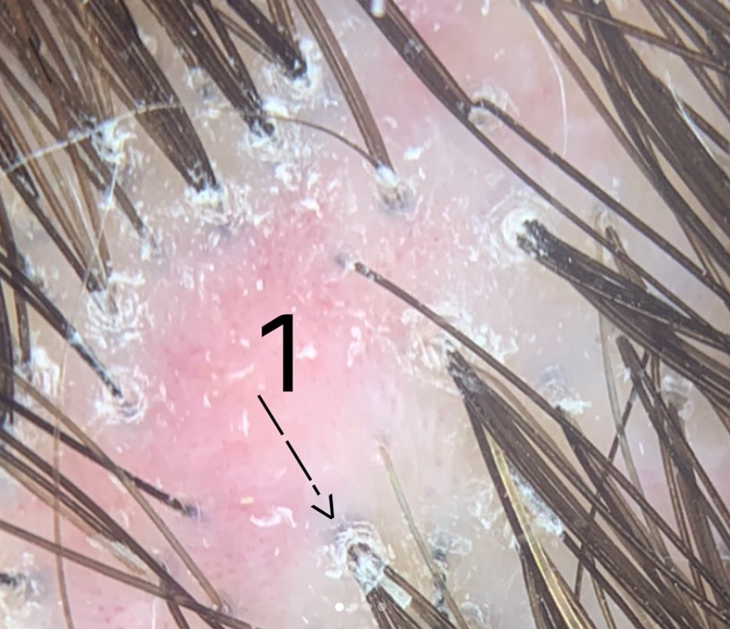
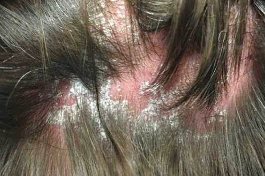
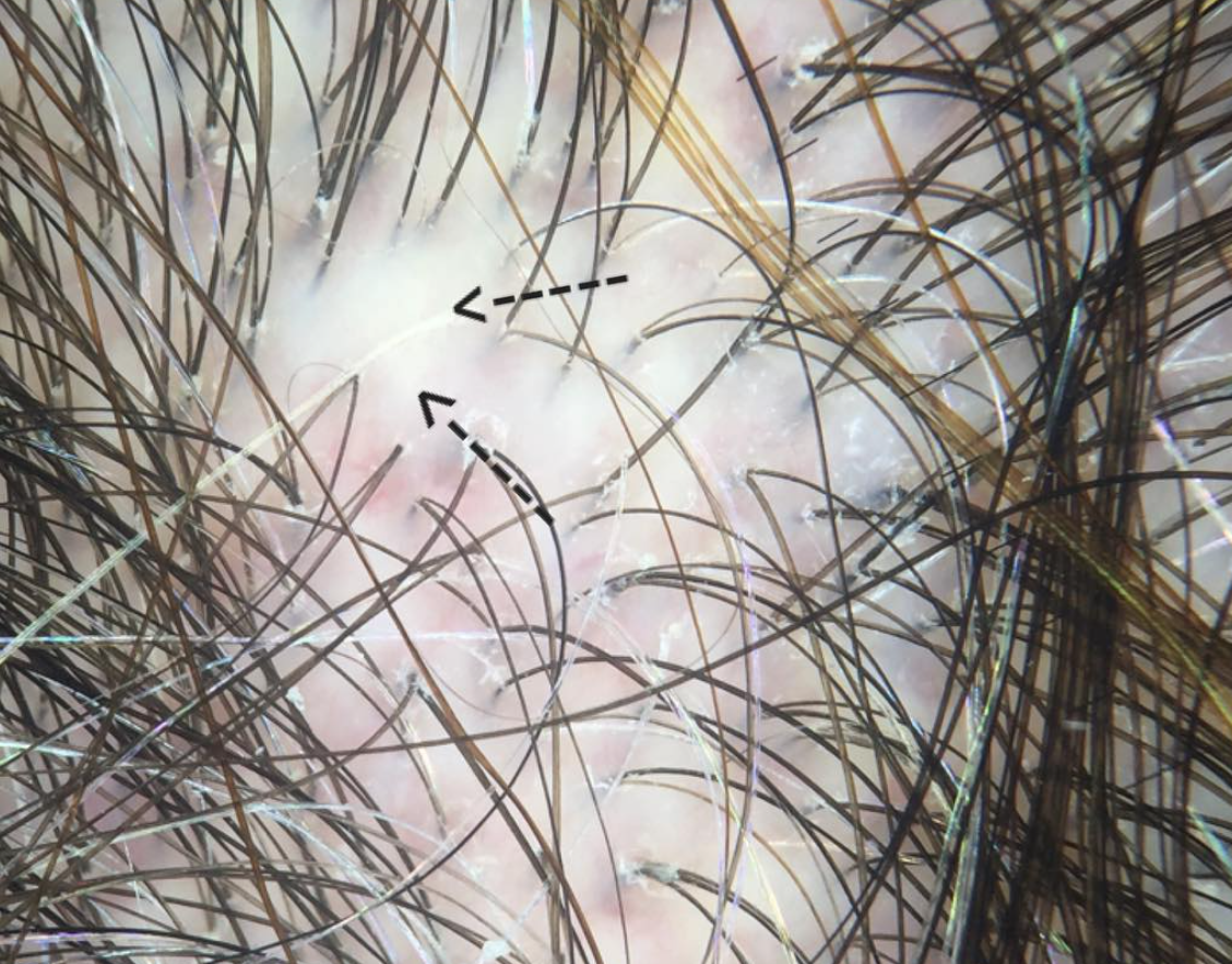
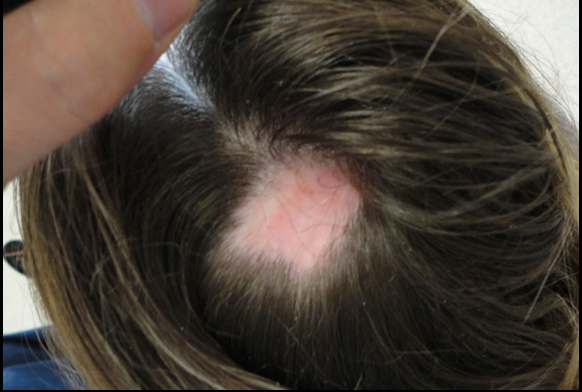
Hair dyes, highlights and bleaching can rarely lead to chemical injury. It's not common of course but the story is always the same: within seconds to minutes of applying a hair dye or highlights, the patient complains of intense burning and/or pain and requests the product to be removed. Hours to days later hair loss starts and within a week or two the patient has permanent hair loss (such as shown in the figure to the right). I have seen this type of scalp injury phenomenon many, many times and I do even feel that it is increasing world-wide. 


Management Chemical Injury to the Scalp

The most important thing to do in these situations of potential chemical irritation is remove whatever chemical could be causing the reaction. The dye or highlight must be removed, neutralized and rinsed off.  In my opinion a dermatologist should be consulted for management and monitoring. Rarely skin necrosis can occur from ehuberant reactions. One can not predict on day 1 whether the patient will have hair loss and whether any hair loss will be permanent. This will not be clear until day 14-28. In the short term one must management skin health, prevent infection, and limit and control inflammation. These are within the skills of a dermatologist.  A biopsy may be considered to determined the type of inflammation and evaluate for scarring if it is unclear. 


Hair transplantation or Surgical Correction: Best methods for Camouflaging Chemical Injury

Too often I hear it said in these scarring alopecias that a biopsy was done and because the biopsy said the disease was inactive the patient proceeded to surgery. Keep in mind that we determine if a scarring alopecia is inactive by simply following what it does over time. Relying on a biopsy alone to determine if it is acitve is not a good idea for most people. If the area of hair loss has not changed at all in any way shape or form (same size area) and is not itchy and has no burning or pain thena biopsy supports it is inactive.  Even if a biopsy says the scarring alopecia is inactive but the area is expanding over time and is itchy or red... it is not inactive. This is a common scenario and a common error in managing scarring alopecia.

One needs to wait 12-24 months for a scarring alopecia before surgery. Photos need to be done every 2-3 months in my opinion even for chemical burn related hair loss. If the photos look the same when placed side by side over a one year period, one can say the scarring alopecia is probably quiet.  Rarely, this can be shortened to 6 months for chemical injury but one year is a safer waiting period to be confident there is no evidence of a slowly progressive scarring alopecia in evolution. 


Is waiting really necessary when planning surgery in scarring alopecia?

All this background waiting and monitoring needs to be done before surgery. It sounds excessive and time consuming and unnecessary- but it is far from it. Surgery for scarring alopecia can be highly successful provided it's done in the right patient. Too often, it is not done on the right patient... and then it does not work well or does not work at all and physicians, patients and the medical community as a whole loses confidence in the value of surgical restoration options.


Options for Restoration

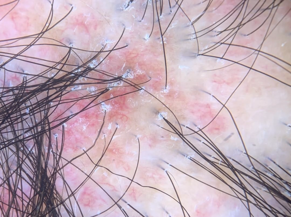
 The only way to restore the appearance is surgical. Medical options do not help improve density once the area is permanently scarred. If the area is small surgery via a plastic surgeon can be a great option. Many burns from hair dyes are in the form of small coin shaped patches. A flap (rotational flap etc) can work wonders and may be superior to hair transplanting. For this a surgeon is needed with skill in such flaps. The above patient would be a good candidate for a flap.

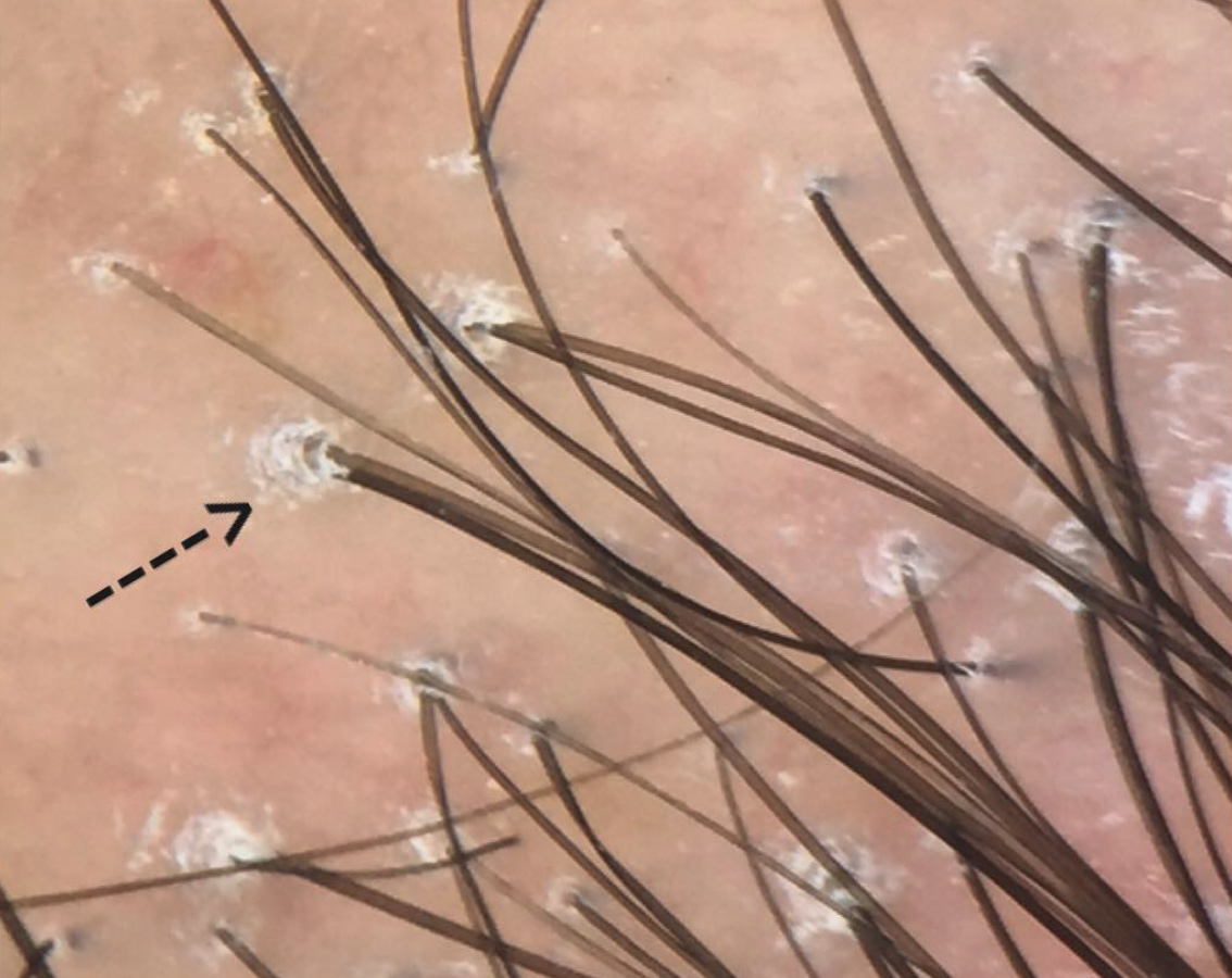
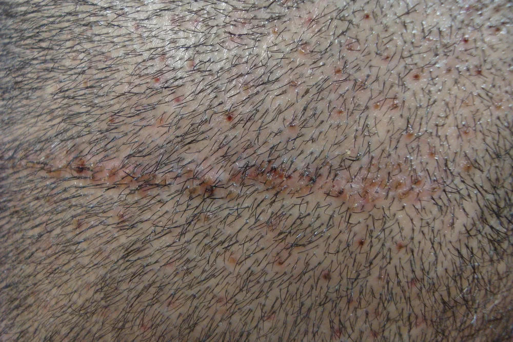
For hair loss that occurs more diffusely (and not in the above mentioned classic hair dye chemical burn patches), hair transplants can sometimes ca a good option. In my opinion, the key factor in choosing a surgeon is their experience and dedication to hair transplantation. The actual credentials is not so important to me and some of the world's top surgeons are a range of family physicians, dermatologists, plastic surgeons, former emergency room physicians. If her or she is dedicated soley to hair transplanting and has performed a large number surgeries and has been doing it for many years and has a good before and after album of scar procedures, then it may be worth a visit to speak to that surgeon. 


This article was written by Dr. Jeff Donovan, a Canadian and US board certified dermatologist specializing exclusively in hair loss.
Share This
No Comments

Lichen planopilaris: A closer look at "follicular hyperkeratosis"

Follicular hyperkeratosis = Perifollicular scale

Lichen planopilaris (also known as "LPP") is a scarring hair loss condition. Individuls affected by the condition develop hair loss, increased hair shedding along with scalp itching, burning and pain.


Scaling in LPP

Screen Shot 2017-10-14 at 8.24.15 AM.png

An up close examination with dermoscopy is shown here and shows classic features including white scale around hairs in early stages. This scale is known as "perifollicular scale" or "follicular hyperkeratosis." This scale is often prominent in active stages of the disease. It can be reduced by treatment and even reduced to some extent by a good shampooing of the scalp.


Treatments for LPP

Treatments include topical steroids, steroid injections, topical calcineurin inhibitors, and oral treatments such as doxycycline, hydroxychloroquine (Plaquenil), methotrexate, mycophenolate mofetil, cyclosporine, and others. Lasers, including the 308 nm excimer laser may also provide benefit.


This article was written by Dr. Jeff Donovan, a Canadian and US board certified dermatologist specializing exclusively in hair loss.
Share This
No Comments

Partially Treated Lichen Planopilaris

 Scale Gone, Redness Remains

Lichen planopilaris (LPP) is a scarring hair loss condition. The goal of treating LPP is to stop the condition. Successful treatment is associated with a halting of hair loss but also with an improvement in the symptoms and signs of the disease. Patients will notice a reduction in itching and burning and clinically there will be an improvement in scaling and redness around hairs. Sometimes scaling is the first to improve and improvements in redness happen later. This picture shows a patient with partially treated lichen planopilaris. The disease is still active although scaling has improved. The patient's itching has also improved.


This article was written by Dr. Jeff Donovan, a Canadian and US board certified dermatologist specializing exclusively in hair loss.
Share This
No Comments

AGA or LPP: Who is right?

In many fields of medicine, the pathology report provides the final answer as to a patient's diagnosis. We're most familiar with this for example with cancer diagnoses. It comes as a surprise for many patients that scalp biopsy reports are sometimes not so definitive.


Differentiating AGA and LPP

A great example is the diagnosis of early androgenetic alopecia (AGA and early lichen planopilaris (LPP). Sometimes it is pretty clear cut - but not always. Sometimes a diagnosis of LPP is made and the patient really has AGA. Sometimes (although much less commonly) a diagnosis of AGA is made and the patient really has LPP.


LPP: Brief Overview

Lichen planopilaris (LPP) is a scarring alopecia that typically starts with scalp symptoms such as itching and burning. Sometimes the scalp is quite tender in areas. Shedding is often present as well. LPP affects similar areas to androgenetic alopecia (female pattern thinning) so it is a close mimicker. In the early stages, some scalp redness may be present and inflammation may be seen around the hairs clinically. 


AGA: Brief Overview

Androgenetic alopecia (AGA) also starts with shedding. There can be a hint of itching/tingling but not too often. Usually the front of the scalp is more affected by hair loss than the back. 


Biopsies: Helpful or not?

A biopsy can be very helpful provided it is read by an experienced dermatopathologist. Traditionally we have thought of AGA as "non inflammatory" and "non scarring" so one might not think that inflammation and scarring should be present on the biopsy. We know now that's not completely true.  Inflammatory infiltrates are present in AGA in the upper hair follicle and so is loose perifollicular fibrosis. In LPP biopsies, inflammation is also present in the upper hair follicle but it specifically appears to be attacking the hair follicle outer root sheath. (We call this "lichenoid" change). To differentiate AGA and LPP one needs to direct their attention to this specific change in the actual hair follicle. When this specific immune attack is seen, one needs to consider LPP over AGA. Also the amount of perifollicular fibrosis is usually greater as LPP advances. LPP may have other changes in the skin as well that help differentiate it from AGA.

So by biopsy,  androgenetic alopecia and LPP can be confused as both can have inflammation (perifollicular inflammation in the isthmus) and both can have scarring (perifollicular fibrosis).  An experienced dermatopathologist can sort this out. 


So how does one resolve this? Does the patient have AGA or LPP?

One needs to take into account the patient's entire story. If a physician just biopsies every patient that comes into the office, I can guarantee one will make a whole lot more diagnoses of LPP than truly are present. I'm a big believer in this - even though LPP is under diagnosed in the world!  But by listening to the patient's entire story, and examining the scalp and reviewing what the biopsy shows (not just the final read out on the bottom line), one can usually get a fairly good sense. However in rare cases - time is the best judge as a missed case of LPP will likely declare itself over time.


This article was written by Dr. Jeff Donovan, a Canadian and US board certified dermatologist specializing exclusively in hair loss.
Share This
2 Comments

Hormonal Changes in LPP and FFA

The Clevland clinic performed a new study that  showed that hormone abnormalities can be common in lichen planopilaris (LPP) and frontal fibrosing alopecia (FFA).

A proportion of patients with LPP were found to have "androgen excess" (increased levels of the male hormones). However there was a portion of patients with FFA that were shown to have "androgen deficiency." This did not prove to be true of everyone - but was a trend seen in a large proportion.

This study is surprising, especially when considering that antiandrogens are helpful in FFA. It may however provide insight into differences between LPP and FFA. More studies are needed. For now, I agree with the authors conclusions that hormone levels are important to order in women with these scarring alopecias.


Reference
Ranasinghe GC, et al.Prevalence of hormonal and endocrine dysfunction in patients with lichen planopilaris (LPP): A retrospective data analysis of 168 patients.  J Am Acad Dermatol. 2017.


This article was written by Dr. Jeff Donovan, a Canadian and US board certified dermatologist specializing exclusively in hair loss.
Share This
No Comments

Can alopecia areata occur at the location of a hair transplant?

Can alopecia areata develop at the site of a hair transplant?
 

Alopecia areata affects 2 % of the world. It is an autoimmune hair loss condition whereby the immune system targets the hair follicle causing it to fall out. Alopecia areata can develop anywhere on the scalp - and anywhere on the body where there is hair such as eyebrows, lashes, etc. 

In previous published reports, alopecia has been documented to occur at the site of a hair transplant. However, proving there is a direct link between the two is challenging. Alopecia areata usually develops in most people without trauma or injury.
 

Is a link plausible for some?

It is certainly not impossible that some sort of a more direct link could exist between alopecia and injury. I have many patients with autoimmune type reactions in the donor area following hair transplantation - including alopecia areata and lichen planopilaris. It's just really difficult to prove a direct association.

This photo show "black dots" and vellus hairs that are typical of the scalp in patients with alopecia areata. The photo also shows the scar from a previous hair transplant done using follicular unit strip surgery (FUSS).


This article was written by Dr. Jeff Donovan, a Canadian and US board certified dermatologist specializing exclusively in hair loss.
Share This
No Comments

Lichen planopilaris - a type of scarring hair loss condition

Lichen planopilaris (LPP) is a type of scarring hair loss condition. The cause is unknown although several treatments are possible to try to stop the condition from progressively destroying hair follicles.


This article was written by Dr. Jeff Donovan, a Canadian and US board certified dermatologist specializing exclusively in hair loss.
Share This
No Comments

Hair Transplantation in Scars - Is it Possible?

New hair follicles transplanted into a scar.

New hair follicles transplanted into a scar.

Hair Transplantation in Scars

A small number of men and women have scars in the scalp. This can be scars from an previous accident, scars from a previous surgery in the scalp, scars from a previous hair transplant surgery or scars from a scarring hair loss problem. Regardless of the cause of scars, patients visiting the office want to know:

Is it possible to transplant hairs into a scar to improve the appearance of the scar?

In many cases, the answer is "yes" and certainly we've transplanted a wide variety of scarring issues.  But the decision as to whether someone is a good candidate for repairing a scar with a hair transplant depends on a number of factors:

1.  Thickness of the scar. Sometimes scars can be very thick and this compromises the ability of hairs to grow in the scar. A number of techniques can be used to improve a thick scar and make it more receptive to receiving new hair follicles. This includes thinning the scar slightly with steroid injections and debulking the scar by removing bits of the scar before putting in new hair follicles. 

2. Thinness of the scar. As surprising as it sounds, some scars are too thin to be successfully transplanted. The medical term for such thinning is 'atrophy.'  Some scars are too atrophic to receive new hair follicles. When we do decide to proceed and transplant atrophic scars, we use a number of techniques to improve the growth and 'uptake' of transplanted hairs but some severely atrophic scars are challenging to transplant. 

3. Blood supply to the scar. Before a transplant is performed in a scar, I usually test the blood supply. Sometimes a scarring process in the scar can reduce the blood supply to area and in turn reduce the chances of having successful uptake of hair follicles. Certain techniques, such as pre-operative and post-operative use of minoxidil to improve blood flow into a scarred area may be beneficial and is something we often recommend.

In general, our experience has been that transplanting scars can be very successful and patients are really happy to have their scars less noticeable.  It does require experience and a certain degree of art to best camouflage scars with a hair transplants.  We frequently perform smaller sessions (fewer grafts), space the grafts out a bit further and wait longer between sessions if another session is required.  A few other modifications are done during the actual procedures as well. All of these considerations are important and help improve the likelihood that individuals with a scar will achieve outstanding results.


This article was written by Dr. Jeff Donovan, a Canadian and US board certified dermatologist specializing exclusively in hair loss.
Share This
No Comments

Can Patients with Trichotillomania have a Hair Transplant?

Trichotillomania: Is it possible to have a Hair Transplant?

Trichotillomania is a hair loss disorder whereby individuals pull out their own hair.  About 1-2 percent of the population meet the diagnostic criteria at some point in their lives.  The condition is classified as an impulse control disorder.  A previous blog discussed the features of this condition.

In the early stages of the condition, hair regrowth is possible if the patient can be helped (either with medications or psychotherapy) to stop pulling.  If the pulling goes on long enough, the resultant hair loss may be permanent.  This is because scars develop around the damaged hair follicles and these scars block further hair growth.

Patients with trichotillomania often ask if a hair transplant is possible.  In some cases it can ben possible, but certainly not in all cases.   Generally, I look for four features to be present in order to determine if a patient with trichotillomania can have a transplant:


Candidacy for Hair Transplantation in Patients with Trichotillomania

1. The patient has not had the compulsion to pull their hair for at least 1 year

2. Patient has no ongoing scalp symptoms like itching, burning, pain or tingling

3. The area of hair loss has not enlarged over a 1-2 year period.

4. The patient is medically fit, has a good donor supply of hair, and is over 18 years of age.


These are general guidelines that I use in my practice which have been very helpful. Patients with ongoing symptoms like itching in the scalp and who have ongoing compulusion to pull, twist of pluck hairs are not good candidates because the the transplanted hair may be ultimately pulled out again.  


This article was written by Dr. Jeff Donovan, a Canadian and US board certified dermatologist specializing exclusively in hair loss.
Share This
2 Comments

Hair Transplantation in Scars


Hair loss can sometimes be associated with scarring.  For example, some patients I see have hair loss from previous cosmetic surgery, including facelifts. Some patients have scarring from previous burns or trauma to the scalp.  In addition, there is an entire group of hair diseases known as “scarring alopecias.”  Can a hair transplant be performed into scars? Will the transplant be successful?

One of miraculous aspects of hair transplantation is that hair follicles can grow in scar tissue.  For patients with scars from cosmetic surgery, I will restore hair density in a single session.  For those patients with scar tissue from scalp disease, or other complex scarring processes, I will wait 2 years after the disease has been declared quiet or "burnt out."  At this point, I will generally recommend placing a few test grafts in to an area and observing the survival of these grafts over a 6 month period.  If the graft survival rate is high, I will proceed with surgery.  If the graft survival is poor, I will either declare the patient not to be a candidate for surgery, or place the patient on additional treatment to ensure the disease has truly become burnt out.


This article was written by Dr. Jeff Donovan, a Canadian and US board certified dermatologist specializing exclusively in hair loss.
Share This
No Comments


Share This
-->