h1.qusth1 { display: none !important; }

QUESTION OF THE WEEK

Dr. Donovan's Articles

QUESTION OF HAIR BLOGS

Filtering by Category: Biopsies


Cicatricial Marginal Alopecia: Your traction alopecia patients will thank you!

Not all ‘Traction Alopecia’ is Actually Traction Alopecia

Traction alopecia is a form of hair loss that occurs due to pulling of hair. Diagnosing traction alopecia sounds easy but surprisingly there are a great number of mimicking conditions that can fool the hair specialist.

Frontal traction alopecia refers to hair loss in the frontal hairline that is due to traction. Often the temples are affected but any part of the frontal hairline, temples and area around the ears can be affected. Often the hairs in the very frontal hairline are unaffected leading to the appearance of a so called “fringe” sign:

Classic ‘fringe’ sign in a patient with traction alopecia. The fringe refers to the fringe of hair in the frontal hairline.

Classic ‘fringe’ sign in a patient with traction alopecia. The fringe refers to the fringe of hair in the frontal hairline.

Cicatricial Marginal Alopecia (CMA)

There are times when patients who present with what seems to be traction alopecia tell us that they couldn’t possibly have traction alopecia. These are the patients who tell us that they have worn their hair fairly natural for years and that a diagnosis of traction alopecia just makes no sense to them. These are the patients that politely stare at us when we tell them to be careful how they style their hair and to be carefully to avoid heat or chemicals. When a hair specialist wants to make a diagnosis of traction alopecia but realizes the patient’s story just does not add up to give a convincing story of traction alopecia - the diagnosis of cicatricial marginal alopecia (CMA) must be considered.

The Differential Diagnosis of Frontal Hair Loss: What’s a specialist to consider anyways?

Of course, the diligent hair specialist considers many things in the differential of frontal traction alopecia like presentations including

1. Traction alopecia

2. Cicatricial Marginal Alopecia

3. Frontal fibrosing alopecia

4. Discoid lupus

5. Androgenetic alopecia

6. Telogen effluvium

7. Alopecia Areata

8. Trichotillomania

Cicatricial Marginal Alopecia: A Traction Alopecia Like Alopecia Without A Traction History

It was Dr Lynn Goldberg in Boston who put forth the notion of cicatricial marginal alopecia. She described 15 patients who presented with hair loss in a typical traction alopecia like pattern. Information pertaining to whether or not the patient relaxed or straightened the hair was available In 12 patients. 6 of the 12 patients gave a history of relaxing the hair or straightening the hair. For the other 6 other patients there was no such history. In other words, in 50 % of patients with frontal 'traction alopecia-like” hair loss a history of true traction styling practices were not present. These patients still had some degree of scarring on their biopsies indicating that this too could be a scarring type of hair loss. 

Treatment of CMA involves topical or oral minoxidil combined with topical and/or intralesional steroids. In some patients use of agents like oral doxycycline or topical tacrolimus can be helpful.

Summary and Key Lessons

As soon as we let open our mouths to pronounce the words traction alopecia, we must say in the same breath “or a traction alopecia like mimickers.” Could my patient have traction alopecia or a “traction alopecia like mimicker.”

Cicatricial Marginal Alopecia is one of these closely related mimickers. I like to refer to it as cicatrical marginal alopecia to honour my great colleague Dr Goldberg and so this is what I write in all my letters and consultation notes to other physicians. In my mind, I say the patient has a Traction Alopecia Like Alopecia Without a Traction History because it helps me remember the key elements of this presentation.

Reference

Goldberg L. Cicatricial Marginal Alopecia: Is It All Traction? Br J Dermatol 2009 Jan;160(1):62-8.


This article was written by Dr. Jeff Donovan, a Canadian and US board certified dermatologist specializing exclusively in hair loss.
Share This
No Comments

What cancers metastasize to the scalp?

Lung cancer tops the list of cancers that metastasize to the scalp


Focal alopecia refers to hair loss in a single area of the scalp. Common causes of focal alopecia include alopecia areata, tine capitis, infections, trauma, trichotillomania. Cancer is another cause of focal alopecia - albeit an uncommon one.

Scalp metastases refer to cancer the started in another organ and then the cancer spread to other parts of the body, including the scalp. Less than 2 % of patients that are known to have metastatic cancer will experience scalp metastases.


What are the most common cancers that metastasize to the scalp?

Lung cancer is the most common cancer that is associated with scalp metastases. Of all metastases to the scalp, lung cancer is the most common at 24 % followed by colon (11 %), liver (8 %) and breast (8%). Kidney and ovary remain other causes on the list. In 30 % of cases, the exact origin can’t be precisely determined. There are many types of ‘lung cancer’ and it remains debated as to which of the types is really the most likely contributor to scalp metastases (adenocarcinoma, large cell, small cell, squamous).


What are the clues that an area of hair loss may actually represent a metastasis?

Scalp metastases can be challenging to diagnose in the early stages. Sometimes they aren't large and sometimes they get overlooked. They may appear as papule or nodules, or firm indurated plaques or ulcers or as an area closely mimicking alopecia areata. They may simply appear as an area that looks like an infection. In many cases, they are red from dilated blood vessels - and sometimes hemorrhagic from localized blood clots in the area.  “Alopecia neoplastica” is a term that refers to hair loss associated with destruction of hair follicles. It is accompanied by red, indurated skin with dilated blood vessels and sometimes ulceration. In most cases, alopecia neoplastica represents hematologenous spread (spread in the blood stream) of a breast cancer. In some cases of metastastic scalp lesions, the areas of involvement are associated with no symptoms which in turn adds to the delay in diagnosis. In fact, there can be a delay in diagnosis for many patients with some studies showing a delay in proper diagnosis of 4–10 months following the time they are first noticed.

The identification of a scalp metastasis can rarely be the very first indication that the patient has a cancer inside the body. This is not common and in most cases it is already known that the patient has a cancer somewhere in the body. Overall, a study by Lookingbill and colleagues of 7316 cancer patients found that 0.8 % of patients had a skin lesion that represented the presenting sign of the cancer inside the body.

Key Conclusions and Summary

Scalp metastases can be challenging to diagnose in the early stages and diagnosis of a scalp metastasis ALWAYS comes from a biopsy. One can never look at a skin lesion and know with 100 % certainty that it is a metastasis from a cancer somewhere in the body. One only reaches that conclusion after a biopsy is done.

It’s often a good idea for a doctor to consider performing scalp biopsy when a patient has a focal areas of hair loss that does not improve over time - especially in patients over 45 years of age. In situations where the diagnosis is completely clea…

It’s often a good idea for a doctor to consider performing scalp biopsy when a patient has a focal areas of hair loss that does not improve over time - especially in patients over 45 years of age. In situations where the diagnosis is completely clear simply by examining the scalp, a biopsy is not necessary.


In general, a biopsy should at least be considered in the following situations:

1) A patient with a history of CANCER AT ANYTIME IN THE PAST who presents with a solitary area of hair loss (i.e. affecting a single localized area of the scalp).

2) A patient with CURRENT CANCER DIAGNOSIS who presents with a solitary area of hair loss (affecting a single localized area of the scalp).

3) A patient over 45 years of age who presents with a solitary LOCALIZED, RED PATCH OF HAIR LOSS ON THE SCALP that has persisted for 3 or more months.


Most patients with previous cancer diagnoses or who are dealing with cancer at the present time do not have a scalp metastasis as the reason for their hair problems when they present to the hair doctor’s office. However, it must always be on the doctor’s radar - especially when the patient has a solitary patch of hair loss (a single spot).

Persistent solitary patches of hair loss, especially when red, are extremely important to consider biopsying in anyone over 45.


Reference

Chiu CS, Lin CY, Kuo TT, et al: Malignant cutaneous tumors of the scalp: a study of demographic characteristics and histologic distributions of 398 Taiwanese patients. J Am Acad Dermatol. 56:448–452. 2007.

Frey L, Vetter-Kauczok C, Gesierich A, Bröcker EB and Ugurel S: Cutaneous metastases as the first clinical sign of metastatic gastric carcinoma. J Dtsch Dermatol Ges. 7:893–895. 2009

Kim HJ, Min HG and Lee ES: Alopecia neoplastica in a patient with gastric carcinoma. Br J Dermatol. 141:1122–1124. 1999.  

Lifshitz OH, Berlin JM, Taylor JS and Bergfeld WF: Metastatic gastric adenocarcinoma presenting as an enlarging plaque on the scalp. Cutis. 76:194–196. 2005

Lookingbill D.P., N. Spangler, F.M. SextonSkin involvement as the presenting sign of internal carcinoma. A retrospective study of 7316 cancer patients. J. Am. Acad. Dermatol., 22 (1) (1990), pp. 19-26


This article was written by Dr. Jeff Donovan, a Canadian and US board certified dermatologist specializing exclusively in hair loss.
Share This
No Comments

The Essential Components of the Scalp Biopsy Report

What should pathologists be including in the biopsy report?

Interpreting scalp biopsies is not easy. It requires specific expertise and I’m fortunate to work with amazing pathologists. Every day, I receive biopsy reports from around the world for patients that want advice with their hair. Some reports are so clear, I’m mesmerized and feel like I’m right there sitting at the microscope. Some reports are so nice I want to print them out and frame them. 

And then there are reports that look nice but aren’t nice. They have a lot information in them but lack the key information to help you decide as a clinician what the real diagnosis is. Last week, I read a long report where the pathologist was trying to decide whether the case was a case of lichen planopilaris …. or not!! The report discussed a lot of good information - but lacked information about the presence or absence of sebaceous glands which probably would have solidified the diagnosis. I felt frustrated but hopefully when I see the patient in a few weeks we’ll know if it’s a scarring alopecia simply by looking at the scalp. Sometimes in these types of cases, a biopsy was not even needed. Biopsy reports that are lacking key information don’t matter if the diagnosis is obvious. But these sorts of biopsies are frustrating in cases where the diagnosis is challenging. It’s like reading a good book and being three-quarters of the way through the book only to find that the publisher of the book forgot to print a few chapters. 

So what makes a good report, anyways?

There’s one main criteria that makes up a good report. The report must be helpful to the clinician and helps rule out other issues.Below, I’ll outline the key parts of a scalp biopsy.

SECTION 1: What’s the quality of the biopsy? and How was the biopsy processed?

The first section of the biopsy usually begins by confirming the size of the biopsy and whether the biopsy was taken properly by the doctor and was deep enough and whether it was taken parallel to the hairs or whether the clinician took it in a manner that cut through all the hairs. When I read reports from other physicians, I like knowing about the size of the biopsy. If a 2 mm biopsy was taken instead of the standard 4 mm biopsy, this is important to know. A tiny 2 mm biopsy might be taken to try to limit the patient’s chance of developing a visible scar - but that tiny biopsy is going to be limited in the amount of information it can give. 

The report then describes whether the biopsy was obtained with vertical or horizontal sections by the lab. That’s helpful to put things into context for the reader of the report. If the biopsy was taken using horizontal sections, it’s going to be possible to count the number of hairs fairly accurately and so I’ll be waiting for some information on hair follicle numbers as I read more. If the biopsy was processed using vertical sections, this information is simply not possible and I won’t be expecting this as a read the report. 

SECTION 2: What’s the pathologist’s feeling about the hair density? 

It’s nice to get a sense from the start if the pathologist feels hair numbers are reduced compared to normal. If horizontal sections were used, the pathologist may actually count the total number of hairs he or she sees and make a comment about density. If vertical sections are used, accurate measurements of density are not going to be possible, but the astute pathologist may still make a comment if density appears reduced. 

SECTION 3. Is there INFLAMMATION in the biopsy? If so, where exactly is it .. and where exactly is it not?

Inflammation can be very relevant to the diagnosis. But the key information needed from the pathologist is where exactly is this inflammation. 

a) Is the inflammation perifollicular? and if so is found at the level of the isthmus, infundibulum or bulb .. or even deeper? 

Perifollicular inflammation is common in many conditions and in and of itself doesn’t mean much. Perifollicular inflammation around the isthmus can be seen in scarring alopecias and in androgenetic alopecia too. Inflammation around the bulb is often seen in alopecia areata. Deeper inflammation (below the bulb) can be seen in dissecting cellulitis, various infiltrative conditions and in the diseases of the fat. 

b) If there is inflammation, is any ‘lichenoid’ in nature?

if the pathologists does. find inflammation at the level of the isthmus, it’s helpful to know if that inflammation is causing hair follicle keratinocytes to die or not. Evidence of such “lichenoid” involvement is important. It’s not always seen even in cases of lichen planopilaris - but the presence of epithelial cell necrosis is important to know about if it’s present. 

c) How much inflammation is present?

It’s helpful to get a sense if there is just a bit of inflammation or whether the biopsy is plugged tight with inflammation. In the case of a scarring alopecia, severe degree of inflammation are probably best handled with systemic treatments (although more studies are needed in that regard). 

d) Is there any other inflammation seen anywhere else?

The pathologist may not comment on other inflammation but we hope he or she has looked for it. Inflammation around blood vessel (perivascular inflammation) and around eccrine glands (perieccrine inflammation) is helpful in some autoimmune issues. 

SECTION 4. Is there FIBROSIS (scarring) in the biopsy? If so, where exactly is it .. and where exactly is it not?

It’s important to have a sense of whether or not the biopsy shows evidence of increased scar tissue. We call. such scarring fibrosis. In scarring alopecias like lichen planopilaris, the scar tissue is initially just around the hair follicles and the majority of the dermis is not involved with any scarring whatsoever. In other scarring  alopecias like folliculitis decalvans, the areas between the follicles are also involved with scarring. 

SECTION 5. Are the sebaceous glands reduced or affected in any way?

Unfortunately, comments about the sebaceous glands are frequently omitted from many scalp biopsy reports I see. Sometimes it does not matter. The biopsy is a biopsy of scarring alopecia and all the other features in the report go together to say the same thing - this patient has scarring alopecia. 

In many cases (particularly tough cases!), however , information about the sebaceous glands would have helped a lot to figure out challenging cases of hair loss. 

Reduction in sebaceous glands is very much a feature of many of the scarring alopecias. Perifollicular fibrosis and perifollicular inflammation can be seen in both scarring alopecia and in androgenetic alopecia but only the scarring alopecias are going to show loss and reduction of sebaceous glands. 

SECTION 6. Is there miniaturization of hair follicles? If so, it is a spectrum of miniaturization or are all the thin hairs mainly one size? If the lab used horizontal sectioning, what is the T:V ratio?

The findings of a spectrum of hairs of different calibers that are rooted to different depths in the skin (i.e. miniaturized, vellus-like follicles) often signals that some degree of androgenetic alopecia might be present. The finding of miniaturized vellus hairs that are all one caliber (rather than a spectrum of different calibers) and all one depth (rather than a spectrum of different depths) in the skin may in some cases suggest a diagnosis of alopecia areata. 

Miniaturized, vellus-like follicles can be seen with specimens processed with both vertical sections and horizontal sections. The benefit of horizonal sections is that the pathologist can add up all the terminal hairs and add up all the vellus hairs and provide an estimate of the so called terminal to vellus ratio. 

The normal T:V ratio is between 6:1 and 8:1. A T:V ratio less than 4:1 is suggestive of a diagnosis of androgenetic alopecia. A T:V ratio that is above 8:1 is suggestive of a diagnosis of chronic telogen effluvium. 

SECTION 7. If the lab used horizontal sectioning, what is the percentage of hairs in catagen and telogen phases?

It’s often very helpful to have information on the proportion of hairs in telogen phase.Often the numbers can be normal even if clinically the patient seems to have a telogen effluvium. Nevertheless, if the proportion of hairs in telogen is increased well above 15 % , this is indicative that a telogen effluvium is present. In addition, one needs to look carefully at information the pathologist provides about the proportion of catagen and telogen hairs. An increased proportion of hairs in the catagen and telogen phase is often seen in alopecia areata as well. 

SECTION 8. Is the epidermis normal? 

The epidermis or the very top of the patient’s scalp can also be affected by a variety of issues and inflammation about what’s happening in the epidermis is very important. Information about psoriasis and seborrheic dermatitis and other infectious issues can come from careful review of the findings of the epidermis. 

SECTION 9. Are any special stains needed? What were the results?

There are several special stains which can be very helpful in challenging cases, including PAS stain (Periodic Acid Schiff) , Verhoeff stain and Alcian Blue. The PAS stain can assist with identifying dermatophyte infections in the epidermis as well as basement membrane thickening in the case of autoimmune disease. Alcian Blue stains can identify patterns of mucin staining and highlight interstitial mucin that is typical of autoimmune diseases like dermatomyositis and lupus. Perifollicular mucin is often seen in and around the perifollicular fibrosis in lichen planopilaris, but not in the interstitial dermis. Verhoeff van Giesen stains can help identify scars from follicular streamers and from from the normal surrounding dermis. This stain can also identify patterns of elastin staining and fragmentation of elastin fibres that is seen in the scarring alopecias.

CONCLUSION

Scalp biopsies can sometimes be incredibly helpful when the diagnosis is not clear. But one must remember that they are samples of 30-40 hairs out of the 100,000 or so that were originally on the scalp. Biopsies are the the most helpful when the reports that come from interpreting the biopsy contain the right information.


This article was written by Dr. Jeff Donovan, a Canadian and US board certified dermatologist specializing exclusively in hair loss.
Share This
No Comments

Scalp Biopsies For LPP: Wonderful Tool When Used Properly

LPP

Scalp biopsies are wonderful tools but they must be interpreted properly. Getting a scalp biopsy “just because” it sounds like a good idea is rarely every never a good idea. One needs to have a purpose of doing a biopsy - to rule in a disease or rule out a disease.

There is much confusion when it comes to diagnosing LPP and AGA. Every year I see at least 30 patients who come into through my office with a diagnosis of LPP and leave my office with a diagnosis of AGA. It's not some treatment I did that changed the diagnosis, it's the diagnosis that changed. It’s a pretty remarkable and sometimes emotional consult.


How’s this even possible? How can a diagnosis be wrong?

AGA

First off, let me say that most people who come into the office with a diagnosis of LPP actually have LPP. So what we are talking about here is something specific.

There is, however, tendency to overcall or overdiagnose LPP on account of a failure to recognize a few points. First, perifollicular inflammation and fibrosis is common in AGA. In fact, nearly 75 % of patients with AGA have perifollicular fibrosis and 30-40 % have perifollicular inflammation. So these alone are certainly not criteria for LPP! What needs to be properly recognized is that LPP is associated with “lichenoid change” in the outer root sheath and death of hair follicles keratinocytes.

LPP2

The other cardinal feature of scarring alopecia is loss of the sebaceous glands. These latter two features need to be the focus of the pathologist’s attention and not solely the perifollicular fibrosis and inflammation. As simple as it sounds, many lives can be altered be understanding these principles.


REFERENCES

Evaluation of Perifollicular Inflammation of Donor Area during Hair Transplantation in Androgenetic Alopecia and its Comparison with Controls.
Nirmal B, et al. Int J Trichology. 2013.

Diagnostic and predictive value of horizontal sections of scalp biopsy specimens in male pattern androgenetic alopecia.
Whiting DA. J Am Acad Dermatol. 1993.


This article was written by Dr. Jeff Donovan, a Canadian and US board certified dermatologist specializing exclusively in hair loss.
Share This
2 Comments

Punch Biopsies & Pencil Erasers

Scalp Punch Biopsies

bx size.png

Punch biopsies are performed when the diagnosis of a patient’s hair loss is not clear.

Patients frequently ask “how big of a piece are you going to take?” A punch biopsy for hair loss should normally be 4 mm in size - which is a bit smaller than the typical 5 mm pencil eraser. 


This article was written by Dr. Jeff Donovan, a Canadian and US board certified dermatologist specializing exclusively in hair loss.
Share This
No Comments

Do I need a scalp biopsy?

Scalp Biopsies Helpful to Confirm or Refute Suspicions

Scalp biopsies are very brief procedures that result in a tiny core of tissue being taken from the scalp and tend off to a pathologist for analysis. Scalp biopsies are not necessary for every patient with hair loss since most of the time the diagnosis of a patient's hair loss can be made by listening to their story, examining their scalp up close and reviewing relevant blood tests.  

When one can't be certain of the diagnosis, a biopsy might be considered. There are many hair loss conditions that resemble each other and a biopsy is helpful to differentiate between these mimickers.  For example, some forms of lichen planopilaris (a scarring hair loss condition) can look nearly identical to some forms of androgenetic alopecia (a non scarring hair loss condition). In addition, some forms of diffuse alopecia areata (an autoimmune condition) can resemble telogen effluvium (a hair shedding condition). A biopsy can help sort things out. 


Reasons for a Scalp Biopsy

In general, I perform a biopsy in about 1 out of 7 patients that come into my office. One can see that the number is not 7 out of 7 and not even 5 or 6 out of 7.  A scalp biopsy is performed for many reasons 

1. To differentiate between two "look-alike" conditions

or

2. To refute the current diagnosis that the patient is thought to have


Using a Biopsy to Refute the Current Diagnosis

I sometimes perform a scalp biopsy to refute a diagnosis that the patient or his or her doctor thinks they have (i.e. the current so called "working diagnosis"). There are times, where a diagnosis just doesn't make sense. It simply doesn't fit. A biopsy can be helpful in these situations to re-direct the patient and their physicians to the proper diagnosis. Interestingly, in some of these situations a biopsy has already been done - and I still won't agree with the results. Let's take a look at a few examples of how a biopsy can be used to "refute" a diagnosis. 


1.  I frequently perform a scalp biopsy in young men under 25 who have been given a diagnosis of telogen effluvium or chronic telogen effluvium. 

Telogen effluvium and chronic telogen effluvium are hair shedding conditions. Telogen effluvium can occur in men and  certainly does occur in men. However, it is far less common than in women.  When I see a young male patient who thinks they have telogen effluvium (or whose physician thinks they have telogen effluvium), I'm always on high alert. I need to answer three questions before the patient leaves:

Does the patient really have a "TE"?

Does the patient have "TE" PLUS another diagnosis (like androgenetic alopecia, "AGA")

Does the patient not have "TE" but rather really a diagnosis of "AGA" (male balding) ?

It is not well known that there are many mimickers or 'lookalike' conditions that frequently lead to incorrect diagnoses of telogen effluvium in men. A good example of this is the early stages of male balding  (AGA) in men. Men with early balding experience increased hair shedding which looks very similar to a telogen effluvium. Many such men are given a diagnosis of TE when in fact the correct diagnosis is AGA. To complicate matters slightly, some men have an initial TE that ultimately speeds up their arrival of genetic hair loss. The most important question that should be asked in any male with a diagnosis of  suspected telogen effluvium is: does this patient actually have androgenetic alopecia instead of telogen effluvium OR does this patient ALSO have androgenetic alopecia together with telogen effluvium?

I would like to point out that telogen effluvium can occur as a sole diagnosis in men. However, more times than not in the patients I see, TE is not the only diagnosis that is present in a young male patient. 


2.  I frequently perform a scalp biopsy in individuals under 40 who have been given a diagnosis of "scarring alopecia" if clinically there does not appear to be a scarring alopecia. 

It comes as a surprise to many people that a biopsy is not the the final answer in the field of hair medicine. It's not the ultimate 'last step' in diagnosing hair loss. That would be nice. But it's not. In some parts of medicine (like cancer diagnoses for example), a biopsy is frequently the last step en route to solidifying a diagnosis (whatever the pathologist says in his or her report is the final answer). 

A biopsy is a tool that given power information. But the patient's story itself also represents a powerful tool - and details of their story can sometimes trump a biopsy result. One should never perform a biopsy "just to perform a biopsy." This often leads to confusion with the final diagnosis.  In the same line of thinking, one should never hand over their biopsy to a pathologist unless that pathologist has significant experience in diagnosis hair loss by histology. That too leads to confusion. I have filing cabinets full of examples of these examples, but I'll share a good example here. 

Consider the 37 year old woman who comes to see me devastated about a diagnosis of scarring alopecia that was made by biopsy. I first listen to her story. There is a history of slow and steady hair loss. There is a bit of itching in the scalp. There's no real scalp burning or scalp pain. She experiences more hair shedding that normal - and there's a bit more hair on her bathroom floor than normal. Her blood tests are pretty normal. When I look at her scalp I see that she's lost most of the hair in the centre of the scalp. It's a bit red in areas, but nothing too unusual. When I use by dermatoscope to look at her scalp, I see hairs of all different diameters. But nothing else is all too concerning.  I pause and read the biopsy report.  It shows reduced density with inflammation in the scalp. There is scarring (perifollicular fibrosis) around the hair follicles. A diagnosis of scarring alopecia is noticed in the report. 

This situation is not so uncommon. At first glance it seems pretty straight forward. A biopsy shows scarring and so a patient is given a diagnosis of scarring alopecia. What I'd like readers to appreciate today is that many of these patients don't actually have a diagnosis of scarring alopecia. Some of course do.

What is often  not appreciated is that biopsies of patients with androgenetic alopecia (male balding and female hair loss) frequently shows scarring (i.e. perifollicular fibrosis) and frequently shows the presence of inflammation. There's more to diagnosing scarring alopecia than just these two points. These two pieces of information alone do not give a diagnosis of scarring alopecia. What I need to read in the report, and what the pathologist needs to comment on - is features that would sway the diagnosis more towards a definite diagnosis of scarring alopecia. These include a) reduced density of sebaceous glands b) specific changes in the hair follicle (necrotic keratinocytes, lichenoid change in the case of LPP and FFA), as well as other features too (presence or absence of mucin, presence or absence of inflammation in the blood vessels). 

In summary, one can immediate tell if a biopsy report they are reading is a 'good' biopsy report or a 'poor' biopsy report. A poor biopsy report can still be right, but is less likely to be right if it just doesn't fit with the patient's clinical features.  If I see a patient whose history, examination and blood test suggests that what we are dealing with is unlikely to be a scarring alopecia, I perform a second biopsy even if a first biopsy is suggesting a diagnosis of scarring alopecia. 


3.  I frequently perform a scalp biopsy in patients who have been given a diagnosis of telogen effluvium, chronic telogen effluvium or androgenetic alopecia if they have marked scalp symptoms such as itching, burning or pain regardless of whether I see scarring on on the scalp. 

The early stages of scarring alopecia happen beneath the scalp and then 'show up' on the surface a bit later. It's not the other way around. The earliest stages of scarring alopecia are often associated with the immune system becoming active deep under the scalp followed by the  development of scar tissue deep under the scalp. Soon into the course of a scarring alopecia, it comes difficult for hairs to grow and so many hairs shed. Later on the presence of deposits of scar tissue act like cement and make it impossible for a hair to push back up. The result for the patient is permanent hair loss and patches of scarring on the surface of the scalp.

The key point here is that the very earliest stages of scarring alopecia don't look strikingly like a scarring alopecia. They can look either normal, or be associated with a bit of increased shedding, (i.e. an trigger a physician to think it's a diagnosis of telogen effluvium). In some cases the whole set of events brings the patient's hair loss to focus where they may never have noticed any hair loss before. The patient may have had some minor degree of balding (androgenetic alopecia) present for years without them even knowing. Not surprisingly when they now go to visit the doctor, the immediate response is "Oh, you have some genetic balding." It will take a bit longer for the patient to actually receive the correct diagnosis "You have some genetic balding as well as some scarring alopecia."

The presence of scalp symptoms like burning and tenderness should cause all patients and their physicians to take note. There are many many reasons for these types of symptoms and certainly not all represent scarring alopecias. But unless one recognizes the possibility that a patient with symptoms of scalp burning and scalp tenderness 'could' have a scarring alopecia - these conditions will continue to be under-diagnosed and under-recongized around the world.


Conclusion

There are many situations where a biopsy is appropriate. But a biopsy should never be performed 'just because.' It's a bad idea to perform a biopsy 'just because' a friend also had one. It's a bad idea to have a biopsy 'just because' one feels it completes the work up. A biopsy should be done with a purpose. A biopsy result is not a stand alone result. It needs to be interpreted with the entire clinical picture. For this reason, I never accept a consult for a patient who 'want me to look a their biopsy result and tell me what I think." A biopsy result can only be interpret fully when I know everything about a patient!

A biopsy is a great tool to refute a diagnosis. When a 22 year old male walks in and tells me they have been told by 4 dermatologists they have telogen effluvium, by first thought is "wow that would be uncommon but let's see if he's right." My second thought is frequently ... "perhaps we need a biopsy to get back on track."  This is the same line of  thinking with many patients who come through the door with biopsies showing 'scarring alopecia' when it's clear that it just does not make sense. 

Biopsies are a wonderful tool, but need to be used properly. 


This article was written by Dr. Jeff Donovan, a Canadian and US board certified dermatologist specializing exclusively in hair loss.
Share This
No Comments

PERFORMING A SCALP BIOPSY

Trimming Hairs

BX-TRIM HAIR.png

Scalp biopsies are valuable when they are taken from the correct area of the scalp, submitted for both vertical and horizontal sectioning and the biopsy is interpreted by an expert dermatopathologist.

The biopsy specimen needs to be taken properly as well. A 4 mm punch biopsy tool should always be used. I always clip the hairs a few mm with scissors before biopsying in order to have a better sense of the angle to place the punch biopsy tool into the skin. One must always place the biopsy tool into the skin at the same angle that the hairs emerge from the scalp ... rather than simply take a sample perpendicular to the skin. Failure to appreciate the correct angle of the hairs as they emerge from the skin will lead to incorrect placement of the punch and many transected hairs in the specimen. In turn, this will create several unique challenges in interpreting the specimen under the microscope.


This article was written by Dr. Jeff Donovan, a Canadian and US board certified dermatologist specializing exclusively in hair loss.
Share This
No Comments

Normal vs Compound Follicles

What is the difference?

lpp-vs-fd.png

Hairs emerge from the scalp through pores or hair follicle openings. Some pores have just one hair, but most normally have two or three hairs emerging through a single hair follicle opening. This is completely normal. 
It’s important to be able to quickly spot when something is not quite right. Most hair loss conditions lead to a reduction in the number of hairs coming out of each pore. Instead of seeing the plentiful bundles of two and three hairs one starts to see pores with either no hairs at all or just a single hair. 
Some scarring alopecias are associated an unusual feature- and that is an increase in the number of hairs coming out of the pores. When six or more hairs come out of a single opening we refer to this as a “compound” follicle. The scarring alopecias which frequently show compound follicles include folliculitis decalvans (tufted folliculitis) and sometimes acne keloidalis. It tends to be the scarring alopecias associated with neutrophils that are associated with formation of compound follicles.

Compound follicles occur because of the destructive enzymes released from the inflammatory process. These enzymes destroy tissue and promote fusion of follicles together. The photos here show compound follicles in folliculitis decalvans and single haired follicles in lichen planopilaris. 

SINGLE HAIRS IN LICHEN PLANOPILARIS 

SINGLE HAIRS IN LICHEN PLANOPILARIS


COMPOUND HAIRS IN FOLLICULITIS DECALVANS

COMPOUND HAIRS IN FOLLICULITIS DECALVANS


This article was written by Dr. Jeff Donovan, a Canadian and US board certified dermatologist specializing exclusively in hair loss.
Share This
2 Comments

Scalp Burning: Many reasons but diagnosis is essential

Scalp Burning: Before talking treatment, talk diagnosis

In our clinic, many patients present with concerns about scalp burning. There are many reasons for scalp burning and the precise treatment depends entirely on the diagnosis of the burning. A carefully obtained history, along with an examination of the scalp is needed. Some patients with burning also have itching and some have pain. 


Causes of Scalp Burning


1.  Diseases/Disorders of the scalp

Individuals with scalp burning needs a thorough examination to evaluate for underlying scalp disease. A variety of inflammatory scalp disorders can trigger burning including scalp psoriasis, seborrheic dermatitis, scarring alopecia, dermatomyositis, tinea capitis, sunburns, irritant and allergic contact dermatitis. Common hair loss conditions such as androgenetic alopecia, telogen effluvium and scarring alopecia are also associated with burning in some cases.


2.  Dysesthesias

The scalp dysesthesias, as described by Hoss and Segal in 1998, are a group of conditions that give physical symptoms in the scalp without any other unusual findings at the time of examination (normal scalp examination). In addition to scalp burning, many patients with scalp dysesthesias have itching and pain.

The cause of scalp dysesthesias is not clear. One study (reference below) suggested that a high proportion of women with scalp dysesthesias had cervical spine disease. It seems that patients worsen with stress and improve with anti-depressants (venlafaxine, amitrytyline). Many respond to topical or oral gabapentin.

The burning scalp syndrome (similar to burning mouth syndrome) is a variant of scalp dysesthesia. Sensitive scalp syndrome may also be as well.


3. Depression and Other Psychological Issues. 

There is a well known relationship between the brain and the skin and this has been referred to as the 'brain-skin' axis. Stressful life events are a well known trigger to scalp burning. Burning is more common in patients with a host of psychological and psychiatric diagnoses including anxiety, depression,  post traumatic stress disorder, schizophrenia.


4. Drugs

Drugs can trigger scalp burning, both topical drugs and oral medications. Topical medications containing alcohol are frequent triggers or scalp burning. Topical calcipotriol, topical steroids, and a host of anti-dandruff shampoos can trigger burning. Oral medications, including cyclophosphamide can trigger scalp burning.


5.  Damaged Nerves and Small fiber neuropathies

Scalp burning may be a result of damage to nerves. As mentioned above, cervical spine disease may be one such condition. But diabetes, multiple sclerosis, and stroke can all give scalp symptoms.

Many issues affecting the tiny nerves of the scalp can cause scalp burning. This is seen in many of the autoimmune diseases including Sjogren’s syndrome.


6. Sleep Deprivation  

Sleep deprivation has been associated with many cutaneous symptoms including scalp burning.


Treatment for Burning Scalp

The treatment of burning scalp will depend on the diagnosis. For patients with scarring alopecia, treatments such as topical steroids, steroid injections and oral anti-inflammatory mediation such as doxycycline or hydroxychloroquine will frequently help stop the scarring alopecia itself as well as the burning. For burning due to psoriasis, a variety of topical steroids, topical vitamin D analogues can help.  The scalp dysesthesias are frequently more challenging to treat but options include topical steroids, oral gabapentin, topical gabapentin, oral amitryptyline, and topical capsaicin. Avoiding harsh shampoos is important. 

Breathing, exercise and scalp exercises are also important as outlined in prior posts.


Conclusion 

In summary, there are many reasons for a patient to present with concerns about burning scalp. A careful and detailed history along with a scalp examination is important. Many times, a scalp biopsy is needed.


This article was written by Dr. Jeff Donovan, a Canadian and US board certified dermatologist specializing exclusively in hair loss.
Share This
No Comments

The Self-Diagnosis of Hair Loss: A DIY Project to Avoid

ON SELF-DIAGNOSIS  

DIY.jpg

Nowadays, do-it-yourself (DIY) projects are very popular. A "DIY" Project is an activity that one undertakes without directly seeking the help of an expert.  You're probably well aware that DIY is particularly popular among individuals interested in home renovation. There are countless numbers number of books and videos to guide the lay person to renovate their home from top to bottom, inside and out. DIY need not be limited to home decor; the concept of DIY extends broadly into so many aspects of our lives nowadays.  The internet is full of DIY projects.

On account of the internet, DIY also extends into medicine. The public is increasingly looking to take control of their health and in the same light to have more autonomy to decide how they receive advice on their health. We are all familiar with the power of modern search engines. My patients often joke about what they learned form "Dr. Google". However, the reality is clear: patients are spending more and more time looking for reliable information from the internet.  We know that a vast majority of patients search for health information online. Various studies have suggested that 25-50 % of individuals seek the internet for self diagnosis.

I'm not a great fan of self diagnosis (i.e. DIY diagnosis) when it comes to hair loss. In my opinion, attempting to self diagnose the cause of one's own hair loss is rarely wise.  Unless an individual has had a comprehensive review of their medical history, a full examination of their scalp (including use of dermoscopy) and had blood tests, they have not yet begun to properly investigate their hair loss. 

.

Why are we self-diagnosing anyways?

There are many reasons why individuals want to diagnose their own medical health, including hair loss. It's certainly much faster and more convenient to look on the internet than take the time to go to a doctor.  For many, it may also present a considerable cost savings as well. For others, there is a tremendous amount of fear that surfaces when one needs to face their own health issues with a doctor. It's easier for a person with hair loss to avoid this by trying to solve one's health issues themselves.  For others, looking at information on the internet provides a much needed sense of autonomy over health-related decision making. The public increasingly wants control over their health and how decisions get made and who makes them.  When it comes to hair loss, however, it's often a false sense of control.


Why I don't encourage self-diagnosis   

I realize that patients generally want to try to figure out the cause of their own hair loss by themselves. Fundamentally, this is good.  Most of us are naturally interested to understand our own health. What concerns me is when individuals attempt to diagnosis their hair loss without the help of an expert (by definition, a DIY Project). There are several considerations I'd like patients to think about as they consider going down one of these paths to self diagnosis. 


DIY Consideration 1. All Humans have Bias

'Bias' is a term that means that we have a tendency to make certain decisions based on our previous life experiences. Essentially, the way our brains are wired based on all our collective life experiences and previous knowledge makes it more likely for us to make certain decisions than others. For example, when you see your child fall to the ground crying while playing with a group of children, your instinct may gear you up to look for the exact child that could have pushed your child. This is bias. The reality could be that your child could have tripped on an untied shoelace. We are wired to think certain ways.

When it comes to thinking about the causes of our hair loss, we have biases. For example, most of us really don't want to be diagnosed with genetic hair loss, so our bias could easily cause some of us to consider other diagnoses as the top choice.  For example, you remember a family member with balding or someone you saw in the grocery store with hair loss, and your own emotions tell you that this is not what you want to have as a diagnosis.  You have a natural bias to think you might have another diagnosis than genetic hair loss.


"There is absolutely no one in my family with balding, so my hair loss must be from stress"

This is bias.  Bias is all around us.   We are all humans and we are all filled with bias. But as a physician, I am likely filled with less bias when examining a patient's hair loss than the patient is


Bias often leads to Forgotten Information

It's normal to be biased because after all, we're human. Bias can sometimes be a good thing, and certainly the instinctive reactions that come with bias sometimes do help us. In the example above, the bias we have when we hear our children cry helps us protect our children. But time and time again, I have witnessed how bias leads to some components of information to be forgotten. 

The the past few years, I have made it a priority to have patients in my clinic complete a very detailed questionnaire about their hair loss long before meeting me. Most of my patients complete this questionnaire in the comfort of their own home, days or weeks before their appointment date. My hope in doing so is to reduce the bias that comes from face-to-face meetings with a physician. Although it's true that 'white coat syndrome' itself can influence what patients remember about their health, there many other aspects of the visit to the clinic introduce bias.

Since introducing the questionnaire some years ago, there has been a  significant reduction in phone calls and emails after the appointment from patient's that 'forgot' to tell me certain pieces of information.  My hope is that, by using this questionnaire, we're cutting down on bias that exists in standard patient-doctor interviews.


Physicians Have Bias Too

I too am filled with bias. When a close friend asks me about their hair loss, my 'gut reaction' is that I don't want them to have some systemic illness that is contributing to their hair loss. I don't want the strands of hair they pull from their scalp to be due to some serious disease. I need to recognize that I have bias and do my best to remove this from the decision making algorithms.

In fact, when discussing hair loss with friends and family, it's really best that I remove myself from the situation altogether. Legally and ethically, most physicians are not permitted to offer diagnostic or treatment advice to their closest friends and family. Medicine has recognized that physicians have bias and they are not able to properly evaluate an individual who close to the physician. If one of my family of friends has hair loss, I'm supposed to ask them to see a colleague. Sounds strange, but real bias is real!


DIY Consideration 2: Not everyone knows about the 'zebras'   

Self diagnosis (hair loss DIY diagnosis) is also dangerous because there is often an assumption by the individual that they appreciate all the various causes of hair loss that exist and simply need to choose which one fits best. To the individual, they mistakenly feel that all the cards are on the table and they just need to pick the right one. The reality is that most patients considering their own diagnosis do not in fact appreciate all the 100 causes of hair loss. Instead of choosing from a list of 100 causes, many individuals with hair loss are trying to choose from a short list of common causes of hair loss. Fortunately, many patients will get their diagnosis right (because common hair loss conditions are common), but unfortunately too many patients get it wrong.

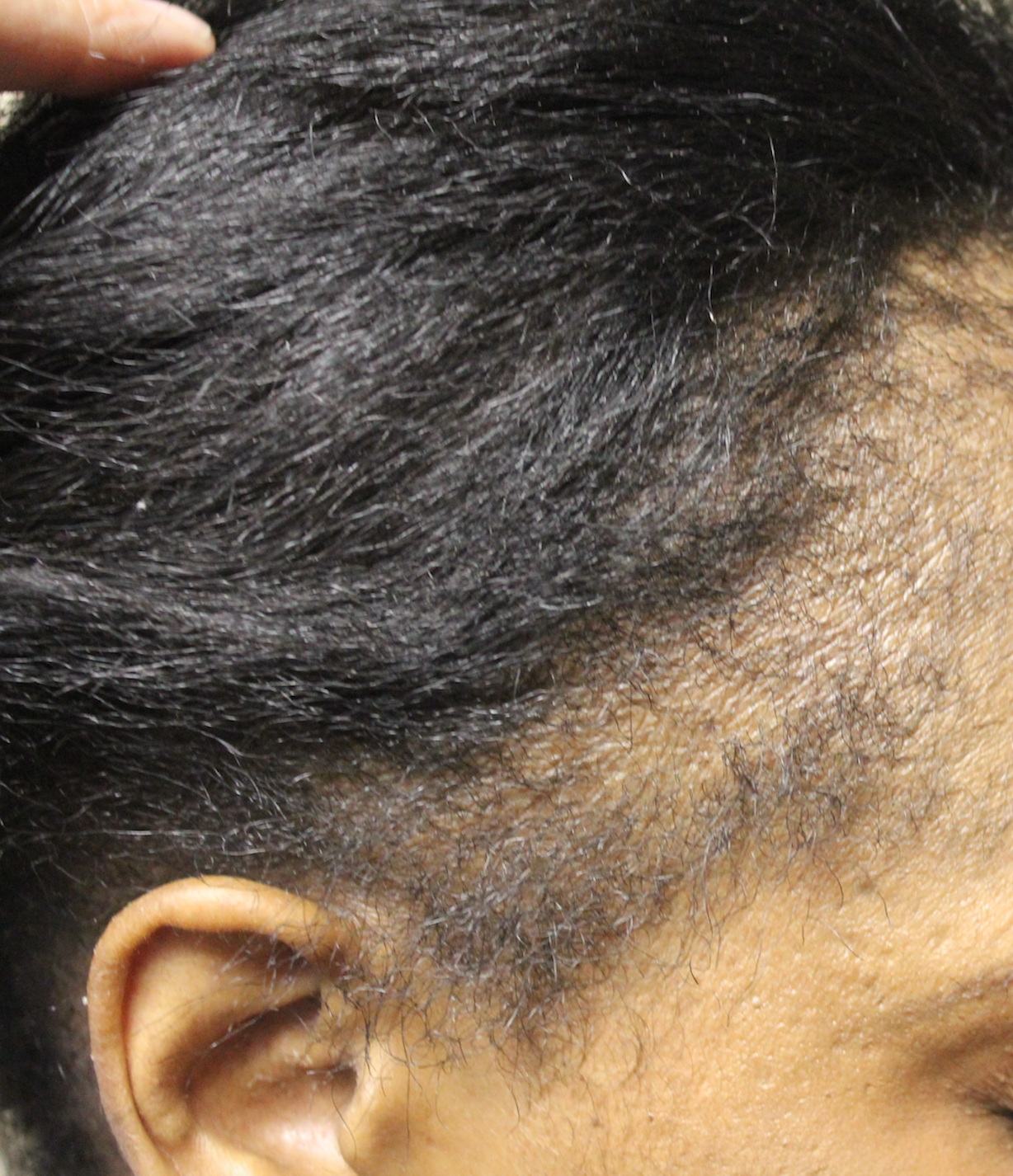
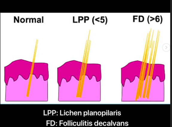
if you've never seen a zebra before, you'll be quite likely to call it some type of horse.  Perhaps a striped horse. Dog owners with more exotic breeds will tell you that few people ever get their dog's breed correct. If you've never seen or heard of the hair loss condition 'pseudopelade', you're likely to incorrectly call a circular patch of hair loss 'alopecia areata'.  If a patient has never seen folliculitis decalvans, then they are likely to go on battling what they think is stubborn folliculitis for a long time. If a patient has never heard of the condition lichen planopilaris, they may be continually searching for newer strategies for treating their stubborn dandruff or eczema.

Attempting to self diagnosis one's hair loss can be challenging if one does not know all the potential entities on the list. 


DIY Consideration 3:  Most are On the Lookout for Only 'One' Diagnosis

In my experience, everyone with hair loss is on the lookout for the cause of their hair loss.  However, very few people take the time to consider that they could have more than one cause for their hair loss.   For many patients, it's not so much of a decision as to whether they have androgenetic alopecia or telogen effluvium but rather what proportion of the patient's hair loss is from androgenetic alopecia and what proportion if from telogen effluvium. Many patients have both! It's possible to have one, two three, four or even five causes contributing to one's hair loss. 


DIY Consideration 4:  The Hair Follicle is More Complex Than Your Car and Few of us Venture to Fix our Own Cars

The hair follicle is incredible.  It's also incredibly complex.  There are 20 different cell types in the hair follicle and 100,000 hairs on the scalp (2-4 million on the entire body). Added together there are trillions and trillions of cells working together in the scalp.  Can one really try to sort through the actions (or inactions) of trillions and trillions of cells themselves?

It's difficult to think in terms of such large numbers, so let's go smaller. A car, they say, has over 30,000 parts when you consider all the tiny components. Few of us venture to even guess what's gone wrong when our car breaks down. We (hopefully) seek an expert.  I often say to patients that if my car was making strange, strange noises and blowing black smoke out the back and front, I would likely be told to get an automobile mechanic to check things over. Any attempt at self-diagnosis and fixing the car oneself would not be wise, at least for most people. It certainly would not be wise for me. When my car breaks down, I need someone who works with cars and  knows exactly how things go wrong. I need an expert.


DIY Consideration 5: Some diagnoses can only come from advanced tests meaning that an individual in such cases will never determine their diagnosis themselves.

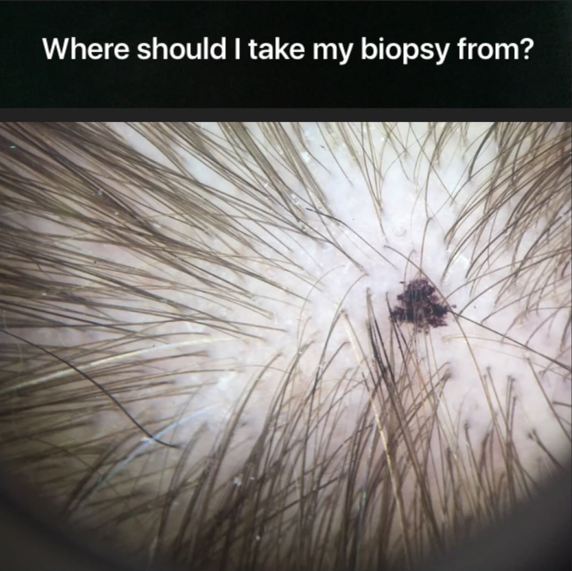
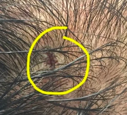
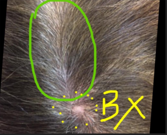
It is important to keep in mind that some diagnoses can only come from a biopsy and some diagnoses can only come from blood tests. Individuals attempting to diagnose the cause of their own hair loss may never reach their diagnosis unless they have these specific tests performed. A patient with hair shedding may think they have a vitamin deficiency or some other cause of telogen effluvium. However, a biopsy could reveal that the hair loss was actually from a scarring alopecia. Similarly, a patient with hair shedding may think their hair loss is simply from stress when the reality is that they have low iron levels from a life-threatening bleeding stomach ulcer. 

In these cases, any attempt to try to self diagnose one's hair loss simply leads to a delay in getting the appropriate medical attention that is needed.  Rarely, this delay in diagnosis can have serious consequences.


DIY Consideration 6. Patients who self diagnosis often fall prey to online marketing

Finally, a high proportion of individuals who attempt self diagnosis end up spending hundreds and sometimes thousands of dollars on products and supplements they have seen online. The patient's vulnerable state makes them very susceptible to consider buying anything that could help. Online marketing is remarkably powerful. Supplements, pills, shampoos, vitamins, tonics can all be bought with a click of a button. And often are. 


Conclusion

I've read many Do-it-Yourself books on various topics and I'm all for liberating people to take on new challenges.    DIY self diagnosis, however, is rarely a good idea. 

I'm not one to judge how patients want to access their health care. Certainly patients are spending more and more time looking for reliable information from the internet and there are many reasons why individuals want to self diagnose the reasons for their own hair loss.  However,  properly diagnose hair loss one needs the full story, not the abbreviated version. And to properly diagnosis hair loss one needs the story told with as little bias as possible. Second, a full examination of their scalp (including use of dermoscopy) is needed. There are many mimickers of hair loss and one needs to know all the potential mimickers to make a proper diagnosis. Finally, one needs to have blood tests to complete the basic work up.  


Reference

1; Danielle Ofri, MD What Patients Say, What Doctors Hear (Beacon Press, 2017). Reprinted with permission from Beacon Press.

2. Jerome Groopman. How Doctors Think. Houghton Mifflin 2007


This article was written by Dr. Jeff Donovan, a Canadian and US board certified dermatologist specializing exclusively in hair loss.
Share This
No Comments

Challenging Cases of Hair Loss: Practical Tips When Nothing Seems to Help

What to do when a patient's hair loss refuses to improve? 

tips


Every now and then there are some unusually challenging cases of hair loss that cause me to sit quietly at the end of the day and rethink the best means to treat me it. I'm talking about patients with alopecia unversalis who do not improve with any treatment, including the most potent of oral immunosuppressives. I'm talking about patients with scarring alopecia who continue to have symptoms and lose hair despite the most aggressive treatments. I'm talking about patients with early onset androgenetic alopecia who progress despite anti-androgens, minoxidil, laser and more. Is there anything we can do in these situations? Fortunately there usually is. Here are some practical tips.


Practical Tips


1. If the diagnosis is at all in question, a scalp biopsy should be done and possibly two. Blood tests should have been checked prior to the appointment but if not, basic screens are appropriate.

2. If a patient's diet is poor, one might look at ways to improve it. 


3. If stress and emotional issues are high, it might be worthwhile to address these. Stress is clearly relevant for some people.

4. Consideration needs to be given to whether a current treatment is actually causing the hair loss to worsen. Stopping treatment for a period may be useful in some situations.

5. A complete health check should be done by the patient's regular physician. Routine screening exams (mammograms, colonoscopies) should be up to date according to age appropriate screening.

6. One should always at least ask if patients are using their recommended treatment. Every now and then there are some incredible surprises.

7. If a different route of administration is possible this should be considered. Some oral drugs might be compounded topically. Some topicals may be available in oral form.
 

Conclusion

If a physician sees enough patients with hair loss, he or she will encounter cases of hair loss that don't seem to respond to anything. An organized approach in these situations is needed. Every so often some surprising improvements can finally occur!


This article was written by Dr. Jeff Donovan, a Canadian and US board certified dermatologist specializing exclusively in hair loss.
Share This
No Comments

Where should a scalp biopsy be taken from?

Choosing (Wisely) A Site to Biopsy

bx-90

Scalp biopsies are a brief 10-15 minute procedure that allows the clinician to obtain a sample for analysis under the microscope.

Great care is needed to choose the spot or location to sample. Random biopsies are rarely helpful. The site should ideally be the site where the most changes representative of the potential disease in question are seen by the clinician or the most symptoms are felt by the patient (ie itching, burning and pain).

Far too often I evaluate biopsies form patients whereby a biopsy was taken from an area that is not truly representative of the disease that is believed to be present. Biopsies are often taken from an area that will be 'hidden' in the event that a scar forms. However, a biopsy should always be taken form a representative area or one should not take the biopsy at all. Too many biopsies for androgenetic alopecia are taken from the lateral (side) parts of the scalp rather than the central regions. This often yields information that is not always helpful. If a decision is made to take a biopsy, it must be taken from an area that is most likely to capture the disease in question. Otherwise my personal opinion is not to do it. 

Once the site is chosen, the area is marked with a dot (as shown in the photo). The scalp in this area is anesthetized ("frozen") with local anesthetic medications such as lidocaine. Then, a punch biopsy instrument is used to obtain a sample about 4 mm in diameter. The area where the sample was removed is then stitched with suture. The punch biopsy specimen is then sent to a dermatopathology lab for processing.  Results may take 2-6 weeks depending on the type of processing needed. 


This article was written by Dr. Jeff Donovan, a Canadian and US board certified dermatologist specializing exclusively in hair loss.
Share This
No Comments

Scalp Biopsies: Always 4 mm in size

Scalp Biopsies

biopsy

Scalp biopsies are performed when the diagnosis of an individual's hair loss is not clear from their story and from examination of the scalp.

A biopsy is performed under local freezing and involves removal of a tiny sample 4 mm in diameter.

The sample is sent off the the dermatopathologist for review under the microscope


This article was written by Dr. Jeff Donovan, a Canadian and US board certified dermatologist specializing exclusively in hair loss.
Share This
No Comments

Will a scalp biopsy site be immediately visible to others?

Will a scalp biopsy site be immediately visible to others?

BX Scalp.jpg

Scalp biopsies are extremely important when performed in the right patient. They can help exclude a variety of causes of hair loss - especially various inflammatory and scarring alopecias. The procedure is a brief 5-10 minute procedure done with local freezing (anesthesia). A stitch is placed in the scalp at the end. For most patients the stitch will not be noticeable to others especially if the sample is taken in an area where neighboring hair can help cover it. If a biopsy is taken from an area which is rather devoid of hair, the stitches may be visible to others for a few weeks.

This photo shows the scalp of a patient who has just finished a biopsy. The area is quite hidden. As the patient leaves the office, nobody would know a biopsy had been performed. The patient can even return to work. Stitches here are dissolving stitches. After a few weeks the area will heal with a small scar. But that scar too should be relatively hidden by neighboring hair.


This article was written by Dr. Jeff Donovan, a Canadian and US board certified dermatologist specializing exclusively in hair loss.
Share This
No Comments

Can a biopsy showing AGA be wrong?

Can a biopsy of AGA be wrong?

I'm often asked if a biopsy can be 'wrong.' The short answer is that yes, a biopsy can potentially be wrong. Biopsies are simply "samples" and used to represent a larger area. However,  the long answer is that if a biopsy is done properly and from the right area, and read by a good pathologist, them no, it is very likely that if a biopsy returns showing that androgenetic alopecia is one of the diagnoses that this is correct. The identification of vellus hairs and a terminal to vellus hair ratio of less than 4:1 with telogen hairs less than 15% is typical of androgenetic alopecia. Many individuals (especially women) with androgenetic alopecia do not have a family history so this fact should not be given too much emphasis.


A biopsy can "sometimes" be wrong in these situations:

1. Diagnoses of Scarring Alopecia (Lichen planopilaris, Folliculitis Decalvans)

2. Some diagnoses of Telogen Effluvium

3. Some diagnoses of Alopecia Areata

Most of the time, of course, a biopsy is correct in these situations. However, there are many cases alopecia areata, telogen effluvium and scarring alopecia that are challenging. A biopsy is just a piece of the puzzle and one must put together all the facts from the clinical history. examination, blood test results to come up with the diagnosis. 


A biopsy is less likely to be 'wrong' in these situations

1. Androgenetic alopecia (especially with use of horizontal sections)

2. Tinea Capitis

3. Trichotillomania

4. Skin Cancers and Metastases


This article was written by Dr. Jeff Donovan, a Canadian and US board certified dermatologist specializing exclusively in hair loss.
Share This
3 Comments

Scalp Biopsies

Avoid the Central Part If Possible

Scalp biopsies are  helpful in situations where the diagnosis is not clear. If a biopsy needs to be performed, the sample should first and foremost be obtained from an area on the scalp that is most likely to display the histological features of the disease in question. However, one must also try to take a biopsy from an area that will hide any scar that may eventually form.

One of the easiest places to do a scalp biopsy is the central scalp right on the "central part line." This is rarely ever a good idea as the patient is likely to have a visible scar in the area for the rest of his or her life. Except in rare circumstances, there is usually no need to take a biopsy exactly on the central part.

A site 1-2 cm away from the area often serves equally well. If a biopsy must be taken from the central part, the patient should be warned about the potential for extreme visibility of the scar that will form in the area. The patient in this picture had a biopsy (BX) taken years ago from the central part (shown in green circle). The scar is obvious and regardless of the hairstyle chosen, it is challenging to hide.


This article was written by Dr. Jeff Donovan, a Canadian and US board certified dermatologist specializing exclusively in hair loss.
Share This
No Comments

Scalp Biopsies

Where Should the Sample Come From?

Scalp biopsies are very helpful in situations where the diagnosis is not clear. A 4 mm punch is the minimum size and the specimen should always be obtained from a region on the scalp that has the highest chance of showing diagnostic features under the microscope.

The use of dermoscopy (trichoscopy) greatly increases the chance that a biopsy will lead the clinician to the correct diagnosis. The blue dot in the picture shows an area that was selected for a biopsy.


This article was written by Dr. Jeff Donovan, a Canadian and US board certified dermatologist specializing exclusively in hair loss.
Share This
No Comments

Where should I get my scalp biopsy?

Random Biopsies are Rarely Helpful

Where on the scalp should my doctor take a biopsy sample?

This is an important question as random biopsies from "just anywhere" are generally not very helpful. First, it must be stated that biopsies are not needed in most patients with hair loss. Secondly, it must also be stated that not enough patients have scalp biopsies! A biopsy should be taken from the area that is undergoing the most change. If it is decided that a biopsy is needed to exclude androgenetic alopecia (AGA), biopsies should be taken from the front, middle or top (depending on where the most change is happening). A biopsy from the very back might show changes of AGA if present - but more significant degrees of change would always be up front rather than in the very back. In suspected scarring alopecia, a biopsy should first and foremost be taken from where the dermatologist sees evidence of the condition. This is best assessed with an instrument called a "dermatoscope." Changes of scarring alopecia include redness around hairs, scaling, scarred areas, sometimes pustules and crust. The biopsy must always contain hairs as biopsies of completely scarred areas are useless. For conditions that affect the entire scalp (like acute and chronic telogen effluvium) the biopsy should be taken from the mid scalp to better assess hair cycle characteristics (anagen and telogen ratios) and to detect conditions that mimic CTE including androgenetic alopecia and lichen planopilaris (LPP).   The three "X's" in the picture show where I would typically take a biopsy to assess for CTE. Note that these biopsies are not taken from the temples - even if the patient states this is where most of the hair loss has occurred. (A separate biopsy from the temple could be considered). This allows the dermatpathologist to best assess the terminal and vellus ratios (T:V ratios). A T:V ratio above 8:1 taken from the mid scalp is highly suggestive of a diagnosis of chronic telogen effluvium. A T:V ratio less than 4:1 is more in keeping with androgenetic alopecia.

Biopsies always leave a small scar. One should consider performing a biopsy away from the central "part" if at all possible as many women make use of a central part to style their hair. 


This article was written by Dr. Jeff Donovan, a Canadian and US board certified dermatologist specializing exclusively in hair loss.
Share This
No Comments

Which hairs should be taken during a scalp biopsy?

What is a scalp biopsy? Which hairs should be taken ?


A scalp biopsy is a 15-20 minute procedure that helps the dermatologist obtain a small piece of tissue for analysis.  The sample is processed in a history laboratory into thin sections and mounted on glass slides for review by a pathologist under the microscope.  A scalp biopsy is not required for most patients with hair loss. When the diagnosis is uncertain, it can be very helpful.

FOR DETAILS ON THE SCALP BIOPSY, CLICK HERE
 

Which hairs should be included?
 

First, an area of the scalp containing hair follicles should always be included in a biopsy. A scalp biopsy specimen from a completely bald area is not helpful!

Second, the hair follicles obtained should be taken from an area showing the specific abnormalities thought to be in keeping with the hair loss condition in question. If no such features are present, hair follicles might be obtained from an area of the scalp where the hairs are easily extractable (positive pull test), or from an area having the most symptoms (itching, burning or pain).

The key point is that obtained a scalp biopsy from a random area on the scalp does not usually yield useful information.


This article was written by Dr. Jeff Donovan, a Canadian and US board certified dermatologist specializing exclusively in hair loss.
Share This
No Comments

Scalp biopsies for hair loss: Is it always needed?

Is a scalp biopsy always needed to determine the cause of an individual's hair loss?


The answer of course is no. A biopsy is only needed if the diagnosis is uncertain. 
In tough hair loss cases, I often like my patients to go a few days without shampooing the scalp. This allows the highest chance of some of the key features of the "suspected" condition to be present on the scalp (and not washed away by a recent shampooing).

Where does one biopsy?

I always biopsy an area which has the most features of the particular hair loss condition I suspect. For scarring alopecias, I often try to biopsy areas that cause the patient's scalp to be itchy, have burning or areas that cause pain.


This article was written by Dr. Jeff Donovan, a Canadian and US board certified dermatologist specializing exclusively in hair loss.
Share This
No Comments

Blogs by Topic


Share This
-->