h1.qusth1 { display: none !important; }

QUESTION OF THE WEEK

Dr. Donovan's Articles

QUESTION OF HAIR BLOGS


 Meta-Analysis Highlights Good Short Term Safety of JAK Inhibitors for Dermatologic Issues.

 No Increased Risk in Blood Clots, Cardiac Events or Death Identified

Janus kinase inhibitors have entered onto the scene as helpful treatments for many diseases in dermatology including alopecia areata, atopic dermatitis and vitiligo. As we’ve talked about many times over the years, there remains a “boxed warning” from the FDA on JAK inhibitors warning the public and physicians about the possible increased risk of major adverse cardiovascular events (MACE), venous thromboembolism (VTE), serious infections, malignant neoplasm, and death.

This boxed warning comes about from past studies like the ORAL Surveillance Study which showed that patients with rheumatoid arthritis who were 50 years of age and older and who had at least 1 cardiovascular risk had increased risk for major adverse cardiac events, infections, cancer venous thromboembolism and death from using the JAK inhibitor tofacitinib. We have reviewed the ORAL Surveillance Trial in the past:


As JAK inhibitors continue to be studied and continue to be approved for a variety of dermatologic conditions, the question that dermatologists ask themselves over and over is “do these ORAL Surveillance trial results really and truly apply to patients with skin diseases like it does to patients with rheumatologic diseases? Does a patient with alopecia areata or vitiligo or atopic dermatitis also have an increased risks for things like blood clots and heart disease?

Ingrassia JP et al 2023

Authors of a new systematic review and meta-analysis set out to determine the risk of all-cause mortality, MACE, and VTE in patients specifically with dermatologic conditions who used JAK inhibitors.

The authors included dermatology patients who were participating in phase 3 randomized clinical trials of an approved JAK inhibitor for the treatment of an immune mediated inflammatory skin disease - provided the trial also had a placebo/active comparator group. Studies without a comparison group, case reports, observational studies, and review articles were excluded.

The primary outcome was major adverse cardiac events (MACE) and all-cause mortality, and VTE.


The authors included in their meta-analysis a total of 35 RCTs (8 with baricitinib, 6 with abrocitinib, 2 delgocitinib cream, 4 ruxolitinib cream, 7 with tofacitinib, 1 with ritlecitinib, and 7 with upadacitinib). The analysis included 20 651 patients (mean age, 38.5 years; male, 54%). The mean follow up time was quite short age 4.9 months.

The trials enrolled a total of 20 651 patients. This included 13 597 patients who were randomized to a JAK inhibitors and 7054 to placebo/active comparators. Dermatologic diseases included atopic dermatitis (21 trials), alopecia areata (3 trials), psoriasis (including psoriatic arthritis) (9 trials), and vitiligo (2 trials).

All in all, the authors’ findings did not show a significant difference between JAK inhibitors and placebo/active comparator in composite MACE and all-cause mortality (OR, 0.83; 95% CI, 0.44-1.57) or VTE (OR, 0.52; 95% CI, 0.26-1.04).


Comments

This is a helpful study for all of us in dermatology who use JAK inhibitors to treatment various autoimmune skin diseases.

What I particularly liked in this paper was the honest and straightforward discussion. The authors point out that there are many differences between the studies included in this meta-analysis and the ORAL SURVEILLANCE TRIAL.


The authors point out to readers that

a)    The patients in this study with dermatologic conditions were younger (mean age, 38.5 years) than the patients with RA (mean age, 61 years). This fact alone could explain the low overall incidence of MACE, all-cause mortality, and VTE among patients in this meta-analysis (of the dermatology clinical trials).

b)     Patients in the ORAL Surveillance had higher cardiovascular risk than the patients in the meta-analysis because the ORAL surveillance trial only enrolled patients with RA with at least 1 cardiovascular risk factor.

c)    Patients in the ORAL SURVEILLANCE TRIALS received  methotrexate and some were also receiving systemic corticosteroids. This is very different than patients included in dermatology clinical trials, who were receiving JAK inhibitor monotherapy.

d)    The dermatology meta-analysis here included trials with short duration (4.9 months). The ORAL SURVEILLANCE STUDY followed patients for 4 years whereas this meta-analysis followed patients for a far shorter period

e)    In addition, most of the new JAK inhibitors used in this particular meta-analysis were JAK 1/2 inhibitors rather than JAK 1-3 inhibitors like (tofacitinib)

f)      Finally, the authors point out to readers that in this study, they did not focus on all the boxed warnings but rather 3 of the 5 possible boxed warning: MACE, VTE, and death. The authors did not examined infections and cancer in this study ..


For now, what we can say is that in these 35 trials of fairly short duration of follow up, there is no major signal for venous thromboembolism and major adverse cardiac events. This is good news so far for all of us who use these medications!


REFERENCE

Ingrassia JP et al.   Cardiovascular and Venous Thromboembolic Risk With JAK Inhibitors in Immune-Mediated Inflammatory Skin Diseases A Systematic Review and Meta-Analysis. JAMA Dermatol. 2023 Nov 1:e234090.

Ytterberg SR et al. Cardiovascular and Cancer Risk with Tofacitinib in Rheumatoid Arthritis. N Engl J Med. 2022 Jan 27;386(4):316-326. doi: 10.1056/NEJMoa2109927.PMID: 35081280 Clinical Trial.


This article was written by Dr. Jeff Donovan, a Canadian and US board certified dermatologist specializing exclusively in hair loss.
Share This
No Comments

Vaccine Excipients and Spike Protein May Trigger Alopecia Areata

New Study Identifies Mechanisms of COVID-related Alopecia Areata

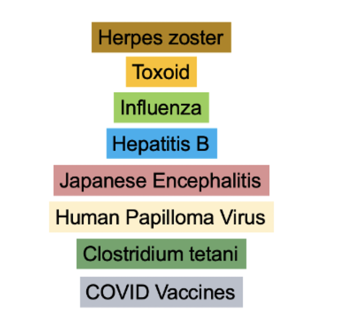
It’s clear that many different vaccines can potentially trigger alopecia areata. Vaccinations have previously been suggested as a possible trigger for immune-mediated alopecia, such as hepatitis B, herpes zoster, quadrivalent HPV, influenza, and Japanese encephalitis vaccines. COVID vaccines are now on these lists too.

It has been proposed that epitopes derived from the COVID19 virus or the COVID19 vaccine resemble antigen epitopes of the human and can trigger alopecia.


Wang et al 2023

In a new study, authors set out to better understand the potential mechanisms of COVID-19 vaccine-related alopecia areata (AA).

The authors recruited patients presenting with COVID-19 vaccine-related alopecia areata (AA) during the period 2021 to 2022. The authors compared their results to control patients who did not have alopecia areata from vaccinations.

There were 27 new-onset of vaccine related alopecia areata patients  and 106 vaccines-tolerant individuals. The mean age of the patients with vaccine-related AA was 38.2 while that of the tolerant controls was 43.7 years. Approximately 63.0% of the vaccine-related AA patients were women. There was no significant difference in sex and age between the groups.


Of the 27 patients with vaccine related AA, the Moderna vaccine (mRNA-1273 vaccine) was the culprit in 40.7% of patients. The Pfizer vaccine (BNT162b2 vaccine) was used in 29.6% of patients and Astrazeneca vaccine (AZD1222 vaccine) was used in 25.9% of patients. Other vaccines (such as the MVC-COV1901 vaccine) were used in 3.7%.

Most patients (59.6%) had symptoms occurring after the second dose of the vaccine. In 22.2 % of patients it was the very first dose that triggered alopecia areata and in 18.5 % symptoms occurred after the third and fourth doses of the vaccine.


Elevated Total IgG, Positive ANA and Elevated Eosinophils Associated with Vaccine AA

The authors found that there were three blood test results that were much more likely to be elevated in COVID – AA patients than controls. These included positive ANA, total IgE levels and eosinophilia. In this study, 20 % of COVID – AA patients had positive ANA compared to 3.8% of controls. (p=0.013). 45.5 % of COVID – AA patients had increased total IgE levels compared to 10.2 % of controls (p=0.00019). 28.6 % of COVID – AA patients had eosinophilia compared to 10.2 % of controls (p=0.027).


Serum CCL18 and Granulysin Associated with Vaccine AA

The authors assessed the serum levels of 23 different types of cytokines, chemokines, and inflammatory proteins. The patients with COVID-19 vaccines-related AA had significantly higher levels of PARC/CCL18 and granulysin  than controls.

For those not aware, CCL18 is produced by antigen presenting cells. It is involved in the innate and adaptive immune system and plays a role in attracting immune cells. Granulysin is a pro-inflammatory molecule expressed by NK cells and activated T cells.  


Lymphocyte Activation Tests Point to Role Excipients and Spike Protein

Some of the key experiments done by the authors were the lymphocyte activation tests.

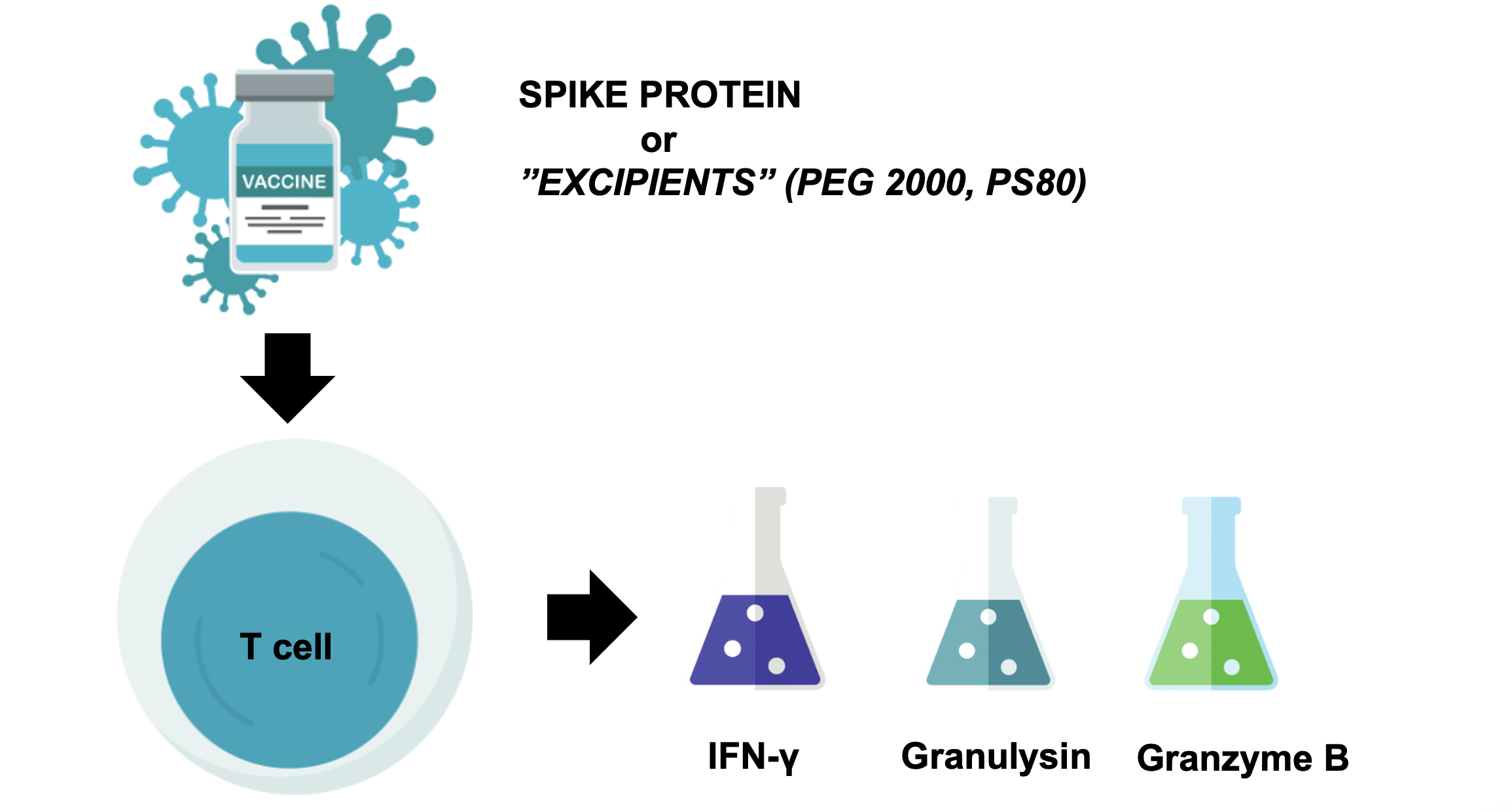
The authors showed using an in vitro lymphocyte activation test (LAT) that granulysin, granzyme B, and IFN-γ released from the T cells of COVID-19 vaccines-related AA patients could be significantly increased by COVID-19 vaccine excipients (polyethylene glycol 2000 and polysorbate 80) or spike protein (P = 0.002–0.04).

All in all, the authors concluded that Spike protein and excipients of COVID-19 vaccines (like PEG2000 and polysorbate 80) could trigger T cell-mediated cytotoxicity, which in turn contributes to the pathogenesis of immune-mediated alopecia associated with COVID-19 vaccines.


What are excipients?

Before we go on, it’s helpful to talk a bit about excipients. Excipients are added to a vaccine for a specific purpose. These include things like preservatives to prevent contamination (ie thimersol), adjuvants to stimulate a stronger immune response (ie aluminum salts) and stabilizers to help keep the vaccine functioning optimally during its transportation or stage (ie certain sugars)

Immediate‐type hypersensitivity has been described to several excipients with most reports on polyethylene glycols (PEGs) and the structurally related polysorbates. PEG 2000 is used in the mRNA vaccines from Pfizer/BioNTech  and Moderna and polysorbate 80 in viral vector vaccines from several other companies including AstraZeneca.  

PEG 2000 is really an important excipient for us to know about in the COVID era. It’s found in the mRNA vaccines. The mRNA molecules contained in the Pfizer and Moderna vaccines are stabilized by lipid nanoparticles, which are “PEGylated” to improve delivery to target cells. Now, PEG is obtained from the polymerization of ethylene glycol and can be found at different molecular weights (MW) depending on the chain length. Higher molecular weight PEGs are more likely to cause allergy. The PEG contained in mRNA-based anti-SARS-CoV-2 vaccines has a molecular weight of 2000 kDa (and that’s why it’s called PEG 2000). PEG 2000 is well known to be a potential culprit molecule in hypersensitivity reactions.

Polysorbate is found in the Astrazeneca vaccine. Polysorbates help with the overall solubility of the anti-SARS-CoV-2 vaccine. Recently, increased attention to PS allergy has arisen.


Treatment Outcomes Quite Good in COVID Associated AA

In this paper, the authors also outlined how patients were treated and what sort of hair regrowth they experienced. COVID related AA has been reported to be difficult to treat in other studies. In this study, 52 % had complete response to treatment, 41 % had partial and 7% had no response. Treatments included topical therapies in 100% of patients (minoxidil, DPCP and topical steroids) and 81 % had systemic treatments (methotrexate, cyclosporine, baricitinib, tofacitinib and systemic steroids).  In this study 92.6% of the patients with COVID-19 vaccine- related AA reached either complete or partial remission.


Discussion.

This is an interesting study. It is the largest cohort study to date to provide data on the clinical characteristics and comprehensive immune profiles of patients with alopecia areata due to COVID-19 vaccines.

The authors pinpoint that the vaccine excipients of mRNA COVID-19 vaccines (ie PEG 2000 and polysorbate 80) were regarded as the main culprit.   

Taken together, the authors propose that some patients develop AA after COVID-19 vaccinations through specific CD4+ and CD8+mediated immune reactions triggered by vaccine excipients or spike protein.

The authors found that the ANA level, total IgE, and peripheral eosinophil count were significantly elevated in the patients with COVID-19 vaccine-related AA compared with those in the tolerant controls. It may be possible that these sorts of laboratory data can help physicians in the diagnosis of AA induced by vaccines in the clinical setting.


REFERENCE

Wang C-W et al. Clinical characteristics and immune profiles of patients with immune-mediated alopecia associated with COVID-19 vaccinations. Clin Immunol. 2023 Oct:255:109737.


This article was written by Dr. Jeff Donovan, a Canadian and US board certified dermatologist specializing exclusively in hair loss.
Share This
No Comments

Another Handful of Patients with COVID VACCINE Induced Alopecia Areata

COVID Vaccines Trigger Alopecia Areata in Some Patients, Yet Reduce the Risk in Others

Nobody every said the immune system is simple! Studies have shown that COVID Vaccines can reduce the risk of developing alopecia areata and studies have shown that COVID vaccines can increase the risk of alopecia areata.

Genco et al 2023

Genco et al report 5 more patients to add to the growing list of patients who have experienced new onset or flares of alopecia areata after COVID vaccines. These 5 patients included 4 females and 1 male. Mean age 32 years. All had a prior history of alopecia areata. 4 of the patients had no evidence of alopecia areata before the vaccine was given and 1 had mild disease. Flares of AA occurred in these 5 patients 1-3 weeks of receiving the vaccine. The vaccine was Pfizer in 2 patients and Moderna in 3 patients. In 4 of the 5 patients, subsequent doses did not further flare the alopecia. However, 1 patient had a booster effect with each dose causing more and more hair loss.

Comment

This is a nicely documented report. Not all reports of COVID vaccines induced AA have been in patients with prior histories of AA so it’s interesting that all patients in this report had prior AA. It’s amazing just how fast these AA flares occur. In 3 patients it was just 2 weeks. In 1 patient it was 1 week and in 1 patient it was 3 weeks.

A recent systematic review by Pastukhova et al highlighted the subject in greater detail.

REFERENCE

Genco L et al.  Alopecia Areata after COVID-19 Vaccines. Skin Appendage Disord. 2023 Mar;9(2):141-143.

Pastukhova E et al. Alopecia Areata as a Sequela of COVID-19 Vaccination: A Systematic Review J Cutan Med Surg . 2023 Jan-Feb;27(1):64-65.


This article was written by Dr. Jeff Donovan, a Canadian and US board certified dermatologist specializing exclusively in hair loss.
Share This
No Comments

COVID 19 Infection Increases the Risk of Alopecia Areata and Alopecia Totalis; Vaccination Reduces the Risk

COVID19 Infection Increases the Risk for Alopecia Areata and other Autoimmune Diseases


A number of studies to date have suggest that patients infected with COVID 19 are at risk for several autoimmune diseases.

A new study set out to investigate the incidence and risks of various autoimmune and autoinflammatory diseases after COVID 19.

To do so, the authors performed a retrospective study using patient data from a nationwide database in Korea. Patients included in the study were those with a PCR positive COVID 19 diagnosis between Oct 8 2020 and Dec 31, 2021 and their data was compared to controls without COVID 19.


In total, there were 354, 527 individuals with COVID 19 that were included in the study, with a mean age 52.24 years. Data was compared to 6, 134, 940 control patients that did not have COVID 19.

Both alopecia areata as well as alopecia totalis were increased by COVID 19. The risks of alopecia areata increased 12 % (adjusted hazard ratio [aHR], 1.12; 95% CI, 1.05-1.19), and the risk of alopecia totalis increased 74 % alopecia totalis (aHR, 1.74; 95% CI, 1.39-2.17).

Many Autoimmune Diseases Increased by COVID Infection

Other diseases also were found to be increased in those with COVID19 including Antineutrophil cytoplasmic antibody– associated vasculitis (aHR, 2.76; 95% CI, 1.64-4.65), Crohn disease (aHR, 1.68; 95% CI, 1.31-2.15), and sarcoidosis (aHR, 1.59; 95% CI, 1.00-2.52). However, the risk of SLE was lower (aHR, 0.47; 95% CI, 0.36-0.61) in the COVID-19


Alopecia Areata and Totalis According to Gender

When Data was analyzed according to gender, the authors showed that both males and females with COVID 19 had an increased risk of developing alopecia totalis. In terms of alopecia areata, males had a decreased risk of alopecia areata and females had an increased risk


Alopecia Areata and Totalis According to Age

When Data was analyzed according to age, the authors showed that younger patients (under 40) with COVID 19 had a decreased risk of risk of developing alopecia totalis and no difference in the risk of alopecia areata.

 Patients over the age of 40 had an increased risk of both alopecia areata and totalis.


Alopecia Areata and Totalis According to Severity

When Data was analyzed according to severity, the authors showed that patients who were in the ICU had a much higher chance of developing alopecia totalis than those not in the ICU (2.45 vs 1.46).


Alopecia Areata and Totalis According to Vaccination Status

Patients who were vaccinated had a lower risk to develop alopecia areata and those who had more complete vaccinations had an even lower risk. Patients that were not vaccinated had a 33 % higher risk of developing alopecia areata (aHR, 1.33; 1.11-1.59) and an even higher risk to develop alopecia totalis (aHR, 2.26; 1.29-3.97)   


Discussion

This is an interesting study which again points to the increased risk of autoimmune disease in those previously infected with COVID 19.

The risk of alopecia totalis was increased in those with severe COVID 19, those over 40 and in those who were not vaccinated.

It’s hard to know exactly what to make of all this data as the data doesn’t always follow perfectly the same trends.

For example, patients under 40 infected with COVID 19 seem to have a decreased risk of alopecia totalis but those over 40 seem to have an increased risk. Males with COVID 19 have a decreased risk for Alopecia areata but females with COVID have an increased risk. (Both have an increased risk for totalis). But nevertheless the main point does seem clear – and that is that patients with COVID 19 overall seem to have an increased risk of alopecia areata and alopecia totalis.


Why are autoimmune diseases increased by COVID19 Infection?

The reasons for this increased autoimmunity area really not entirely clear. Some have hypothesized that patients infected with COVID 19 develop a variety of antibodies to SARS-COV-2 epitopes and these antibodies may cross react with tissue antigens in the human body.  Other studies have proposed that the body’s increase in cytokines (ie. the ‘cytokine storm) to try to stop the SARS-COV-2 infections (like IL1 and IL6) may trigger autoimmune reactions

What about COVID VACCINE Induced Alopecia Areata?

This study suggested that patients who are vaccinated have a lower risk of alopecia areata and patients who are not vaccinated have a higher risk to develop alopecia areata.

It’s clear that this does not apply to everyone. It’s clear that alopecia areata can rarely be triggered by vaccines as well so there is a population of patients who actually develop AA from the vaccine. A nice systematic review by Pastukhova E et al on this topic can be found in the references below,

REFERENCE

Lim SG et al. Autoimmune and Autoinflammatory Connective Tissue Disorders Following COVID-19  JAMA Netw Open . 2023 Oct 2;6(10):e2336120.

Pastukhova E et al. Alopecia Areata as a Sequela of COVID-19 Vaccination: A Systematic Review J Cutan Med Surg . 2023 Jan-Feb;27(1):64-65.

Genco L et al.  Alopecia Areata after COVID-19 Vaccines. Skin Appendage Disord. 2023 Mar;9(2):141-143.


This article was written by Dr. Jeff Donovan, a Canadian and US board certified dermatologist specializing exclusively in hair loss.
Share This
No Comments

Increased Risk of Autoimmune Diseases, Including Alopecia Areata, After COVID 19 Infection

New Study Again Points to Increased Risk of Autoimmune Diseases with COVID 19 Infection

It’s becoming increasing clear that patients who have COVID19 infections are at increased risk to develop autoimmune diseases. A few months ago, we reviewed the February 2023 study by Chang et al which showed the patients with COVID19 had an increased risk of developing a variety of autoimmune issues.

The Chang et al study showed an increased risk of rheumatoid arthritis (aHR:2.98, 95% CI:2.78–3.20), ankylosing spondylitis (aHR:3.21, 95% CI:2.50–4.13), systemic lupus erythematosus (aHR:2.99, 95% CI:2.68–3.34), dermatopolymyositis (aHR:1.96, 95% CI:1.47–2.61), systemic sclerosis (aHR:2.58, 95% CI:2.02–3.28), Sjögren's syndrome (aHR:2.62, 95% CI:2.29–3.00), mixed connective tissue disease (aHR:3.14, 95% CI:2.26–4.36), Behçet's disease (aHR:2.32, 95% CI:1.38–3.89), polymyalgia rheumatica (aHR:2.90, 95% CI:2.36–3.57), vasculitis (aHR:1.96, 95% CI:1.74–2.20), psoriasis (aHR:2.91, 95% CI:2.67–3.17), inflammatory bowel disease (aHR:1.78, 95%CI:1.72–1.84), celiac disease (aHR:2.68, 95% CI:2.51–2.85), type 1 diabetes mellitus (aHR:2.68, 95%CI:2.51–2.85).


Tesch et al 2023

A cohort study from Germany showed similar findings to what was shown in the Chang study, namely that patients with COVID19 are at increased risk to develop autoimmune diseases. Tesch et al matched 641,704 patients with COVID-19 in a 1:3 manner with control patients without COVID19. Here, Tesch et al found a 42.63% higher likelihood of acquiring autoimmunity for patients who had suffered from COVID-19. These autoimmune diseases included conditions like Hashimoto thyroiditis, rheumatoid arthritis, and Sjögren syndrome. The highest IRR was observed for autoimmune diseases of the vasculitis group. The risk of developing autoimmune diseases was higher in older individuals than younger individuals and was higher in those who had more severe COVID19 infection (such as being in hospital or the ICU) than those with less severe infection.

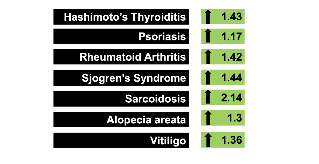
Specific rates included an increased risk of Hashimotos’ thyroiditis 1.42 (95 % CI 1.33-1.59), psoriasis 1.17 (95 % 1.09–1.26), rheumatoid arthritis 1.42 (95 % 1.30-1.56), Sjogren’s syndrome 1.44 (95 % 1.27–1.63), sarcoidosis 2.14 (95 % 1.73–2.65), alopecia areata 1.30 (95 % 1.05–1.61), vitiligo (95 % 1.36 1.05–1.77). The risk was particularly high in Behcet’s (95 % 2.42; 1.10–5.35), Wegener’s (95 % 2.51; 1.42–4.46 and Temporal Arteritis (95 % 1.63; 1.05–2.53).The risk of systemic lupus was not found to be increased in those with COVID 19 in this study.

Conclusion and Discussion


The authors showed that in the first 3-15 months after COVID19 infections there is 43% higher likelihood of developing a first onset autoimmune disease. The authors found that the risk is highest for the “vascular” autoimmune diseases. Risks were highest in this study for Behcet’s, Wegener’s and Temporal Arteritis.


REFERENCE

Tesch  F et al. Incident autoimmune diseases in association with SARS-CoV-2 infection: a matched cohort study. Clin Rheumatol. 2023 Oct;42(10):2905-2914.

Chang R et al. Risk of autoimmune diseases in patients with COVID-19: A retrospective cohort study. EClinicalMedicine. 2023 Feb;56:101783.


This article was written by Dr. Jeff Donovan, a Canadian and US board certified dermatologist specializing exclusively in hair loss.
Share This
No Comments

Calcium Channel Blocks May Reduce Hair Loss in Long COVID Patients

Study from Japan Highlights Potential Benefits of CCB’s on Long COVID

Calcium channel blockers have received significant attention regarding their potential to impact COVID. Several studies suggested that use of calcium channel blockers (CCBs), especially amlodipine and nifedipine, can reduce mortality from coronavirus disease 2019 (COVID-19). A 2021 meta-analysis suggested that the benefits were mainly in those with hypertension.

I was interested in a new study addressing whether use of calcium channel blockers affect long COVID symptom, especially hair loss.

Authors from Japan performed a mutlicenter cohort study,examining patients ≥18 years old diagnosed with COVID-19 from November 2020 to March 2022 and hospitalized at participating medical facilities. The study included 1066 patients (361 female, 620 male). Hypertension was the most common comorbidity (n = 344; 32.5%).

Interestingly, females with hypertension were significantly less likely to develop long COVID symptoms than those without hypertension (odds ratio [OR] 0.51, 95% confidence interval [CI] 0.27-0.98; p = 0.043). In females, calcium channel blocker administration, rather than having hypertension, was associated with a 7 fold reduction in the frequency of hair loss (OR 0.14, 95% CI 0.03-0.67, p = 0.015).

Other long COVID symptoms were also reduced by calcium channel blockers including a 7 fold reduction in memory impairment (OR 0.14, 95% CI 0.02-0.82, p = 0.029), a 6 fold reduction in sleeping disorders (OR 0.17, 95% CI 0.04-0.67, p = 0.012), a 4 fold reduction in tinnitus (OR 0.23, 95% CI 0.05-0.98, p = 0.047), a 3 fold reduction in sputum (OR 0.31, 95% CI 0.10-0.92, p = 0.035), and 3 fold reduction in fever (OR 0.33, 95% CI 0.12-0.93, p = 0.036).

Conclusion

This is an interesting study which again highlights potential benefits of calcium channel blockers in the treatment of COVID 19 related health issues. More studies are needed to understand how calcium channel blockers affect hair related issues caused by the virus.

REFERENCE

Takuya Ozawa et al. Calcium channel blockers may reduce the development of long COVID in females. Hypertens Res. 2023 Nov 17. doi: 10.1038/s41440-023-01501-w. Online ahead of print.

Alsagaff  MY et alAssociation of calcium channel blocker use with clinical outcome of COVID-19: A meta-analysis. Diabetes Metab Syndr. 2021 Sep-Oct;15(5):102210.


This article was written by Dr. Jeff Donovan, a Canadian and US board certified dermatologist specializing exclusively in hair loss.
Share This
No Comments

WNT10A gene variants found in Short Anagen Syndrome

NEW DATA POINTS TO WNT10A GENE VARIANTS IN SHORT ANAGEN SYNDROME (SAS)

Short anagen syndrome was first described in 1991.

Individuals with short anagen syndrome can’t grow long hair. The anagen phase is short ( 9-20 months instead of 6-7 years) and patients can only produce short hair (typically under 6 cm). Patients often come to medical attention around age 2-4 when parent report that their child has never needed a hair cut and tend to be shedding more than what they expect.Parents report “the hair just won’t grow!” or report “she’s never needed a haircut!”

Examination of the hair shows short hair with tapered ends indicating that the hair has not been cut.

SAS It is more commonly noted in white girls with blonde hair but other hair colors and other racial backgrounds have been reported too. This includes thee white children with brown hair, an African American patients and a Hispanic patient.

The condition has been reported to be inherited in an autosomal dominant manner in some families although sporadic inheritance has been suggested. The condition may improve to some degree after puberty - in some but not all individuals.

Many children with SAS are healthy. Associations have been reproted with micronychia and tricho-dental syndrome.

Cesarato  N et al 2023: WNT10A Variants in SAS

A recent study examined possible genetic variants responsible for short anagen syndrome and found that 20 of the 48 individuals (40%) have genetic variations in WNT10A.

Interestingly genetic variations in WNT10A were also found in some patients with male pattern hair loss suggesting some type of overlap.

MAIN REFERENCE

Cesarato  N et al. Short anagen hair syndrome: association with mono- and biallelic variants in WNT10A and a genetic overlap with male pattern hair loss. Br J Dermatol. 2023 Nov 16;189(6):741-749.

OTHER REFERENCES

Antaya RJ, Sideridou E, Olsen EA. Short anagen syndrome. J Am Acad Dermatol. 2005;53:S130–4.

Avashia N, Woolery-Lloyd H, Tosti A, Romanelli P. Short anagen syndrome in an African American woman. J Am Acad Dermatol. 2010;63:1092–3.

Barraud-Klenovsek MM, Trüeb RM. Congenital hypotrichosis due to short anagen. Br J Dermatol. 2000;143:612–7.

Doche I, Donati A, Valente NS, Romiti R, Hordinsky MK. Short anagen syndrome in a girl with curly dark hair and consanguineous parents. J Am Acad Dermatol. 2012;67:e279–80.

Giacomini F, Starace M, Tosti A. Short anagen syndrome. Pediatr Dermatol. 2011;28:133–4.

Jung HD, Kim JE, Kang H. Short anagen syndrome successfully controlled with topical minoxidil and systemic cyclosporine A combination therapy. J Dermatol. 2011;38:1108–10.

Kersey PJ. Tricho-dental syndrome: A disorder with a short hair cycle. Br J Dermatol. 1987;116:259–63.

Thai KE, Sinclair RD. Short anagen hair with persistent synchronized pattern of scalp hair growth. J Am Acad Dermatol. 2003;49:949–51.


This article was written by Dr. Jeff Donovan, a Canadian and US board certified dermatologist specializing exclusively in hair loss.
Share This
No Comments

DIFFERENTIATING ANAGEN AND TELOGEN HAIRS

Being Able to Spot the Difference Between Anagen and Telogen Hairs is Important!

It is important to be able to differentiate anagen hairs from telogen hairs.

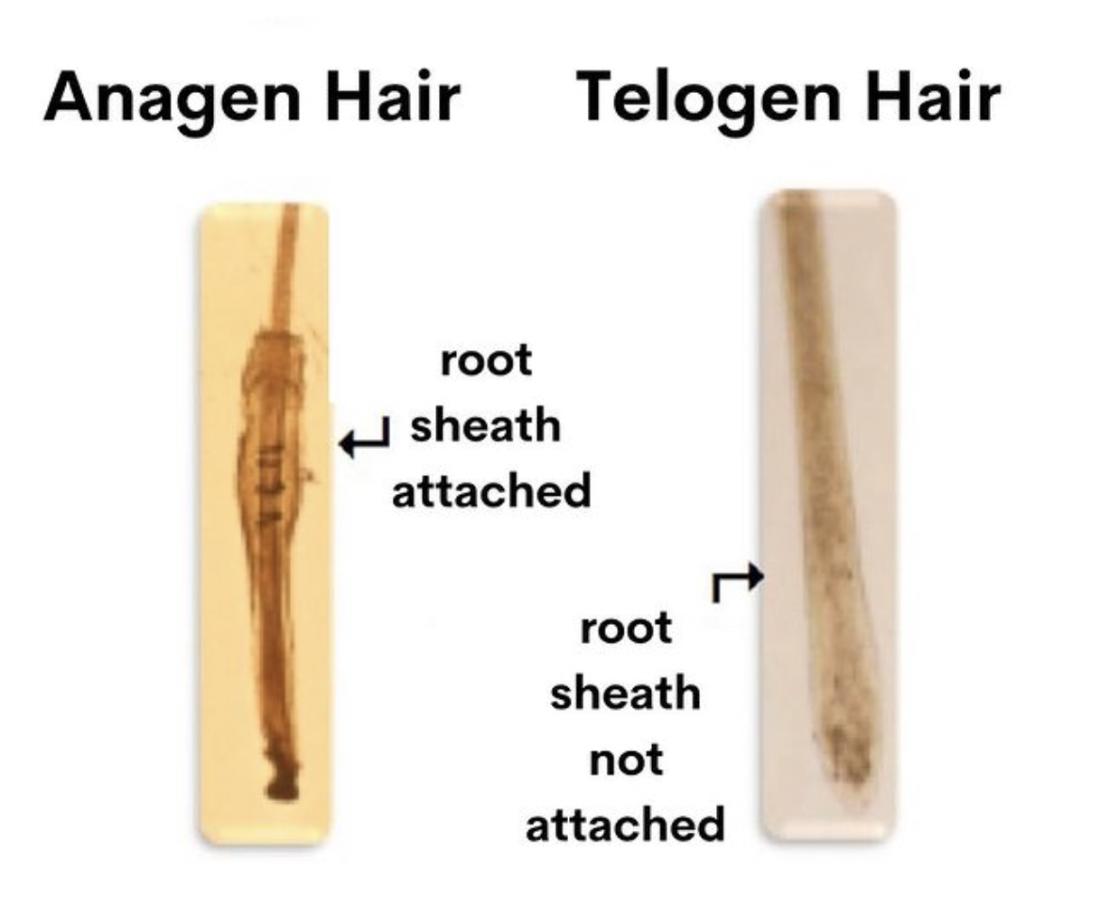
Anagen hairs are actively growing hairs. Most hairs on a person’s scalp are anagen hairs. They are firmly rooted in the scalp and therefore are rarely seen in hairs that routinely fall from the scalp. For most people, the only way to obtain an anagen hair is to forcefully pluck a hair from the scalp. In that case, one sees a hair fiber surrounded by a mass of keratinocytes. The hair is deeply pigmented at the bottom.

The routine retrieval of anagen hairs during a pull test or when reviewing hairs that are shed from the scalp suggests something is wrong. Anagen hair are not supposed to come out easily! Patients with active Scarring alopecias may notice that fairly normal looking anagen hairs are easily removed from the scalp. Patients with alopecia areata, patients undergoing chemotherapy and patients with loose anagen syndrome also lose anagen hairs but these anagen hairs are not really so typical looking like the one shown in the photo. They are special types of anagen hairs.


This article was written by Dr. Jeff Donovan, a Canadian and US board certified dermatologist specializing exclusively in hair loss.
Share This
No Comments

TRICHOSCOPY OF ANDROGENETIC ALOPECIA

Trichoscopy of Androgenetic Alopecia: Brief Overview

Trichoscopy of androgenetic alopecia (AGA) showing a variation in the caliber of hairs that are present. This is known as “anisotrichosis.” Many thick terminal hairs are present in this image and so are many miniaturized hairs and tiny tiny vellus hairs that are difficult to even see.

A few follicular units still contain two hair fibers but most contain just a single hair.

A subtle brown halo is present around some hairs known as the brown peripilar sign (BPPS). This represents a follicle with a bit of inflammation underneath the scalp (in an area know as the isthmus and infundibulum).


This article was written by Dr. Jeff Donovan, a Canadian and US board certified dermatologist specializing exclusively in hair loss.
Share This
No Comments

INVITATION TO THE TOP 20 HAIR RESEARCH STUDIES OF 2023

YOU ARE INVITED!

I would like to invite you to join me for an annual celebration honoring the top hair research studies of the past year.

On Dec 13, 2023 from 5 -7:30 pm PST (8 -10:30 pm EST) I’ll review the top 20 hair loss studies of 2023.

The webinar is followed by a question and answer session. Questions can be submitted during the webinar.

For those unable to join, we’ll record and post the webinar at a later date.

Register at https://donovanmedical.com/webinars


This article was written by Dr. Jeff Donovan, a Canadian and US board certified dermatologist specializing exclusively in hair loss.
Share This
No Comments

Hair thread tourniquet syndrome (HTTS): A Review for the Hair Specialist

A Closer Look at Hair Torniquet Syndrome

What is Hair-Thread Tourniquet Syndrome?

Hair thread tourniquet syndrome (HTS) is an uncommon condition characterized by strangulation of a toe, finger, or other appendages such as uvula or penis by a hair (or thread) wrapped around the appendage. HTTS has been recognized since the 17th century.

The condition goes by many names including Hair tourniquet syndrome (HTS), Hair thread tourniquet syndrome (HTS). When specific sites are involved it may be known by the specific site. For example, Neck Hair Thread tourniquet syndrome (NHTTS) is a subtype of Hair tourniquet syndrome affecting the neck.

The cause may be hair fibers or thread fibers or both. The causes of HTTS were attributed to hair-related factors in 85.2% of cases, thread-related factors in 13.8% of cases, and both factors in 1% of cases.


What sites are often involved in Hair Thread Tourniquet Syndrome?

Areas involved in HTTS include the toe, finger, penis, clitoris, labia, teeth, tongue, neck and uvula.

A 2012 review by Sivathasan Vijayarajan estimated that the common areas were fingers (24%-47%), toes (25%-43%) or penis (44%). However, a 2006 review of all cases by Mat Saad et al propsed that fingers were much less commonly involved than toes. Theses authors found that 40.4% of the cases were reported in toes, 8.57% were reported in fingers, 44.2% were reported in penis, 3.3% were reported in clitoris and less than 1% were reported in labia, mons pubis, uvula and neck.

When toes are affected, Aslanturk et al found it is often the third or fourth toe and less likely the second and fifth toe.

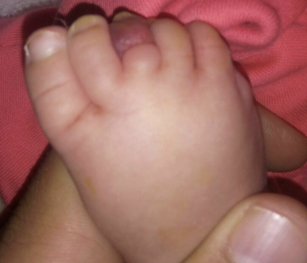
An example of hair thread tourniquet syndrome. FROM: Okan Aslantürk et al. Hair tourniquet syndrome of toes and fingers in infants. Acta Orthop Traumatol Turc. 2019 Jul; 53(4): 306–309. Used with creative commons license.


How often does it occur?

The exact risk of HTTS is not clear. Approximately 100 cases of HTTS are previously described. A 2009 study by Claudet et al estimated the incidence at 0.02 % ( 2 in 10,000).


What are the risk factors for Hair Thread Tourniquet Syndrome?

The risk factors are not completely understood.

Young age is an important risk factor as most affected individuals are under the age of 5 years, with many under the age of 1 year. HTTS of the digits typically affects individuals under the age of 1 years and HTTS of the genitals is more typical of individual over the age of 5

In reported cases of HTTS affecting the toes, the mean time of affection of toes was the fourth month of life according to a review by Mat Saad et al. This correlates with the period of telogen effluvium (hair shedding type loss) that many mothers experience.

Reports by Claudet et al and Aslantürk et al found that most patients were under 6 months.

Thread like materials such as gauze that may be used in babies kept in hospitals are a risk factor.

An infant or child sleeping with a parent or sibling with long hair can increased the risk for neck- hair-thread tourniquet syndrome (NHTTS).

Poor hygeine has been proposed as a risk factor in some studies.


What are the symptoms of Hair Thread Tourniquet Syndrome?

Symptoms will depend on the site involved. For example, oral HTS may present with difficulty swallowing or difficulty breathing. Neck hair-thread tourniquet syndrome (NHTTS) leads to difficulty breathing. Involvement of the toe can lead to redness and swelling and pain.


How serious is Hair Thread Tourniquet Syndrome?

The conditions can be very serious. The condition leads to loss or reduction of blood supply to affected areas which leads to progressive ischemia and eventually necrosis. This condition is often a medical emergency. Loss of the affected area can occur in hours to weeks depending on the specific site and the degree of ischemia.

In 2005, Milkovich et al reported the death of a child from HTTS.

A 2006 review by Mat Saad et al found that the chance of complications will depend on the organ involved. For example, complication developed with a rate of 54.5% in fingers, with a rate of 52.6% in penis and with a rate of 2.3% in toes.


How is it treated?

Urgent treatment may be needed. Removal of the hair is the treatment as well as addressing associated swelling and pain. This may require sedation, anesthesia and/or surgery to remove hair or free hair from the anatomical structure. In some cases, the hair is trapped within tissues as a result of being there for some time. Therefore, it may be more difficult to remove by simply forceps removal and may require surgical removal in a procedure room or operating room.

REFERENCES

Flores JR. Hair tourniquet syndrome in the dental patient. Anesth Prog 2014;61:111–2. 10.2344/0003-3006-61.3.111

Hair-thread tourniquet syndrome in an infant with bony erosion: a case report, literature review, and meta-analysis. Mat Saad AZ, Purcell EM, McCann JJ. Ann Plast Surg. 2006;57:447–452.

Milkovich SM, Owens J, Stool D, Chen X, Beran M. Accidental childhood strangulation by human hair. Int J Pediatr Otorhinolaryngol. 2005;69:1621–1628

Claudet I, Pasian N, Maréchal C, Salanne S, Debuisson C, Grouteau E. Hair-thread tourniquet syndrome. Arch Pediatr. 2010;17:474–9.

Mat Saad AZ, Purcell EM, McCann JJ. Hair-thread tourniquet syndrome in an infant with bony erosion: a case report, literature review, and meta-analysis. Ann Plast Surg. 2006;57:447–52.

Sivathasan N, Vijayarajan L. Hair-thread tourniquet syndrome: a case report and literature review. Case Rep Med. 2012;2012:171368

Klusmann A, Lenard HG. Tourniquet syndrome--accident or abuse? Eur J Pediatr. 2004;163:495–498; discussion 499

Okan Aslantürk et al. Hair tourniquet syndrome of toes and fingers in infants. Acta Orthop Traumatol Turc. 2019 Jul; 53(4): 306–309.

Claudet I., Pasian N., Debuisson C., Salanne S., Rekhroukh H. Tourniquet syndrome: interest of a systematic analysis of families' social conditions to detect neglect situations. Child Abuse Negl. 2009;33:569–572


This article was written by Dr. Jeff Donovan, a Canadian and US board certified dermatologist specializing exclusively in hair loss.
Share This
No Comments

Avoiding Statins in Dermatomyositis/Polymyositis Increases Mortality

Increased Mortality in Dermatomyositis Patients Not Using Statins

Download PDF Version of this Article


I was interested to read a report from authors at Thomas Jefferson University which helped clarify some of the issues regarding whether patients with autoimmune muscle diseases should be given statins. The concern centers, in part, around the possibility that statins increase the risk of myopathy. So using a drug that might cause muscle problems in a patient with muscle problems or at risk for muscle problems is an obvious concern.

The researchers looked back at about 10 years of anonymized US health data from a database. They included patients in their study who were diagnosed with hyperlipidemia after being diagnosed with dermatomyositis or polymyositis or rheumatoid arthritis. The US researchers then compared statin initiation rates and mortality outcomes.


Results

The authors found 33,000 rheumatoid arthritis patients with hyperlipdemia and 1079 dermatomyositis/polymyositis patients with hyperlipidemia. Rheumatoid arthritis patients with high lipids were much more likely to be prescribed a statin than dermatomyositis/polymyositis patients (27.4% vs 17.91 %, p<0.0001). Importantly, the dermatomyositis/polymyositis “statin” users were shown to have a reduced mortality rate (75 deaths/1000/year) compared to “non-statin” users 147 deaths/1000/year (p = 0.0273, HR = 0.515, CI 0.28-0.93). CPK muscle enzymes were similar in dermatomyositis/polymyositis “statin” users and non statin users.


Discussion

This was an interesting and valuable study. I see many patients with dermatomyositis and have become involved in this very discussion about statin initiation many times. Although I’m not generally the doctor prescribing the statin, I’m often asked to weigh in on my thoughts. This paper shows us that we need to address the cardiovascular risk of our dermatomyositis patients a little more aggressively perhaps than we do now. The authors here call for patient-centric cardiovascular guidelines for patients with dermatomyositis and polymyositis who have elevated lipids. Withholding statin therapy due to fear of worsening or triggering muscle disease is probably not the way to go for most patients - and may even increase mortality.


REFERENCE

Fares J et al. Low utilization of statins in patients with dermatomyositis/polymyositis and hyperlipidemia: a multicenter USA-based study (2013-2023). Clin Rheumatol. 2023 Nov 6. doi: 10.1007/s10067-023-06801-7. Online ahead of print.


This article was written by Dr. Jeff Donovan, a Canadian and US board certified dermatologist specializing exclusively in hair loss.
Share This
No Comments

Cutaneous Pili Migrans Developing 2 Days Post Marathon

Another Nice Case of Cutaneous Pili Migrans

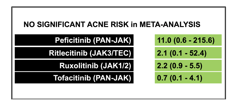
Cutaneous pili migrans is a rare skin condition characterized by a fragment of hair embedded in the epidermis or dermis layers of the skin. Differential diagnosis includes cutaneous larva migrans – as well as calluses and warts (when it occurs on the soles of the feet).

Cutaneous pili migrans (CPMs) was first reported by Yaffee in 1957 and, to date, there have been about 40-50 cases reported. Most patients are from Asia. Body locations in CPM involved include the toe, sole, ankle, breast, cheek, neck and abdomen. They can be asymptomatic or painful. We recently reviewed an interesting case of CPM.

Parker et al, 2023

Authors of a new report share the case of a 50 year old man who developed CPM 2 days after running a marathon. As in so many cases of CPM, he was treated with albendazole out of fear that he may have cutaneous larva migrans. Later, the correct diagnosis of CPM was recognized.

REFERENCE

Parker JJ, Waseh S, Hsu S. The hairy sole sign. JAAD Case Rep. 2023 Sep 24;41:102-103. doi: 10.1016/j.jdcr.2023.09.004. eCollection 2023 Nov


This article was written by Dr. Jeff Donovan, a Canadian and US board certified dermatologist specializing exclusively in hair loss.
Share This
No Comments

The Scalp Biopsy: Angle of the Punch Tool is Important!

Directing the Angle of the Scalp Biopsy

Download PDF Version of this Article

A “punch biopsy” is the preferred way to biopsy the scalp if a biopsy is indeed needed. The biopsy should be 4 mm in diameter and should ideally enter the skin at a very precise angle. The angle of entry of the punch is the angle that the hairs emerge. The angle allows the pathologist to obtain the greatest amount of information when the interpret the biopsy at a later date.

I like to trim the hairs in the area I wish to biopsy to 3 mm. This causes the trimmed hairs to “pop up” and gives me a more precise ability to position my punch biopsy tool directly over top of the hairs at the same angle they emerge. I try not to trim too many hairs as patients do not like having any reduction in perceived hair density after the biopsy. Trimming just a few hairs usually suffices. The biopsy site can be sutured or left open to heal with secondary intention although I prefer to suture.

I’m often asked “What if the biopsy site does not contain hairs?” to which I reply the biopsy site should ideally always contain hairs. If one is performing a biopsy to evaluate for hair loss, the chosen biopsy site should always contain hairs! Biopsies of sites devoid of hair are usually not so helpful.


This article was written by Dr. Jeff Donovan, a Canadian and US board certified dermatologist specializing exclusively in hair loss.
Share This
No Comments

Blocking Allergies Helps Alopecia Areata

Does blocking allergies help IN THE TREATMENT OF alopecia areata?

It’s increasingly clear that blocking allergic responses can be part of an effective treatment strategy for alopecia areata. In 2023, researchers set out to study if allergen immunotherapy (AIT) against house dust mite (HDM) allergy affects disease severity and prognosis for patients with alopecia areata.

69 patients with alopecia areata were studied. All had dust mite allergy. 34 patients received conventional/traditional AA treatment plus allergen immunotherapy against dust mites (group 1) and 35 patients received conventional/traditional AA treatment (group 2) alone.

At the end of the 3-year desensitization course, Group 1 (treated with allergen immunotherapy) presented with better hair density than Group 2 (conventional treatment) - especially for those patients with less severe forms to start with and pre-adolescent alopecia totalis/universalis patients (age ≤ 14).

In patients with elevated total IgE levels before starting allergy immunotherapy, a decrease in total IgE was correlated to a reduced extent of alopecia following completion of the allergy immunotherapy.

Conclusion and Comments

All in all, data continues to emerge that certain environmental triggers are of significance in the pathogenesis of alopecia areata. Reducing their triggers and addressing these triggers helps regrow hair to some extent in patients with alopecia areata.

Practitioners may need to address these triggers as part of a comprehensive management plan.

REFERENCE

Zeng Z et al. Affiliations expandAllergen desensitization reduces the severity of relapsed alopecia areata in dust-mite allergic patients. Exp Dermatol. 2023 Jul;32(7):1108-1119. doi: 10.1111/exd.14819. Epub 2023 Apr 28.


This article was written by Dr. Jeff Donovan, a Canadian and US board certified dermatologist specializing exclusively in hair loss.
Share This
No Comments

What is the risk of acne with JAK inhibitors?

Meta-analysis Highlights Different Risk with Different JAKS. Good News for JAK3

Download PDF Version of this Article

JAK inhibitors are increasingly used for a variety of dermatological conditions. In the hair loss world, we are using JAK inhibitors for treating alopecia areata as well as some scarring alopecias. Acne is a recognized side effect of some of the JAK inhibitors. We have reviewed the risk of acne in the BRAVE-AA baricitinib trials in the past. (See link below)

Do different JAK inhibitors have different risk of causing acne?

Authors of a new study set out to review the risk of acne with JAK inhibitors. To do so, the authors performed a systematic review and meta-analysis of 25 unique phase 2 and phase 3 studies with a sample size of 10,839 subjects. Participants in these studies either received a JAK inhibitor or a control.

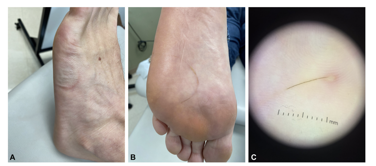
The results showed that acne was a side effect in 6.2% of JAK-treated patients compared to 1.3% of controls. This meant that treatment with a JAK inhibitor was associated with a 3.83 (95% CI, 2.76-5.32) fold increased risk of developing acne compared to controls.


Different JAK Inhibitors Have Different Acne Risk

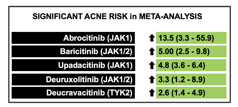
Interestingly, the various JAK inhibitors had different risk for acne. JAK inhibitors with the highest odds ratios for acne development were (in order) abrocitinib, baricitinib, upadacitinib, deuruxolitinib and deucravacitinib. Specific risk via odds ratios is shown below with their confidence intervals. Abrocitinib, for example, was found to have a 13.5 fold increased risk of acne compared to controls.

Some JAK Inhibitors Not Found to have Increased Risk

Some JAK inhibitors were not found to have increased risk across multiple studies. For example, there were no differences in acne incidence in pan-JAK inhibitors like peficitinib and tofacitinib or JAK3-specific inhibitors like ritlecitinib.

Discussion

This is an interesting study. It’s really important that we understand differences in side effects across different JAK inhibitors. Right now, hair specialists tend to do a lot of lumping of class effects when counselling patients about what sort of adverse events they might experience if they were to use a certain JAK inhibitor drug. This study teaches us that some JAK inhibitors probably carry much different risks for acne than others.


REFERENCE

Martinez J et al. Janus Kinase Inhibitors and Adverse Events of Acne: A Systematic Review and Meta-Analysis. JAMA Dermatol. 2023 Oct 18:e233830.


This article was written by Dr. Jeff Donovan, a Canadian and US board certified dermatologist specializing exclusively in hair loss.
Share This
No Comments

Does low dose oral minoxidil affect blood pressure?

Does low dose oral minoxidil affect blood pressure?

Download PDF Version of this Article


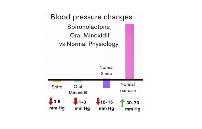
Well, there has been a handful of research studies that have examined whether low dose oral minoxidil affects blood pressure.

Sanabria et al in 2022 showed that 5 mg of oral minoxidil use in 34 males lowered systolic blood pressure by 3 mm Hg and diastolic blood pressure by 2 mm Hg when followed over a long period of time.

Jimenez-Cauhe J et al. showed that 5 mg of oral minoxidil had minimal changes in blood pressure when 10 males were followed closely for 24 hours. However, the only time point where it did have an effect was a 2 hours after intake where diastolic blood pressure dropped from 77 mm Hg to 72 mmHg before recovering completely.


Two new studies this year support the general view that low dose oral minoxidil has minimal effects on blood pressure.

A study by Ong et al in 151 males and females at a mean dose of around 2 mg showed that blood pressure did not really change much at all in users. The one exception was males 35-49 years of age where diastolic blood pressure dropped 3 mm Hg. This was not felt to be clinically significant.

A study by Imhof et al in 25 female patients using a variety of doses showed that systolic blood pressure dropped 2.8 mm Hg and diastolic blood pressure dropped 1.4 mm Hg but for the most part this seemed not all that clinically significant.


Conclusion:

All in all, low dose oral minoxidil can lower blood pressure but if it does … it’s a very minimal amount and for the most part clinically insignificant.
Our blood pressure, as humans, swings 20-60 mm Hg throughout our typical day. Blood pressure drops about 10 mm Hg when we sleep and rises 10-50 mmHg when we exercise. These tiny changes in blood pressure with low dose oral minoxidil are probably not an issue for most patients. Nevertheless, occasional patients may be sensitive to these tiny changes.


Reference

Sanabria et al. J Am Acad Dermatol. 2022
Jimenez-Cauhe J et al. J Am Acad Dermatol. 2022
Ong M et al. J Am Acad Dermatol. 2023
Imhof R et al. JAAD Int. 2023


This article was written by Dr. Jeff Donovan, a Canadian and US board certified dermatologist specializing exclusively in hair loss.
Share This
No Comments

Finasteride Dosing for Male Frontal Fibrosing Alopecia

What is the correct dose of finasteride for treating male FFA?

In this week’s question of the week, I address an interesting question related to dosing of finasteride for frontal fibrosing alopecia in males.


This article was written by Dr. Jeff Donovan, a Canadian and US board certified dermatologist specializing exclusively in hair loss.
Share This
No Comments

Three Weeks to Go Until Applications Close for the EBHF

We are just 3 weeks away from the deadline to apply for the 2024-2025 Evidence Based Hair training fellowship (EBHF). Please contact my team at info@donovanhairacademy.com for more information. The EBHF is a 87 week virtual program for hair loss practitioners around the world who wish to further develop their expertise in hair loss. The program has been developed following many years of discussion with physicians, surgeons, medical educators and allied health professionals about the depth and scope of training that is needed for advanced practitioners who will be the experts of tomorrow.


There are 3 groups of participants. A Wednesday group which meets every Wednesday from 3-6 pm PST, a Thursday group which meets Thursdays 11:30 am - 2:30 pm PST and a group of practitioners which pursue the program without attending live sessions but who complete all assignments and quizzes of the course and watch recordings of the live sessions. The requirements and expectations for successful completion of the training fellowship are slightly different in the various groups.


“Weekly Group” Requirements


For those who are part of the live lecture program on Wednesdays or Thursdays, the requirements are:

1. attendance at 70 % of the sessions (60 or more of the 87 weeks)
2. completion of all weekly MCQ (multiple choice questions).
3. completion of all 13 end of block quizzes
4. completion of 70 % of the weekly assignments


“Parallel Group” Requirements


For those who are not able to attend the live lectures, the requirements are

1. completion of all weekly MCQ (multiple choice questions).
2. completion of all 13 end of block MCQ quizzes
3. completion 100 % of the weekly assignments

Parallel Group participants are provided with access to weekly recordings of the main group’s live sessions (and weekly group participants are also provided links as well). Parallel group participants are always welcome to attend any of the live lectures & weekly sessions but must complete all assignments if fewer than 60 of the 87 meetings are attended.

A participant may switch from the live session to the Parallel group at any time provided all assignments were completed in prior weeks.


This article was written by Dr. Jeff Donovan, a Canadian and US board certified dermatologist specializing exclusively in hair loss.
Share This
No Comments

Finasteride and Hydroxychloroquine in Frontal Fibrosing Alopecia (FFA): Are the really similarly effective?

Finasteride and Hydroxychloroquine in FFA

Download PDF Version of Article

A small study shows that 2.5 mg oral finasteride and “high dose” hydroxychloroquine (above 5 mg /kg) are fairly similar in their ability to halt disease activity in patients with frontal fibrosing alopecia. The typically recommended dose of hydroxychlorqouine was not assessed in this study (ie doses of 5 mg per kg or less).

Thirty-four patients were randomly assigned into two groups: one group received  finasteride 2.5 mg once daily and one group received hydroxychloroquine 200 mg twice daily (400 mg total).

Both groups were also given the same topical treatment which included pimecrolimus (Elidel) twice daily for 5 days a week, mometasone once daily for 2 days a week, and minoxidil (5%) once daily. The use of sunscreens and moisturizing creams was allowed. Follow-up visits were scheduled after 0, 3, and 6 months of treatment.

The activity of the patient’s FFA was assessed using the 2018 Saceda-Corralo et al Frontal Fibrosing Alopecia Severity Score (FFASS) system as well as photography, and trichoscopy. The FFASS evaluates inflammation, redness, scaling and extension. It is described in the March 2018 issue of the Journal of the American Academy of Dermatology. FFASS was measured at baseline and at 3 and 6 months after treatment.

A dermatologist who was blind to the study and familiar with FFASS scored patients at each visit.

Two dermatologists who were blind to group allocation used the Global Aesthetic Improvement Scale (GAIS) a standardized 5-point relative scale range from worse (−1), no change (0), improved (1), much improved (2), and very much improved (3) to interpret the photography.


Results

Baseline characteristics of the two groups were similar including patient age, age of onset, premature menopause, facial papules, eyebrow and eyelash loss, and presence of rosacea.

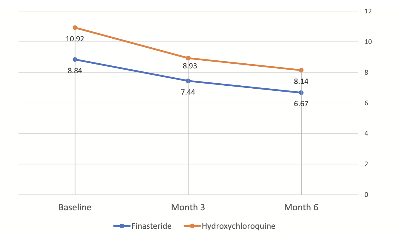
34 patients were enrolled in the study and 32 actually finished the study. This included 15 patients treated with hydroxychloroquine and 17 with finasteride, Both groups exhibited a significant decreasing trend in FFASS. Interestingly,  there was no significant difference between the two groups during the study (p = 0.110)


Figure from: Saber M et al. Clinical effectiveness of finasteride versus hydroxychloroquine in the treatment of frontal fibrosing alopecia: A randomized controlled trial. J Cosmet Dermatol. 2023 Sep 10. Used here with creative commons license.

Trichoscopic signs improved in both groups with 6 months of treatment. This included perifollicular scaling, perifollicular erythema, milky-red area, and the total trichoscopic score.

Overall, there was no significant difference in trichoscopic signs and total trichoscopic score between the finasteride and hydroxychloroquine groups.

A proportion of patients in each group had improvement. In the finasteride group, 6 (35.3%) patients showed no change or worsening of the disease, and 11 (64.7%) patients showed mild to very significant improvement. In the hydroxychloroquine group, five patients (33.3%) exhibited no change or deterioration of the disease while 10 patients (66%) demonstrated improvement ranging from mild to significant. However, statistical analyses showed that there was no significant difference between the two groups.

Table from: Saber M et al. Clinical effectiveness of finasteride versus hydroxychloroquine in the treatment of frontal fibrosing alopecia: A randomized controlled trial. J Cosmet Dermatol. 2023 Sep 10. Used here with creative commons license.

In terms of side effects, finasteride and hydroxychloroquine were both were well- tolerated treatments for patients with FFA.


COMMENTS

I congratulate the authors for performing an RCT. These types of studies are so much needed in our field.

This data here shows that finasteride and “high dose” hydroxychloroquine can both help patients with FFA.

I’m not sure that we can truly conclude that finasteride and regular dose (typical dose) hydroxychloroquine are similarly effective. The dose of hydroxychloroquine in this study was likely too high so many patients probably received a dose that was not typical of what we would normally prescribe.

With an average height of women in Iran of 5 ft 4 and an obesity estimate of 30 %, it’s likely that 70% or more of women in this study received a dose of hydroxychloroquine that was above the recommended 5 mg/kg supported by the recent American Academy of Ophthalmology guidelines.

So all that we can say here is that finasteride 2.5 mg and high dose hydroxychloroquine are likely similar in effectiveness.

We don’t really know if 2.5 mg finasteride and the recommended dose of hydroxychloroquine will be similar in effectiveness. Possibly not.

In my clinic I don’t view hydroxychloroquine as quite the same effectiveness as finasteride or dutasteride. Certainly, this data in the RCT has caused me to question my view. But I’m not convinced with this data that we can really say they are the same. The doses of hydroxychloroquine here are too high.

I agree with the authors that hydroxychloroquine can be a really good option women of reproductive age, including those who intend to become pregnant or have a personal or family history of breast cancer. In can also be a good option for those who experience a mood disturbance with finasteride or those experiencing decreased libido on finasteride.

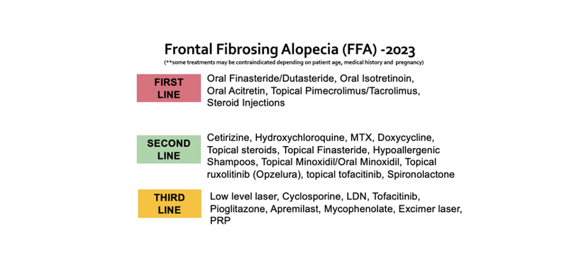
For now, I’m still keeping hydroxychloroquine as a second line agent in FFA. I use it alot and sometimes even combine it with finasteride or dutasteride !


REFERENCE

Saber M et al. Clinical effectiveness of finasteride versus hydroxychloroquine in the treatment of frontal fibrosing alopecia: A randomized controlled trial. J Cosmet Dermatol. 2023 Sep 10.

Saceda-Corralo D et al. “Development and validation of the Frontal Fibrosing Alopecia Severity Score,” Journal of the American Academy of Dermatology. March 2018. DOI: https://doi.org/10.1016/j.jaad.2017.09.034

Melles RB et al. Hydroxychloroquine Dose and Risk for Incident Retinopathy : A Cohort Study. Ann Intern Med. 2023 Jan 17. 

Melles & Marmor. The risk of toxic retinopathy in patients on long-term hydroxychloroquine therapy. . JAMA Ophthalmol. 2014 Dec;132(12):1453-60.


This article was written by Dr. Jeff Donovan, a Canadian and US board certified dermatologist specializing exclusively in hair loss.
Share This
No Comments


Share This
-->