h1.qusth1 { display: none !important; }

QUESTION OF THE WEEK

Dr. Donovan's Articles

QUESTION OF HAIR BLOGS

Filtering by Category: Cicatricial


Do I have Frontal Fibrosing Alopecia?

Do I have FFA or not?

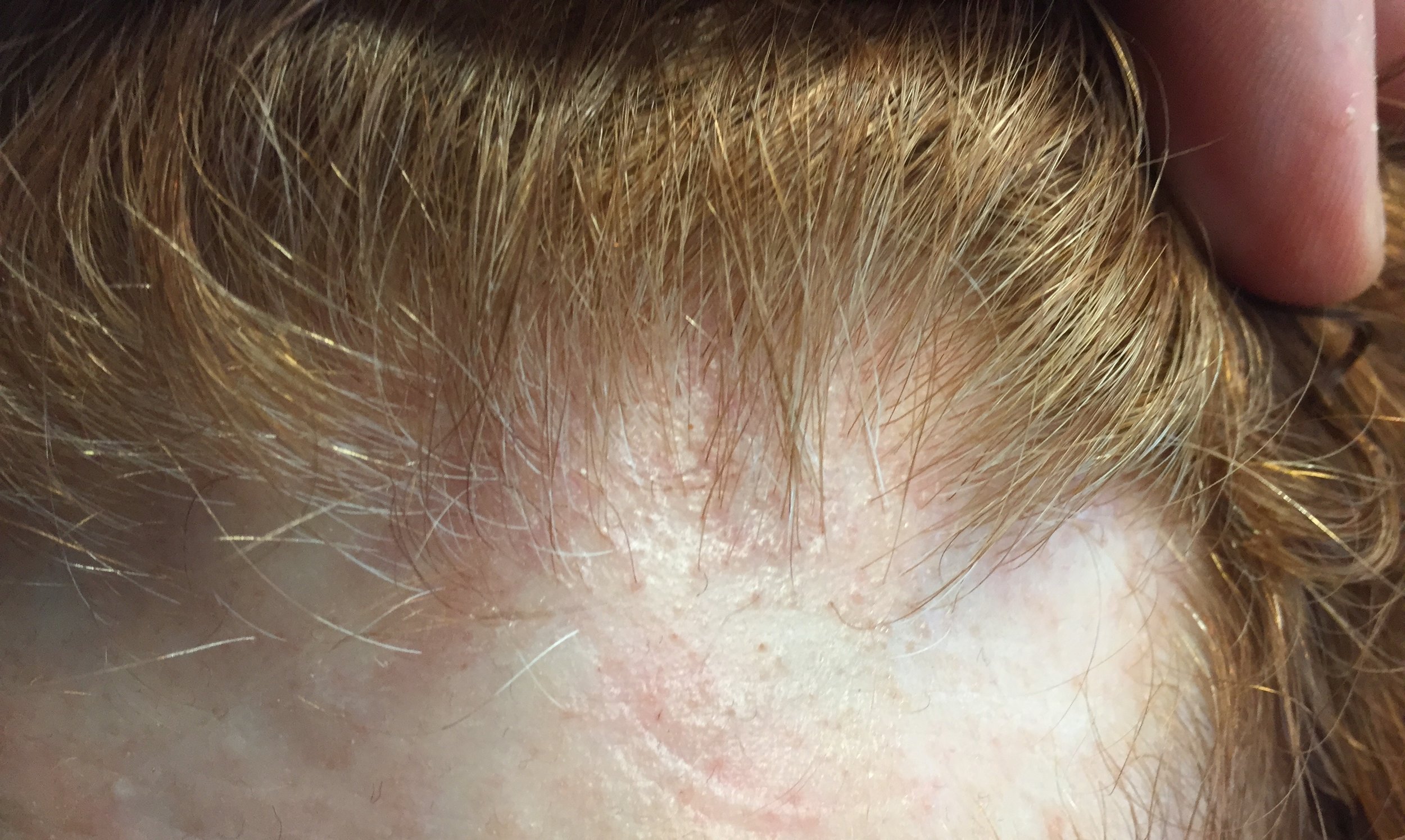
FFA of the frontal hairline. 

FFA of the frontal hairline. 

Frontal fibrosing alopecia is increasingly common. It's still a relatively rare condition overall but there is no doubt that the number of women being diagnosed is increasing. On account of the increase individuals in the general public are much more aware of this otherwise uncommon diagnosis. Patients often want to know if they have this condition. The short answer is that anyone wondering if they have FFA really should see a dermatologist for a comprehensive review and examination of the scalp. 

I enjoy helping patients understand why they do or do not have FFA. Many patients are convinced they have FFA when they clearly do not.  The following are some pieces of information that patients frequently feel points to a diagnosis of FFA when it fact it does not. Following this, I have outlined 5 pieces of information that actually is helpful (and increases the odds somewhat that a true diagnosis of FFA could be present). Exceptions of course exist and nothing replaces an in person review for an up close examination.


5 PATIENT COMMENTS THAT ARE NOT HELPFUL IN DIAGNOSING FFA

In my experience with countless numbers of patients who think they have FFA (but don't).... the following pieces of information are not helpful to making the diagnosis even though patients may think that it is!


Comment 1: "My hairline is changing"

Why is it not helpful? Hairlines change for many reasons including androgenetic alopecia, traction alopecia, alopecia areata and telogen effluvium. Frontal fibrosing alopecia is certainly on that list but there are too many reasons for frontal hairloss to make the observation of hairline changes a key feature for diagnosing FFA. A patient with hairline changes could have FFA but could have other conditions too.


Comment 2: "I see 'lonely' hairs"

Not everyone knows what a lonely hair is. But a patient who understand what is meant by a "lonely hair" has generally done a lot of reading and are extremely knowledgeable about hair loss! The reality however is that most "lonely hairs" that most people find in the scalp are not what really constitutes the gist of what these hairs are all about. My feeling is that the presence of more than 6 hairs across the frontal hairline at a distance of greater than 1.5 cm from the main hairline probably does in fact raise suspicion for true 'lonely hair' phenomenon.


Comment 3: "I have redness and scarring in my scalp" 

Redness alone does not constitute a diagnosis of FFA. In fact, FFA tends to be more pinkish and subtly red than overtly red. There are simply too many scalp conditions that cause redness to give this observation any significance.

I'm always concerned when patients feel they can "see scarring" because one can't truly see scar tissue as it is mostly under the scalp. In advanced scarring alopecia, one certainly can see evidence of scarring but these cases are usually fairly advanced. In all fairness, one can however see scalp color changes (mostly to a white color) that would suggest the presence of scarring. However, one can't actually see scarring.


Comment 4: "I now have so many baby hairs"

Baby hairs are not really a feature of FFA. In fact, the presence of baby hairs probably argues against the diagnosis of FFA for most patients than actually favours the diagnosis. This is because FFA tends to be a destructive process that involves the preferential destruction of baby hairs (medically termed vellus hairs). The presence of abundant baby hairs is not a feature of progressive FFA.


Comment 5: "I have no family history of balding so it only makes sense something else is going on"

The argument that an individual must have some other diagnosis other than genetic hair loss on account of the lack of genetic hair loss in the family is never a valid argument. There are women who come to be diagnosed with androgenetic alopecia despite a family history of men and women with thick hair. The patient's account of her family history is not very relevant to most diagnoses of hair loss. Sounds strange but this is true: I frequently diagnose androgenetic alopecia in women who stated their family is comprised of individuals with 'good hair.'


TOP 5 FINDINGS AND COMMENTS THAT ARE HELPFUL IN DIAGNOSING FFA

Nothing can substitute for a careful review of one's story and examination of their scalp with dermoscopy. The following pieces of information are helpful when considering a diagnosis. Not all patients have the following features, but they greatly increase the odds that what we are dealing with is FFA.


FINDING 1. Both sideburns are lost (above the ears). Not thinned but lost. The regions below where the spectacle of the glasses sit has been depleted of hairs.

Loss of the hair frim both sideburns is actually quite an important piece of information when it comes to diagnosing FFA.  This finding is mot present in everyone with FFA but is quite common if one looks.


FINDING 2. Baby hairs are not seen in the frontal hairline but rather many single long hairs are seen. Not all the hairs have redness around them but many do have a faint redness

FFA is a process that destroys fine vellus hairs - but it does so with inflammation. The presence of small amount of inflammation (usually without symptoms) is a key finding.


FINDING 3. If the eyebrows are lost, they are significantly changed

Eyebrow loss is common in FFA and in a large number of individuals with FFA, they are the first finding. Eyebrow loss of course does not happen in all women with FFA and loss of eyebrows may even come after the loss of the frontal hair. However, significant eyebrow loss requiring tattooing, microblading does raise the odds a bit that the presentation fits with possible FFA.  Still, one needs a careful examination before concluding that anyone's eyebrow loss is FFA. Nevertheless, changes in the eyebrows are common to FFA.


FINDING 4. The patient is between 46 and 66

FFA is rare in the 20s and 30s. That's not to say it can't occur as we have patients in their teenage years affected. However, from a statistical point of view, women under 40 who come in with worries that they have FFA actually rarely end up having FFA. There are exceptions of course but it is not a common occurrence. In our clinic, 98 % of patients who receive a diagnosis of FFA are older than 46.


FINDING 5. Veins are seen much easier on the scalp than before and the skin itself just does not look quite normal.

FFA is a complex condition and is probably more than just a 'hair disease'.  It's likely a complex autoimmune disease that affects hair predominantly but also affects the skin as well The skin itself changes in FFA - and does so by thinning. We call this "atrophy." Some women with FFA have minimal to no atrophy but many have significant atrophy which leads to the veins becoming much more visible. Generally, the skin in the area of the hairline does not look the same as the skin of the forehead - the affected area in FFA is lighter in color and smooth.


CONCLUSION

Frontal fibrosing alopecia is a complex condition. There is no 'one way' that it appears. There are 100s of different combinations. Navigating the diagnosis oneself is frequently met with challenging. I've summarized here 5 points that patients place a great deal of emphasis on when looking at their hair - but actually don't carry all that much weight when making the diagnosis. 


This article was written by Dr. Jeff Donovan, a Canadian and US board certified dermatologist specializing exclusively in hair loss.
Share This
12 Comments

Hair dye for patients with LPP: Any Problems?

Hair dye for patients with LPP: Any Problems?

I am frequently asked if patients with lichen planopilaris (LPP) and similar scarring alopecias can dye their hair?For most people with scarring alopecia the use of permament, semipermanent or temporary hair dyes is completely safe. I always advise that patients review with their dermatologist if they feel any change in their scalps whatsoever following the salon visit or home application. Any marked change in scalp itching, burning or even new tenderness in the scalp would cause concern but fortunately this is extremely rare. 

For my patients with minor irritation from hair dye application, I sometimes recommend use of an anti-inflammatory cortisone shampoo (ie clobetasol proprionate (Clobex) shampoo) 1-24 hours before the dye is applied. Some of my patients even bring the shampoo to the salon and have the stylist use and wash it out let the normal instructions. 

All in all, most individuals with LPP don't experience any difficulties with hair dyes and no special precautions are needed.


This article was written by Dr. Jeff Donovan, a Canadian and US board certified dermatologist specializing exclusively in hair loss.
Share This
1 Comment

Scarring Alopecia: Loss of the follicular opening is a hallmark

Scarring Alopecias Cause Scarring

scarring

Scarring alopecias are hair loss conditions that are associated with the development of permanent hair loss. There are dozens of different types of scarring alopecia. 
Some scarring alopecias itch. Some don't. Some are associated with increased shedding. Some aren't. Some are red. Some aren't. Some bleed. Most don't. 


However what is common to all scarring alopecias is the disappearance of the follicular opening or "pore." The development of scar tissue beneath the skin leads to the destruction of the follicular pore opening.

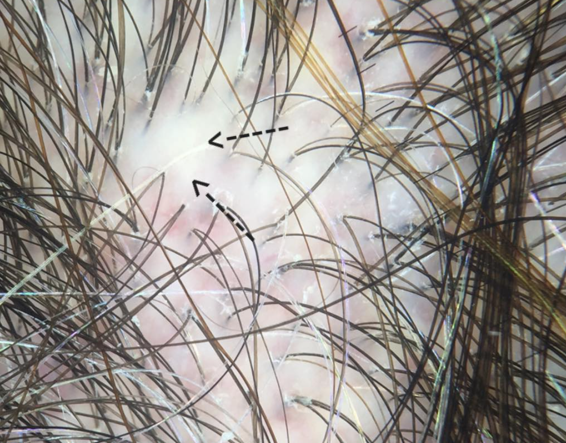
The arrows point to an area of scarring in a subtle early scarring alopecia


This article was written by Dr. Jeff Donovan, a Canadian and US board certified dermatologist specializing exclusively in hair loss.
Share This
No Comments

Am I taking the right amount of hydroxychloroquine (Plaquenil)?

Hydroxychloroquine (Plaquenil): Am I taking too much?

Hydroxychloroquine is an oral medication used in a variety of autoimmune conditions. Side effects have been discussed previously but today we will focus on eye side effects. A number of side effects are possible ranging from vision changes to double vision to asymptomatic changes in various parts of the eye.


The Risk of Retinopathy with Hydroxychloroquine

"Retinopathy" is one of the more worrisome side effects of Hydroxychloroquine. At appropriate doses, studies show that the risk appears to be about 1 % of patients at 5 years of use and 2 % at 10 years. After 20 years, the risk may rise to 20 %. Once the retinal toxicity from hydroxychloroquine occurs, it is believed that the changes in the retina are permanent. Furthermore, the disease can even progress even if hydroxychloroquine is stopped.  


Risk Factor for Retinal Toxicity

Retinal damage can occur in anyone. However, the risk may be increased if the following risk factors are present

  • Longer Duration of use (cumulative dose)
  • Renal or hepatic functional impairment. Compromised kidney and/or liver function can lead to increased accumulation of hydroxychloroquine in the tissues.
  • Age over 60 years.
  • Preexisting retinal disease
  • Concurrent tamoxifen therapy


What dose should I take?

It's clear that taking the appropriate dose reduces (but does not eliminate) the chance of side effects. The optimal dose is 6.5 mg for every kg of lean body weight (not simply what the patient weighs). "Lean body weight" is essentially the patients expected weight for their height and gender - it does not include the "extra" weight that some might carry. Instead of calculating lean body weight, some clinicians advocate simply using the patient's true body weight and multiplying by 5 (instead of 6.5).  In our clinic we typically dose hydroxychloroquine according to the following grid:

Hydroxychloroquine Dosing


Conclusion

The risk of eye related toxicity is low in the first 5-10 years of hydroxychloroquine use provided the dosing is respected. This study has had great importance as it has further helped to define risk and has encouraged changes in screening guidelines. These guidelines now include an initial examination but dedicated yearly screening to begin only after 5 years in otherwise healthy individuals deemed at low risk for eye problems.


Reference

(1) Melles & Marmor. The Risk of Toxic Retinopathy in Patients on Long-term Hydroxychloroquine Therapy. JAMA Ophthalmolol. 2014;132(12):1453–1460.


This article was written by Dr. Jeff Donovan, a Canadian and US board certified dermatologist specializing exclusively in hair loss.
Share This
No Comments

Scarring Alopecia from Hair dyes, highlights and bleach

Chemical injury can lead to scarring alopecia

hair highlights.png

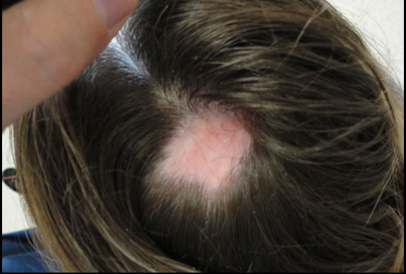
Hair dyes, highlights and bleaching can rarely lead to chemical injury. It's not common of course but the story is always the same: within seconds to minutes of applying a hair dye or highlights, the patient complains of intense burning and/or pain and requests the product to be removed. Hours to days later hair loss starts and within a week or two the patient has permanent hair loss (such as shown in the figure to the right). I have seen this type of scalp injury phenomenon many, many times and I do even feel that it is increasing world-wide. 


Management Chemical Injury to the Scalp

The most important thing to do in these situations of potential chemical irritation is remove whatever chemical could be causing the reaction. The dye or highlight must be removed, neutralized and rinsed off.  In my opinion a dermatologist should be consulted for management and monitoring. Rarely skin necrosis can occur from ehuberant reactions. One can not predict on day 1 whether the patient will have hair loss and whether any hair loss will be permanent. This will not be clear until day 14-28. In the short term one must management skin health, prevent infection, and limit and control inflammation. These are within the skills of a dermatologist.  A biopsy may be considered to determined the type of inflammation and evaluate for scarring if it is unclear. 


Hair transplantation or Surgical Correction: Best methods for Camouflaging Chemical Injury

Too often I hear it said in these scarring alopecias that a biopsy was done and because the biopsy said the disease was inactive the patient proceeded to surgery. Keep in mind that we determine if a scarring alopecia is inactive by simply following what it does over time. Relying on a biopsy alone to determine if it is acitve is not a good idea for most people. If the area of hair loss has not changed at all in any way shape or form (same size area) and is not itchy and has no burning or pain thena biopsy supports it is inactive.  Even if a biopsy says the scarring alopecia is inactive but the area is expanding over time and is itchy or red... it is not inactive. This is a common scenario and a common error in managing scarring alopecia.

One needs to wait 12-24 months for a scarring alopecia before surgery. Photos need to be done every 2-3 months in my opinion even for chemical burn related hair loss. If the photos look the same when placed side by side over a one year period, one can say the scarring alopecia is probably quiet.  Rarely, this can be shortened to 6 months for chemical injury but one year is a safer waiting period to be confident there is no evidence of a slowly progressive scarring alopecia in evolution. 


Is waiting really necessary when planning surgery in scarring alopecia?

All this background waiting and monitoring needs to be done before surgery. It sounds excessive and time consuming and unnecessary- but it is far from it. Surgery for scarring alopecia can be highly successful provided it's done in the right patient. Too often, it is not done on the right patient... and then it does not work well or does not work at all and physicians, patients and the medical community as a whole loses confidence in the value of surgical restoration options.


Options for Restoration

 The only way to restore the appearance is surgical. Medical options do not help improve density once the area is permanently scarred. If the area is small surgery via a plastic surgeon can be a great option. Many burns from hair dyes are in the form of small coin shaped patches. A flap (rotational flap etc) can work wonders and may be superior to hair transplanting. For this a surgeon is needed with skill in such flaps. The above patient would be a good candidate for a flap.

For hair loss that occurs more diffusely (and not in the above mentioned classic hair dye chemical burn patches), hair transplants can sometimes ca a good option. In my opinion, the key factor in choosing a surgeon is their experience and dedication to hair transplantation. The actual credentials is not so important to me and some of the world's top surgeons are a range of family physicians, dermatologists, plastic surgeons, former emergency room physicians. If her or she is dedicated soley to hair transplanting and has performed a large number surgeries and has been doing it for many years and has a good before and after album of scar procedures, then it may be worth a visit to speak to that surgeon. 


This article was written by Dr. Jeff Donovan, a Canadian and US board certified dermatologist specializing exclusively in hair loss.
Share This
No Comments

Hair Systems vs Wigs for Scarring Alopecia: Which should I consider?

Is a hair system possible for someone with active scarring alopecia?

Scarring alopecia is a form of hair loss which can lead to permanent bald areas on the scalp. Treatments focus on helping to stop hair loss but do not improve hair in most. Some individuals elect to purchase a wig, hairpiece or hair system while receiving treatment.  For the purposes of the discussion that follows, I will specifically use the term hair system to refer to a scalp camouflaging method whereby synthetic or human hair is attached to a layer of some sort and glued to the scalp with some type of adhesive. I will use the term wig or hairpiece to include a broad array of similar products that attach via clips, tape or elastic.

Two Key factors to consider

The are quite a few factors that go into deciding what type of wig, hairpiece or hair system is appropriate for someone with scarring alopecia. The two main factors to consider when I am meeting with a patient trying to decide what type of system they should purchase are the following: 

(1) How active is the scarring alopecia right now? 

(2) Does the patient need topical steroids, steroid injections or special shampoos as part of his or her ongoing management strategy? 


(1) How active is the scarring alopecia right now? 

If a patient has a very active scarring alopecia (with many symptoms and/or rapid hair loss), it will be important to consider choosing a hair piece or wig with clips or tape over a hair system that is attached to the scalp with adhesive. A patient with an active scarring alopecia needs to have the scalp examined often to determine if treatment is working and to modify the exact treatment.  For some scarring alopecias like folliculitis decalvans it may be important in some cases to frequently shampoo the scalp with antibacterial agents.  An easily removable wig or hairpiece is preferred.

As the scarring alopecia becomes "quieter" it may be possible to consider  shifting to a hair system and more permanent types of adhesive-based attachments. I generally ask patients to coordinate removal of the hair system at their salon on the same day as their follow up appointment with me or if that is not possible to have good pictures taken at the salon in the day that their system is removed, washed and reapplied (generally referred to as a "servicing).


(2) Does the patient need topical steroids, steroid injections or special shampoos as part of his or her ongoing management strategy? 

For patients who answer yes to the above question, a wig or hairpiece will be preferred over a hair system. Topical steroids and steroid injections can be the mainstay of treatment for many patients with scarring alopecia. Even in relatively quiet scarring alopecias, topical steroids may still be needed every few days. For very active scarring alopecias, steroid injections may be needed monthly. As mentioned above under point (1), for some scarring alopecias like folliculitis decalvans it may be important in some cases to frequently shampoo the scalp with antibacterial agent. Therefore, in such cases where there is active disease, a more permanent hair system becomes either impractical or inconvenient. It needs to be removed for these treatments to be properly administered and this is not easy if a hair system is glued to the scalp. 


Conclusion

All in all, these are the discussions that patients will want to have with their dermatologist. If topical steroids are needed daily and steroid injections are needed monthly for example (for very active disease) use of a hair system with an adhesive is less preferred over wigs or hairpieces with clip attachments or tape. If possible, these are discussions the dermatologist might have with the wig salon itself to best coordinate the proper care of the patient .


This article was written by Dr. Jeff Donovan, a Canadian and US board certified dermatologist specializing exclusively in hair loss.
Share This
1 Comment

Lichen planus of the hair follicle (lichen planopilaris)

LPP-injury

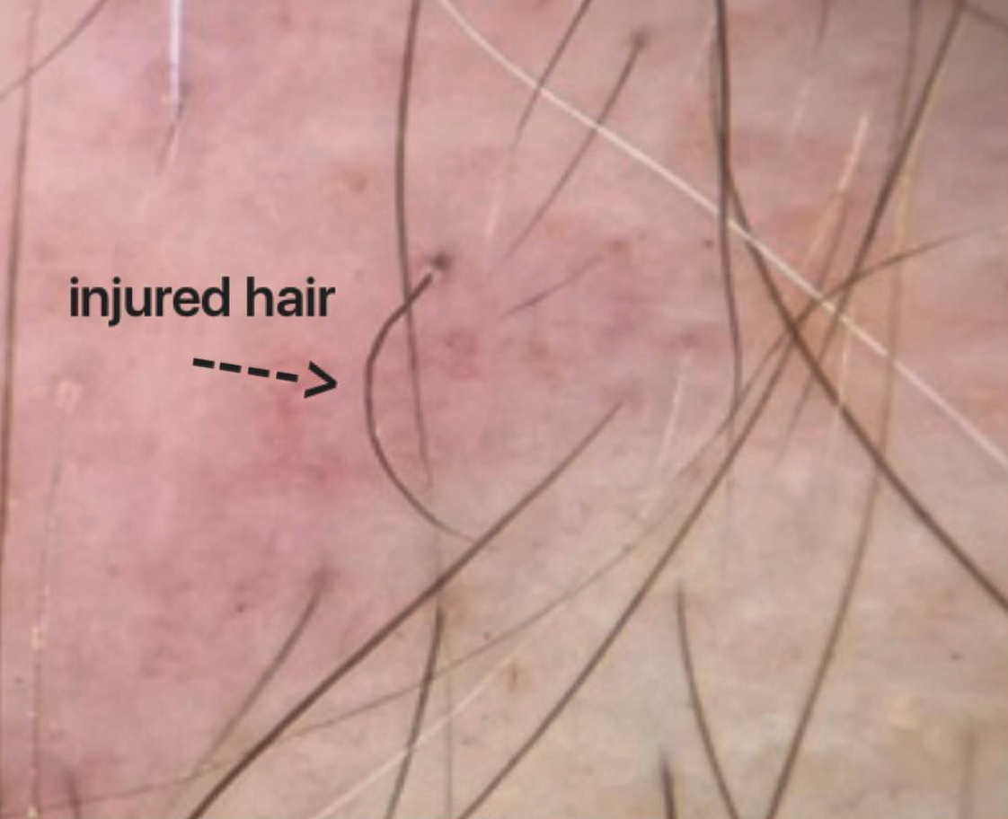
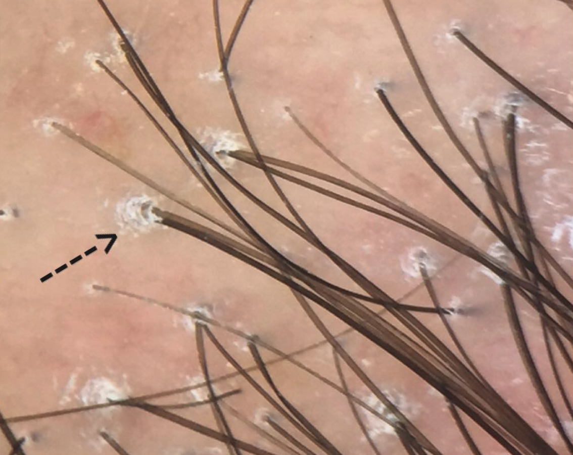
Lichen planopilaris (LPP). In follow up to yesterday's post, here is another example of a hair follicle injured by the inflammatory and scarring reaction that occurs beneath the skin surface in LPP.


This article was written by Dr. Jeff Donovan, a Canadian and US board certified dermatologist specializing exclusively in hair loss.
Share This
No Comments

Injured hairs in Lichen planopilaris (LPP)

LPP: A Scarring Hair Loss Condition

LPP injury.jpg

Lichen planopilaris ("LPP") is a scarring alopecia (scarring hair loss condition). Inflammation first develops around hair follicles which triggers the development of scar tissue (fibrosis) around hairs. This inflammatory reaction causes hairs to eventually die. One such injured hair is shown in the photo.

Many patients first develop scalp itching, burning and tenderness. Once a hair is destroyed, it can not regrow. Early recognition of the disease and aggressive treatment and frequent monitoring is essential.


This article was written by Dr. Jeff Donovan, a Canadian and US board certified dermatologist specializing exclusively in hair loss.
Share This
No Comments

Lichen planopilaris: A closer look at "follicular hyperkeratosis"

Follicular hyperkeratosis = Perifollicular scale

Lichen planopilaris (also known as "LPP") is a scarring hair loss condition. Individuls affected by the condition develop hair loss, increased hair shedding along with scalp itching, burning and pain.


Scaling in LPP

Screen Shot 2017-10-14 at 8.24.15 AM.png

An up close examination with dermoscopy is shown here and shows classic features including white scale around hairs in early stages. This scale is known as "perifollicular scale" or "follicular hyperkeratosis." This scale is often prominent in active stages of the disease. It can be reduced by treatment and even reduced to some extent by a good shampooing of the scalp.


Treatments for LPP

Treatments include topical steroids, steroid injections, topical calcineurin inhibitors, and oral treatments such as doxycycline, hydroxychloroquine (Plaquenil), methotrexate, mycophenolate mofetil, cyclosporine, and others. Lasers, including the 308 nm excimer laser may also provide benefit.


This article was written by Dr. Jeff Donovan, a Canadian and US board certified dermatologist specializing exclusively in hair loss.
Share This
No Comments

Is Lichen Planopilaris Caused By A Fungus?

Lichen planopilaris is the long name given to a type of scarring hair loss condition. It is sometimes referred to as follicular lichen planus. The name "lichen" comes from the skin lesions of lichen planus that some patients with lichen planopilaris also have. The skin lesions are flat just like lichens that one might see walking in the forest. 
Lichen planopilaris is not due to a fungus. It is an autoimmune inflammatory condition that causes permanent hair loss. Treatments include anti-inflammatory agents not antifungal agents.


This article was written by Dr. Jeff Donovan, a Canadian and US board certified dermatologist specializing exclusively in hair loss.
Share This
No Comments

Hair transplants for Scarring Alopecia

How successful are hair transplants in scarring alopecias?

worldwide.jpg


This is an in important question and one that needs good data. I serve as the chair of a committee of the International Society of Hair Restoration Surgery. On Friday we sent out a survey to hair transplant surgeons around the world with the hopes of gathering more information on the successes and failures surgeons have had when transplanting scarring alopecias.

There is no doubt that hair transplantation works wonderfully in some patients with scarring alopecia and does not work well in others. One must always have quiet (inactive) disease for at least 1-2 years before a transplant is attempted.


What are the criteria for transplanting scarring alopecia?

In general, a scarring alopecia must be quiet for 1-2 years before a transplant can be even considered. Several years ago I put forth criteria for determining if an individual with lichen planopilaris is a hair transplant candidate:


Criteria for Hair Transplantation Candidacy in LPP

1.  The PATIENT should be off medications.

Ideally the patient should be off all topical, oral and injection medications to truly know that the disease is burnt out and ‘inactive’. However, in RARE cases, it may be possible to perform a transplant in someone using medications AND who meets criteria 2, 3 and 4 below.  This should only be done on a case by case basis and in rare circumstances as the risk for disease reactivation is high.  A patient using medications to suppress disease activity is at high risk for reactivation following hair transplant surgery. It is a last resort in a well-informed patient.

2. The PATIENT must not report symptoms related to the LPP in the past 12 months, (and ideally 24 months).

The patient must have no significant itching, burning or pain. One must always keep in mind that the absence of symptoms does not prove the disease is quiet but the presence of symptoms certainly raises suspicion the disease could be active.  Even the periodic development of itching or burning from time to time could indicate the disease has triggers that cause a flare and that the patient is not a candidate for surgery. The patient who dabs a bit of clobetasol now and then on the scalp to control a bit of itching may also have disease that is not completely quiet. 

3. The PHYSICIAN must make note of no clinical evidence of active LPP in the past 12 months, (and ideally 24 months).

There must be no scalp clinical evidence of active LPP such as perifollicular erythema, perifollicular scale (follicular hyperkeratosis). In addition, the pull test must be negative. 

4. Both the PATIENT and PHYSICIAN must show no evidence of ongoing hair loss over the past 12 months (and ideally 24 months). 

There must be no further hair loss over a period of 24 months of monitoring off the previous hair loss treatment medications. This general includes the patient and physician's perception that there has been no further loss as well as serial photographs every 6-12 months showing no changes.  As discussed above, the 12 month waiting time is the standard of care as an accepted definition for hair transplant candidacy.

5. The patient must have sufficient donor hair for the transplant.

Not all patients with LPP maintain sufficient donor hair even if the disease has become quiet. 


Disease Reactivation Following Surgery

My research has focused on the chances of reactivation of LPP after surgery. It is important to be aware that ANY patient with LPP is at risk for reactivation or a 'flare' of their LPP after surgery.  The risk, I estimate, is as follows:


LPP Reactivation Risk (Donovan, J, unpublished data)

i)               A patient with active LPP before their transplant is nearly guaranteed to have a flare of his or her LPP if a hair transplant is done. (estimate 90-100 % chance of flare within 2 years post transplant)

ii)             A patient with partially active LPP before their transplant is very likely to have a flare if a hair transplant is done. (estimate 70-90 % chance of flare within 2 years post transplant)

iii)            A patient with medication induced inactive LPP before their transplant has a moderate chance of a flare if a hair transplant is done (estimate 50-70 % chance of flare within 2 years post transplant)

iv)            A patient with inactive LPP off all medications for 1 year before their transplant has a low chance of a flare if a hair transplant is done (estimate 10-25 % chance of flare within 2 years post transplant)

v)             A patient with inactive LPP off all medications for 2 years before their transplant has a low but definite chance of a flare if a hair transplant is done (estimate less than 10% chance of flare within 2 years post transplant)


This article was written by Dr. Jeff Donovan, a Canadian and US board certified dermatologist specializing exclusively in hair loss.
Share This
No Comments

Atrophy in FFA: Is it from my topical steroids?

Atrophy in FFA is often from the disease itself 


A common concern from patients with FFA is that their steroids caused atrophy. By atrophy we mean thinning of the skin. Patients with atrophy have thin skin, visible veins. In FFA atrophy leads to blue veins becoming easy to see throughout the frontal scalp and especially at the temples. Patients want new options for treating the disease because they are worried about the atrophy.


FFA Causes Atrophy

There is one assumption that is often wrong here - and that is that steroids are the sole cause of atrophy in FFA. MOST of the time the steroids are not the main cause of the atrophy ! It is very important to keep in mind that the disease itself causes atrophy and visible veins. It is certainly very true that the steroids can cause atrophy too. But FFA itself is usually the leading cause of atrophy in patients with FFA. Many many patients with FFA who have never used steroids can have atrophy - some severe. In fact, severe atrophy is one of the so called poor prognosis signs in FFA. 


Treatment Considerations for Patients with Marked Atrophy

When patients show a considerable amount of atrophy, I usually try to limit this by using non steroids instead of steroid. Non steroids such as pimecrolimus (Elidel) and tacrolimus (Protopic) do not cause atrophy. They seem equivalent although no comparison studies have been done.  My previous research has also shown that finasteride and dutasteride may actually reverse atrophy in a proportion of patients. 


This article was written by Dr. Jeff Donovan, a Canadian and US board certified dermatologist specializing exclusively in hair loss.
Share This
No Comments

Frontal Fibrosing Alopecia (FFA)

Other than the Front, What is Affected?

Frontal fibrosing alopecia (FFA) is a scarring alopecia that affect women to a much greater extent than men. In FFA the frontal scalp is typically affected. However, the name does not capture the full extent of the hair loss. Patients with FFA frequently develop hair loss around the back of the scalp (behind the ears and very back above the neck), and frequently in the middle of the scalp as well. Eyebrows, eyelashes, arm hair, leg hair, underarm hair and pubic hair are frequently affected.


This article was written by Dr. Jeff Donovan, a Canadian and US board certified dermatologist specializing exclusively in hair loss.
Share This
No Comments

Partially Treated Lichen Planopilaris

 Scale Gone, Redness Remains

Lichen planopilaris (LPP) is a scarring hair loss condition. The goal of treating LPP is to stop the condition. Successful treatment is associated with a halting of hair loss but also with an improvement in the symptoms and signs of the disease. Patients will notice a reduction in itching and burning and clinically there will be an improvement in scaling and redness around hairs. Sometimes scaling is the first to improve and improvements in redness happen later. This picture shows a patient with partially treated lichen planopilaris. The disease is still active although scaling has improved. The patient's itching has also improved.


This article was written by Dr. Jeff Donovan, a Canadian and US board certified dermatologist specializing exclusively in hair loss.
Share This
No Comments

Sea of Singles (SOS): A Potential Sign of Scarring Alopecia

Lichen planopilaris (LPP) is a type of scarring hair loss condition. Patients frequently present with scalp itching, and sometimes scalp burning and tenderness. Increased hair shedding is common in the early stages. Hair loss is generally permanent and treatment helps stop the disease or at least slow down progression.

Clinically, dermoscopy (trichoscopy) of LPP often shows perifollicular erythema and perifollicular scale (follicular keratosis).

These findings are not present in all forms of LPP. A less common presentation of LPP is shown in the photo. Patients have hair loss with scalp itching. However, by dermoscopy they have many single hair follicles growing in a base of redness. This is what I have termed the "sea of singles" (SOS) appearance to describe the numerous single hairs and absence of hair follicle units containing 2 and 3 hairs. This form of LPP is similar to Abbasi's subtype described in 2016 and fibrosing aloepcia in a pattern distribution described by Zinkernagel in 2000. The "SOS" trichoscopic appearance is important to remember and provides a clue that the patient may have a scarring alopecia.


Reference

Zinkernagel MS et al. Arch Dermatol 2000

Abassi A et al. Dermatol Surg. 2016.


This article was written by Dr. Jeff Donovan, a Canadian and US board certified dermatologist specializing exclusively in hair loss.
Share This
No Comments

Oral Immunosuppressants for Lichen planopilaris: should I increase my dose?

Dosing oral immunosuppressants for Lichen planopilaris (LPP)

There are many different immunosuppressants and immune modulators that can be used for treating lichen planopilaris. Examples include doxycycline, hydroxychloroquine, methotrexate, mycophenolate, cyclosporine.  I'm often asked what dose a patient should be using? 


What dose should a patient be using? 

When it comes to immunosuppressant medications, I always try to keep patients on the lowest possible dose that controls their disease. Generally I start at fairly standard doses of immunosuppressants and observe what happens to the patient's hair loss. For example, this might be 200 or 400 mg of hydroxychloroquine (Plaquenil) daily, 15-20 mg of methotrexate weekly, 150-300 mg of cyclosporine, 500-1000 mg of mycophenolate mofetil, 100 mg of doxycycline. If the disease is vastly improved after a few months, we may consider going down on the dose or staying at the same dose for a few more months. If the disease is getting worse, we might consider going up on the dose is their is room to go up or changing the immunosuppressant altogether. 


This article was written by Dr. Jeff Donovan, a Canadian and US board certified dermatologist specializing exclusively in hair loss.
Share This
No Comments

Nausea with Doxycycline: What strategies can help reduce nausea?

Doxycycline and Nausea

Doxycycline is an antibiotic. It's used of course in treating infections but it is commonly used for a variety of scarring alopecias including lichen planopilaris, frontal fibrosing alopecia, central centrifugal cicatricial alopecia, folliculitis decalvans and sometimes dissecting cellulitis.

The drugs has two important properties: it stops infection and reduces inflammation. For some conditions such as lichen planopilaris, it's the anti-inflammatory properties that are useful. For other conditions such as folliculitis decalvans, it's the anti-bacterial and anti-inflammatory properties that are key. 

The drug has a number of potential side effects even though it is generally well tolerated for most. It can cause nausea, vomitting, sun sensitivity, headaches, increased chance of yeast infections in women, rash. 


Doxycycline and Nausea

Some patients developed considerable nausea with doxycycline. Some will even vomit.  This can be a short term issue for some users which improves over time. For others it is something that continues and may even require the patient to stop the medication.  Anyone with nausea from doxycycline should speak to their prescriber for advice on how to reduce the nausea. 


Tips to reduce nausea

1.  Take doxycycline with food. Unlike tetracycline, doxycycline still gets absorbed quite well into the blood stream if the patient takes it with food. The food intake really helps to reduce nausea and this should be encouraged

2.  Avoid spicy foods with the doxycycline. Anything that upsets the stomach has the potential to makes things worse with doxycycline. I generally recommend avoiding spicy foods with doxycycline. 

3. Take Gravol.  If nausea continues despite food intake, dimenhydrate (Gravol) can be used 1 hours before the doxycycline is taken. I generally recommend starting with 25 mg Gravol and then 50 mg and then 100 to see what dose can help reduce the nausea. Gravol can make people drowsy and sleepy so this needs to be considered if one is driving or doing anything that requires focus. 

4. Use Ginger. Ginger is also a helpful anti-nausea treatment. There are a number of candies, lozenges on the market that contain ginger and can be used prior to the patient taking the doxycycline. The company that makes Gravol also has a product "Ginger-Gravol" which can be very helpful. this does not contain Gravol and therefore does not cause drowsiness.

5. Reducing the doxycycline dose. For some users, the nausea is dose related. Reducing the dose can help.


This article was written by Dr. Jeff Donovan, a Canadian and US board certified dermatologist specializing exclusively in hair loss.
Share This
18 Comments

Tetracyclines for Scarring Alopecia: Which one should I use?

Tetracycline Antibiotics for Scarring Alopecia

Tetracycline, Doxycyline and Minocycline are members of the tetracycline family of antibiotics. These drugs are commonly used to fight infection but are also frequently used for their anti-inflammatory effects and are therefore used in a variety of scarring alopecias including lichen planopilaris, frontal fibrosing alopecia, and pseudopelade. 

These medications have several well known mechanisms for halting inflammation: they inhibit matrix metalloproteinases, they inhibit angiogenesis, they have antioxidant effects and they block the production of various pro-inflammatory cytokines. 

In terms of treatments for lymphoctic scarring alopecias, all these drugs are fairly similar in terms of efficacy but good studies have yet to be published. Personally I prefer doxycycline over others. 

Doxycycline is the most commonly prescribed tetracycline family member. It can be taken with food and tends to have the least overall chances of side effects compared to minocycline and tetracycline. That is not to say of course it does not have side effects because it certainly does. Doxycycline can cause upset stomach, headaches and tends to be the most photosensitizing.  Headaches and raised intracranial pressure are a rare side effect but nevertheless must be respected. Immediate cessation of the drug and medical attention is needed in anyone with persistent headaches on doxycycline. Women using doxycycline are at increased risk for vaginal yeast infections. The dose is 100 mg once to twice daily. The medication should be taken while seated upright and with plenty of water to avoid heartburn and esophagitis. Doxycycline is safer than tetracycline (see below) for use in those with kidney disease.

Recently, the possible use of low dose sub-antimicrobial doses of doxycycline have emerged on the market. This is sometimes referred to as "SD" or sub-antimicrobial dosing." Such medications are frequently used for inflammatory conditions such as rosacea. I frequently use these drugs in patients with scarring alopecia who I want to transition off higher doses doxycycline. Doxycycline formulation Oracea was approved by the FDA in 2006. Side effects are less than 100 mg conventional doxycycline but may not be appropriate as a first line off label treatment for active scarring alopecias.

Tetracycline is less expensive than doxycycline which is great but it needs to be taken on an empty stomach.  This makes it less convenient. The dose is typically 500 mg twice daily for typical dosing in lichen planopilaris but this can sometimes be increased to three times daily. It must not be used in those with kidney disease and used only with extreme caution in those with liver disease. It must never be used during pregnancy and never in children (particularly under 8 years due to effects on teeth and bones). Tetracycline is less photosensitizing than doxycycline but caution is still needed. Tetracycline is more photosensitizing than minocycline.

Minocycline can sometimes be associated side effects that are not seen commonly with other tetracycline members including a serious lupus like phenomenon and other side effects like malaise and joint pains. Minocycline is a much more frequent cause of serious reactions like hypersensitivity reactions, serum sickness like reactions and single organ dysfunction. Pigmentation issues are also possible. It is less photosensitizing compared to doxycycline and tetracycline. The dose is 100 mg daily and doses up to twice daily may also be considered. Similar to tetracycline and doxycycline, minocycline must never be used in pregnancy.

All in all, one should speak with his or her dermatologist about other specific side effects of the tetracycline group. These medications may be taken for many months to even several years in those with scarring alopecia. Patients should not use retnoid medications while using tetracyclines. Moreover one should not consume iron, magnesium, calcium or aluminum at the same time as their tetracycline as these bind to the tetracycline and block absorption. Tetracyclines (all 3 members) must never be used during pregnancy and never by children under 8 due to teeth discoloration.


This article was written by Dr. Jeff Donovan, a Canadian and US board certified dermatologist specializing exclusively in hair loss.
Share This
No Comments

Dissecting Cellulitis (DSC)-Healed Sinus Tracts

Dissecting Cellulitis (DSC), is a rare scarring hair loss condition that is characterized by deep inflammation and leads to the formation of draining sinus tracts (especially tunnels that allow pus and inflammation to escape). The diagnosis of DSC in advanced stages is easy as these openings (sinus tracts) can be seen all over the scalp. In early stages, up close exam and use of a dermatoscope can prove extremely helpful.

Early DSC is characterized on dermoscopy by large yellow dots, thin vellus hairs within the area, broken hairs and healing (covered) or open sinus tracts. This picture shows a sinus tract at an earlier stage than the picutre yesterday (panel 4 in our 5 day series). There is inflammation in the skin which gives a red color.


This article was written by Dr. Jeff Donovan, a Canadian and US board certified dermatologist specializing exclusively in hair loss.
Share This
No Comments

Dissecting Celluliti(DSC)

Healed Sinus Tracts

We will continue our week's theme of Dissecting cellulitis (DSC), a rare scarring hair loss condition. It is characterized by deep inflammation and leads to the formation of draining sinus tracts (especially tunnels that allow pus and inflammation to escape). The diagnosis of DSC in advanced stages is easy as these openings (sinus tracts) can be seen all over the scalp. In early stages, up close exam and use of a dermatoscope can prove to be extremely helpful.

As seen yesterday, early DSC is characterized on dermoscopy by large yellow dots, thin vellus hairs within the area, broken hairs and healing (covered) or open sinus tracts. This picture shows a healed sinus tract (arrow).


This article was written by Dr. Jeff Donovan, a Canadian and US board certified dermatologist specializing exclusively in hair loss.
Share This
No Comments

Blogs by Topic


Share This
-->