h1.qusth1 { display: none !important; }

QUESTION OF THE WEEK

Dr. Donovan's Articles

QUESTION OF HAIR BLOGS


Trichotillomania and White Hairs: Can white hairs be permanent?

Development of Permanent White Hairs from Plucking

The process by which a hair gets its pigment is very complex. Melanin needs to be transferred from the melanocytes into the developing hair follicle in the anagen phase. A variety of genetic and inflammatory processes can disrupt this process. The autoimmune disease alopecia areata is a classic example of a condition that causes white hairs to grow.

The development of white hairs is seen in patients who pull their hair. This includes trichotillomania and hair pulling conditions along this spectrum. Any white hairs that are produced typically grow back with color. However, extensive damage to the hair follicle apparatus has the potential to cause the hair to lose its ability to properly pigment hairs.

In 2010, Tan and colleagues published a study in the Journal of Cutaneous Medicine and Surgery where they reported an adolescent who developed permanent white hairs after repetitive plucking. This paper is a nice reminder of how delicate the hair follicle machinery is and how, in some cases, extensive damage to the hair follicle may impair the ability of the hair to properly pigment hairs in the future.

Reference

Permanent poliosis following repetitive plucking in an adolescent.

Tan C, et al. J Cutan Med Surg. 2010 Jul-Aug.


This article was written by Dr. Jeff Donovan, a Canadian and US board certified dermatologist specializing exclusively in hair loss.
Share This
No Comments

FOLLICULR NEOGENESIS: Humans are born with 100,000 hairs, making more is NOT possible

Humans are born with 100,000 hairs, making more is NOT possible

follicular neogenesis.png

Follicular "neogenesis" refers to the generation of brand new follicles in areas where follicles did not exist before.

Humans are born with 100,000-120,000 follicles but no new follicles develop after birth. The only possibility is replacement of follicles that get shed. In other words, follicular neogenesis does not appear to occur in humans at the present time. Existing follicles can shed and regrow but no new ones can be produced.

The hope is that someday follicular neogenesis might occur in humans someday as research in the area advances. In mice, brand new follicles can be made after birth. A landmark study was published in 2007 by Ito et al which showed that mice that are wounded can develop brand new hairs in the area surrounding the wound. These were not just hairs that replaced previous ones but were completely new. This study provided hope that follicular neogenesis might someday occur in humans.
 

Reference
 

Ito M et al, Wnt-dependent de novo hair follicle regeneration in adult mouse skin after wounding.  Nature. 2007.


This article was written by Dr. Jeff Donovan, a Canadian and US board certified dermatologist specializing exclusively in hair loss.
Share This
1 Comment

Why Do T:V Ratios increase in Chronic Telogen Effluvium? (Pool Party Analogy)

Terminal to Vellus (T:V) Hair Ratios (Pool Party Analogy)

cte.png

Today, we’ll talk about a slightly more advanced topic of terminal to vellus (T:V) hair ratios. Terminal hairs are thick (big hairs) and vellus hairs are tiny (thin) hairs. The normal ratio is typically between 4:1 and 7:1 in the healthy scalp. A T:V ratio of less than 4:1 is quite typical of a diagnosis of androgenetic alopecia.

But can this ratio ever rise higher? ... above 7:1? It comes as a surprise to many physicians and patients that the ratio of terminal hairs to vellus hairs increases in biopsies from patients with chronic telogen effluvium (CTE). It can rise above 8:1 in biopsies from CTE.

It’s difficult for many to understand this concept ... even the physicians I teach! The analogy I often use is a kids pool party on a hot summer day. Let’s say for the sake of argument that there are 15 children invited to the party and 3 adults supervising. That’s a children to adult ratio of 5:1, right? Let’s just call this the normal ratio of children to adults on the poolside deck. Now let’s imagine that it’s so incredibly hot outside that the only way anyone can deal with it is to periodically hop in the pool to cool off - that includes the children and the supervising adults. But not everyone wants to swim because there’s alot of fun and games on the deck as well. So kids may enter the pool at random times to cool off and the adults do too! There are periodically so many people entering and exiting the pool.

Now when we look at the deck at any given time we may see 12 children on the deck and 2 adults (a ratio of 6:1) or perhaps when it’s super hot we might even see 9 children on deck and 1 adult (a ratio of 9:1). One can easily see that the weather change has triggered an increase in the children to adult ratio on the deck (from 5:1 to 9:1). This is precisely what happens in CTE! The “trigger” of shedding leads to both terminal hairs and vellus hairs from leaving the scalp and an increase in the T:V ratio. Few people realize the power of measuring the T:V ratio. But now you do! A T: V ratio less than 4:1 is typical of androgenetic alopecia and above 8:1 is typical of pure CTE.


This article was written by Dr. Jeff Donovan, a Canadian and US board certified dermatologist specializing exclusively in hair loss.
Share This
No Comments

Female Pattern Hair Loss: Very Front Hairline Often Unaffected

Very Front Hairline Often Unaffected

fphl front hairline.png

Female genetic hair loss looks different than male genetic hair loss. In men, the frontal hairline progressively moves back. The crown is often affected too. In women with genetic hair loss, the very frontal hairline is usually not completely lost. A band of hair is seen in the front and more prominent hair loss occurs behind that band.


This article was written by Dr. Jeff Donovan, a Canadian and US board certified dermatologist specializing exclusively in hair loss.
Share This
No Comments

Heat and Hair: Hot Combs/Thermal Pressing

heat.png

Hot Combs and Thermal Pressing

Heat has a wonderful ability to modify the structure of hair. The application of heat to hair can break hydrogen bonds (chemical bonds) that were responsible for the original shape of the hair. Unless the hair comes into contact with water, the hair will maintain the new shape. 
Hot combing involves application of temperatures 300-450 F. A pressing oil may be applied prior to the hot combing. The practice is safe for many individuals and can provide remarkable straightening effects. It is not however without side effects.

Damage to the hair shaft and scalp burns are among the potential risks. The temperate settings can be adjusted depending on a patient's specific hair qualities. This does not however completely eliminate risk.

Hair shaft damage (called trichorrhexis nodosa) is commonly seen no matter how careful one is. The combination of heat styling to “chemically relaxed” hair is more likely to lead to hair damage.

In general, limiting heat to 350 F and application once per week or less is a recommended starting routine for those who do wish to use heat. It does not eliminate risk but reduces it considerably. The safest way to treat hair is to do absolutely nothing to it. But we are only human and simply being practical goes a long way.


This article was written by Dr. Jeff Donovan, a Canadian and US board certified dermatologist specializing exclusively in hair loss.
Share This
No Comments

Pumpkin Seeds and Oil: What is the latest research?

What is the latest research?

PUMPKIN SEEDS.png

There’s little doubt that pumpkin seeds and pumpkin seed oil are good for most humans in modest amounts. However, there is still some debate as to whether pumpkin seeds and pumpkin seed oil (PSO) are beneficial for hair loss and how much exactly one should consume.

A good deal of excitement surfaced in 2014 when a study by Cho and colleagues showed that 40 % of men using 400 mg per day of pumpkin seed oil had measureable improvements in their “male balding” (androgenetic alopecia) compared to men in the same study using just placebo.

Surprisingly, good studies of pumpkin seed oil for treating hair loss have not been replicated nor published since that 2014 study first came out and so we are left with some uncertainty as to the true benefits of pumpkin seed oil.

I continue to follow the medical research in the field. I’m interested not only in potential benefits for male balding but the potential for pumpkin seeds to affect the fats and lipids in the skin and of course the hair follicle. Many hair loss conditions, especially scarring alopecias, arise from altered “pro-inflammatory” lipids in the hair follicle. It’s clear that some dietary sources have a remarkable ability to change aspects of our physiology.

Pumpkin seeds are extremely rich in antioxidants and minerals. There is some evidence that they possess 5 alpha reductase abilities and so could impact conditions affected by androgens. Male balding, of course is the prototypical hair loss condition affected by androgens.

But pumpkin seed oil may have other anti-inflammatory properties. They may alter a variety of lipids but studies are limited to date. A recent study in rats showed that pumpkin seed oil at 100 mg/kg affected a variety of lipid metabolic enzymes such as fatty-acid synthase, acetyl CoA carboxylase, carnitine palmitoyl transferase-1, HMG CoA reductase. Pumpkin seed oil also reduced inflammatory markers such as TNF-α and IL-6.

Four years after the Cho research study, I don’t think we’re all that much further ahead in understanding the exact role of pumpkin seed oil when it comes to hair disorders. More research is needed. 


Reference

A K et al. Arch Physiol Biochem. 2018.


This article was written by Dr. Jeff Donovan, a Canadian and US board certified dermatologist specializing exclusively in hair loss.
Share This
No Comments

Scalp Pain (Trichodynia)

Dilated Blood Vessels Frequently Seen in Trichodynia

trichodynia

“Trichodynia” is a somewhat poorly defined term that generally refers to hair pain. To learn what trichodynia really feels like, I encourage physicians I teach to wear a hat (especially a heavy helmet) for many hours and then feel what it’s like when one removes the hat and proceeds to move their hair from one side to the other. The unpleasant experience is ... trichodynia! (and it hurts!). There are many potential causes of trichodynia and so anyone with these symptoms needs careful evaluation. Scarring alopecias, alopecoa areata, telogen effluvium, stress, anxiety, depression all can cause trichodynia.

A study by Willimann and Trueb (in 2002) showed that dilated blood vessels seen in the scalp were strongly associated with trichodynia. The reasons still are not entirely clear but may be related to the release of the neuropeptide known as “substance P.” Substance P is a potent vasodilator and may explain (at least partly) the blood vessel dilation sometimes observed in patients with trichodynia.


REFERENCE

Willimann B, et al. Hair pain (trichodynia): frequency and relationship to hair loss and patient gender. Dermatology. 2002.


This article was written by Dr. Jeff Donovan, a Canadian and US board certified dermatologist specializing exclusively in hair loss.
Share This
9 Comments

Steroid Injections: Helpful for Inflammatory Hair Loss Conditions

Helpful for Inflammatory Hair Loss Conditions

STEROID INJ.png

"Steroid injections" (using triamcinolone acetonide) are helpful for a variety of inflammatory hair loss conditions including alopecia areata, lichen planopilaris, frontal fibrosing alopecia, pseudopelade, discoid lupus, central centrifugal cicatricial alopecia, psoriasis and others.

Steroid injections are one of the most consistently helpful treatments to combat problematic scalp inflammation. When used at the right dose (2.5 to 10 mg/mL for 20 mg max) and at the right frequency (every 5-12 weeks) they have a reasonably good safety profile.

Steroid injections can have a very important role in a well thought out treatment plan for many inflammatory conditions.


This article was written by Dr. Jeff Donovan, a Canadian and US board certified dermatologist specializing exclusively in hair loss.
Share This
No Comments

Hydroxychloroquine (Plaquenil): How does it work?

How does it work?

Plaquenil.png

Hydroxychloroquine (HCQ) is an oral medication widely used for many types of inflammatory and autoimmune type conditions. "Plaquenil" is a popular trade name but there are many generics available. In the world of hair loss, these drugs are commonly used to treat scarring alopecias such as lichen planopilaris, frontal fibrosing alopecia, discoid lupus and pseudopelade.

How does the drug work? Hydroxychloroquine is mildly immunosuppressive. The precise mechanism of action has not been entirely elucidated but dozens of different mechanisms have been uncovered.

HCQ has an unusual property in that it accumulates in a specific area of cells known as “lysosomes” and “endosomes.” By doing so, it changes the pH in these compartments and interferes with the normal jobs that are supposed to go on inside these cellular compartments. Many cells of the immune system, including macrophages, lymphocytes and neutrophils, rely on their lysosomes to function properly.

As a result, HCQ interferes with a range of normal processes of the immune system including interfering with antigen processing.  They are known to 1) inhibit cytokine release of key molecules that activate the immune system (IL) 1, IL-2, IL-6, IL-18, tumor necrosis factor α, interferon γ) 2) decrease the activity of NK cells 3) affect neutrophil function 4) regulate apoptosis and 5) inhibit the activity of T cells.


This article was written by Dr. Jeff Donovan, a Canadian and US board certified dermatologist specializing exclusively in hair loss.
Share This
No Comments

Do patients with LPP have an increased risk of thyroid disease?

Thyroid Disease Present in 1 out of 3 Patients with Lichen Planopilaris


A variety of blood tests are important for all patients with hair loss. The exact tests that are needed vary somewhat depending on the specific type of hair loss in question. For patients with lichen planopilaris, thyroid studies must always be part of a general work-up. 

In 2014, Mesinkovska and colleagues at the Cleveland Clinic set out to evaluate the frequency of thyroid disorders in patients with lichen planopilaris. They evaluated the medical records of 166 patients with LPP. They compared the thyroid lab work in these patients to 81 age- and gender-matched control subjects.

Interestingly, a diagnosis of thyroid disease was present in 34% (n = 57) of the 166 patients with LPP, but just 11% (n = 9) of the control subjects. When confined to hypothyroidism alone, this disease was found in 29% (n = 48) of the patients with LPP and 9% (n = 7) of the control subjects.


Conclusion

Hyperthyroidism is common in lichen planopilaris. This data suggests that about 1 in every 3 patients may have thyroid disease. Screening tests for thyroid abnormalities are essential in all patients with lichen planopilaris.


Reference

Atanaskova Mesinkovska N, et al. Association of lichen planopilaris with thyroid disease: a retrospective case-control study. J Am Acad Dermatol. 2014.


This article was written by Dr. Jeff Donovan, a Canadian and US board certified dermatologist specializing exclusively in hair loss.
Share This
No Comments

Yellow dots in Androgenetic Alopecia

Yellow Dots Represent Sebaceous Material

YD

"Yellow dots" can be seen in many hair loss conditions including alopecia areata and androgenetic alopecia. In androgenetic alopecia, the yellow dots represent hair follicle openings packed with sebum and keratin material. The yellow dots can be easily seen if a patient has not shampooed the scalp for a few days. However, the disappear with washing. In contrast to the yellow dots of alopecia areata, the yellow dots seen in AGA are much more variable in size.


This article was written by Dr. Jeff Donovan, a Canadian and US board certified dermatologist specializing exclusively in hair loss.
Share This
No Comments

Knowing when not to prescribe

To prescribe or not to prescribe

Knowing when not to prescribe a medication is just as important as knowing when to prescribe it.

prescribing


Should be 24 year old male with male balding and severe depression be prescribed finasteride? What about the the 57 year old male with male pattern balding and unstable angina. Can he use minoxidil?  Should the 31 year old female with folliculitis decalvans on isotretinoin also receive doxycycline? Should the 45 year old female with lichen planopilaris and pre-existing retinopathy receive hydroxychloroquine?  The answer to all of these questions is no.

Knowing when not to prescribe a medication is just as important as knowing when to prescribe it.


This article was written by Dr. Jeff Donovan, a Canadian and US board certified dermatologist specializing exclusively in hair loss.
Share This
No Comments

Can hydroxychloroquine (Plaquenil) be prescribed in the setting of G6PD deficiency

Hydroxychloroquine in G6PD Deficiency

Hydroxychloroquine (Plaquenil) is an oral immunomodulating medication commonly used for the the treatment of autoimmune diseases. In the world of hair loss, it is commonly used for treating lichen planopilaris, frontal fibrosing alopecia, discoid lupus and pseudopelade. Some reports have emerged that it may even be useful in alopecia areata. 

For years, physicians have been taught that certain groups of patients should not be prescribed hydroxychloroquine. These include patients with psoriasis, retinal problems, certain psychiatric disorders, porphyria, anemias, neutropenias, and liver problems. In addition, patients with deficiency of an enzyme known as Glucose-6-Phosphate Dehydrogenase (G6PD) were also thought to be ineligible for the mediation given their increased risk of hemolytic anemia. 

About 400 million people have G6PD deficiency. It is more common in the Middle East, Mediterranean, and parts of Africa and Asia.  It is a genetic condition that is present from birth. Without the enzyme, patients experience hemolysis of death of their own red blood cells from triggers like infection, medications, stress and even some foods (i.e. fava beans). The actual severity of the condition varies greatly depending on the specific enzyme that is inherited at birth.

New Studies suggest Plaquenil may be safer in G6PD deficiency than once thought

I was very interested to recently read an abstract by Samya Mohammad and colleagues at Duke University presented at the 2016 meeting of the American College of Rheumatology. The abstract was titled " Hydroxychloroquine Is Not Associated with Hemolytic Anemia in Glucose-6-Phosphate Dehydrogenase (G6PD) Deficient Patients" 

The authors set out to evaluate 275 patients who were prescribed hydroxychloroquine including 11 who were G6PD deficient.   The study did not involve hair patients but rather patients with diagnoses such as lupus (32%), rheumatoid arthritis (29%), and other inflammatory joint problems (14%).  

The G6PD deficient patients had a total of 711 months of exposure to ydroxychloroquine.  In this cohort, no G6PD deficient patients developed hemolytic anemia attributable to the drug during 711 months exposure to the drug.


Conclusion

To date this remains the largest study to date evaluating the frequency of hemolytic anemia in  G6PD deficient patients treated with hydroxychloroquine. Although small in number, the authors felt that their data do not support routine G6PD level measurement prior to initiating HCQ therapy.

Reference

Mohammad S, Clowse MEB, Eudy A, Criscione-Schreiber L. Hydroxychloroquine Is Not Associated with Hemolytic Anemia in Glucose-6-Phosphate Dehydrogenase (G6PD) Deficient Patients [abstract]. Arthritis Rheumatol. 2016; 68 (suppl 10). https://acrabstracts.org/abstract/hydroxychloroquine-is-not-associated-with-hemolytic-anemia-in-glucose-6-phosphate-dehydrogenase-g6pd-deficient-patients/. Accessed June 23, 2018.


This article was written by Dr. Jeff Donovan, a Canadian and US board certified dermatologist specializing exclusively in hair loss.
Share This
No Comments

Focal atrichia in androgenetic alopecia

What is the significance of focal atrichia?

FA-AGA

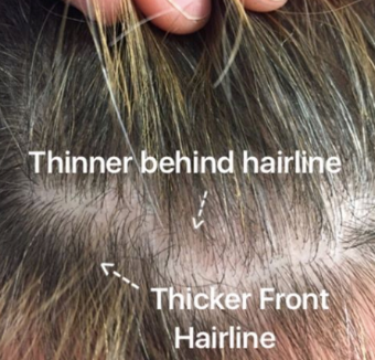
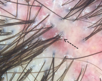
"Focal atrichia" (FA) refers to the absence of hairs is defined areas on the scalp. It is a feature generally seen in androgenetic alopecia whereby small circular areas totally devoid of hair are seen. These areas are typically the size of a pencil eraser and sometimes a bit bigger. Biopsies of these ares in the setting of androgenetic alopecia show accumulation of tiny vellus hairs.


It’s clear that FA is more common in androgenetic alopecia than other types of hair loss. A 2017 study by Olsen and colleagues showed FA was seen in 44 % of patients with FPHL compared to 2 % of other diseases. Those other 2 % may include area of primary scarring alopecia, scars from trauma, and even diffuse alopecia areata. Nevertheless, focal atrichia is most commonly seen in androgenetic alopecia.

There are still a few things that are unclear about whether one sees FA atrichia only in advanced disease. That same 2017 study by Olsen suggested 67 % of women with “late onset” FPHL had FA compared to just 15 % of those with “early onset” AGA. As we continue our discussion one must keep in mind that late onset generally has better prognosis.


Study 1: Olsen and colleagues, 2017

The 2017 Olsen study set out to evaluate the frequency of focal atrichia in various types of hair loss and its histologic characteristics in female androgenetic alopecia. The authors reviewed 250 consecutive female patients seen with hair loss for the presence or absence of FA. Interestingly, FA was identified in 46/104 (44%) of women with female pattern hair loss, including 15 % of early onset and 67% of late onset compared to 3/146 (2%) of those with other hair disorders. The histological (biopsy) findings of FA in FPHL showed mainly a more progressive miniaturization process than that of haired areas of the scalp. Taken together Olsen and her colleagues concluded that FA wasa clinical clue to the diagnosis of FPHL, particularly late onset subtype.


Study 2: Hu and colleagues, 2015

Studies by both Hu and colleagues and Zhang and colleagues both independently showed FA was associated with more severe disease. Additionally, Zhang et al showed that it was also associated with longer duration of disease. These findings seem to be at odds with the Olsen study.

Hu and colleagues performed a case-control observational study to identify the trichoscopic findings of AGA and to evaluate their relationship with the overall severity of the androgenetic aloepcia. The authors performed trichoscopic examination for 750 male AGA patients and 200 female (FPHL) patients, along with 100 male and 50 female normal controls.  In this study, FA was positively related to severity of hair loss (P < 0.05). 


Study 3: Hu and colleagues, 2012

Zhang and colleagues set out to analyze characteristics and investigate associations and clinical and trichoscopic features of female patients with FPHL. The did so by evaluating data from 60 patients with FPHL. FA was positively correlated with the stage and duration of hair loss.
 

Taken together, it is clear that FA is frequently seen in androgenetic alopecia. At first glance, is not uncommon to wonder whether these areas in fact represent scarring alopecia (i.e. tiny patches of Pseudopelade of Brocq or lichen planopilaris) or represent small areas of alopecia areata. However, when taken in context with the miniaturization going on in other areas of the scalp nearby, one can generally be confident these are area of FA. 

The general consensus would be the FA is a negative prognostic factor. 


Reference

Olsen et al. Focal Atrichia: A Diagnostic Clue in Female Pattern Hair Loss.
J Am Acad Dermatol. 2017.

Hu R, et al. Trichoscopic findings of androgenetic alopecia and their association with disease severity. J Dermatol. 2015.

Zhang X, et al. Female pattern hair loss: clinico-laboratory findings and trichoscopy depending on disease severity.  Int J Trichology. 2012.


This article was written by Dr. Jeff Donovan, a Canadian and US board certified dermatologist specializing exclusively in hair loss.
Share This
No Comments

Fusion Follicles: Compound Follicles = More than 6 Hairs

Compound Follicles = More than 6 Hairs

compound follicles 6 hairs.png

Compound Hair Follicles are hair follicles with more than the normal number of hairs emerging from a single hair follicle opening. In this photo, we see some of the earliest signs of a condition known as folliculitis decalvans. The scalp is red and scaly and there is a clear tendency for hair follicles to fuse together. The arrow points to 9 hair follicles all grouped together and emerging from the single opening. This so called compound follicle is commonly seen in many patients with folliculitis decalvans as well as some other scarring alopecias as well.

Treatment with topical antibiotics, oral antibiotics, retinoids, topical and/or intralesional corticosteroids are the best primary options in managing the disease.


This article was written by Dr. Jeff Donovan, a Canadian and US board certified dermatologist specializing exclusively in hair loss.
Share This
No Comments

The Scalp Punch Biopsy: Will anyone notice I had it done?

scalp bx- noticable?.png

Will anyone notice I had it done?

A punch biopsy of the scalp is performed if their is any uncertainty about the diagnosis of a patient’s hair loss. It involves removal of a 4 mm cylindrical core of tissue and placement of a stitch (suture). A punch biopsy should be taken from an area showing the main features of the disease in question. Provided the biopsy is taken from an area that contains a reasonable amount of hair, the stitch and healing site should be relatively unnoticeable. A dissolving suture or non dissolving suture can be placed although I tend to prefer dissolving sutures.


This article was written by Dr. Jeff Donovan, a Canadian and US board certified dermatologist specializing exclusively in hair loss.
Share This
No Comments

Scarring Alopecia: Is it inactive?

Is it inactive?

SA-inactive?.png

There are many misconceptions when it comes to determining if a scarring alopecia is truly “quiet.” Scarring alopecias, are a group of hair conditions whereby the body forms scar tissue in the scalp. This scar tissue ultimately prevents hairs from growing properly.

Here are a few principles that should always be considered when one feels they want to say a scarring alopecia is “quiet” or “inactive” 1) No doctor can tell if a scarring alopecia is quiet by looking at the scalp on a first appointment visit. That sentence deserves reading twice. Some scarring alopecias look quiet but the patient continues to lose hair. You can really only say a scarring alopecia is quiet if you’ve re-examined the scalp 12-24 months after the first appointment. 2) A patient with scalp symptoms like itching, burning or pain probably does not have quiet (inactive) disease. 3) Patients with redness and scaling in the scalp probably have active disease but they may not. Redness can be a sign of active disease but some patients with prolonged use of topical steroids simply have a red scalp. 4) The most important (and too often forgotten) guide that determines if a scarring alopecia is “quiet” is a comparison of scalp photographs taken at two different time points. Provided the interval between photos is greater than 1 year, one can get some pretty good information about whether the condition is quiet.

In my opinion, if there is no change in hair density between 2 photographs taken ONE year apart, the scarring alopecia is “probably” quiet (inactive). If there is no change in hair density between 2 photographs taken TWO years apart, the scarring alopecia is “extremely likely” to be deemed quiet. Despite this inactive appearance, the scarring alopecia STILL carries a risk of reactivation.

If there is no change in hair density between 2 photographs taken THREE years apart, and the patient is OFF MEDICATION during that period of observation, the scarring alopecia is likely burnt out.


This article was written by Dr. Jeff Donovan, a Canadian and US board certified dermatologist specializing exclusively in hair loss.
Share This
2 Comments

Alopecia Areata

Alopecia Areata: A Cause of Non Scarring Hair Loss

AA?.png

Alopecia areata is a form of autoimmune hair loss. Inflammation occuring deep under the scalp causes hair to fall out. The hair loss is non scarring which means the potential exists for hair to grow back. 


This photo shows the scalp of a man with advanced alopecia areata. Yellow dots represent keratin plugged openings of the hair follicles. Despite his widespead hair loss (involving 98% of this patient’s scalp), there are hairs on the scalp that appear completely unaffected by the immune reaction. Why that is remains unknown. Treatments for advanced alopecia areata include steroid injections, diphencyprone, anthralin, methotrexate, sulfasalazine, hydroxycloroquine and tofacitinib 


This article was written by Dr. Jeff Donovan, a Canadian and US board certified dermatologist specializing exclusively in hair loss.
Share This
2 Comments

Lichen Planopilaris: Itching, Burning, Tenderness, Shedding

Itching, Burning, Tenderness, Shedding

LPP symptoms.png

Lichen planopilaris (LPP) is a scarring hair loss condition that leads to permanent hair loss in affected areas without treatment.

Individuals with the condition may initially develop itching, burning or pain. For some, increased amounts of shedding may be the only symptom.

Examination of LPP via up close (“dermatoscopic”) examination is shown here. The scalp is red and scaly. In particular, some of the scale is located around hair follicles (called perifollicular scale). There are distinct whitish areas seen which represent permanently scarred areas of the scalp.

Treatment of LPP includes topical steroids, topical calcineurin inhibitors, steroid injections and a variety of oral immunosuppressive medications such as doxycycline, hydroxychloroquine, mycophenolate, cyclosporine, methotrexate, isotretinoin, tofacitinib and pioglitazone. Low level laser and excimer laser could to be studied and appear to offer benefit for some.


This article was written by Dr. Jeff Donovan, a Canadian and US board certified dermatologist specializing exclusively in hair loss.
Share This
No Comments

Corticosteroid Telangiectasias

Corticosteroid Telangiectasias

corticosteroids.png

Topical steroids have an important role in the treatment of hair loss. Like any medication, they must be respected. 


Dilatation of blood vessels (telangiectasias) can occur with prolonged topical steroid use. This is thought to occur due to stimulation of release of a chemical known as nitric oxide (NO) from dermal vessel endothelial cells which in turn leads to abnormal blood vessel dilatation. This photo shows telangiectasias occurring in a patient with alopecia areata treated with prolonged topical clobetasol. 


This article was written by Dr. Jeff Donovan, a Canadian and US board certified dermatologist specializing exclusively in hair loss.
Share This
2 Comments

Blogs by Topic


Share This
-->