h1.qusth1 { display: none !important; }

QUESTION OF THE WEEK

Dr. Donovan's Articles

QUESTION OF HAIR BLOGS


CSPA Pro-Patient Dermatologist Award

2018 CSPA Pro-Patient Dermatologist Award

I was honoured to be recognized as a 2018 Pro-Patient Dermatologist by the Canadian Skin Patient Alliance (CSPA). The CSPA is a non-profit organization that works hard to improve the quality of life of individuals with a range of dermatological issues.

CSPA

This article was written by Dr. Jeff Donovan, a Canadian and US board certified dermatologist specializing exclusively in hair loss.
Share This
2 Comments

Vaccinations for Patients Using Immunosuppressants for Hair Loss: Which ones do I need?

Vaccinations Before and After Starting Immonsuppressants

Part 1. Vaccines BEFORE starting Immunosuppressive Agents

Immunosuppressive agents are used to treat several hair loss conditions including scarring alopecias and alopecia areata. These include drugs such as methotrexate (higher doses), cyclosporine, mycophenolate, tofacitinib, ruxolitinib, azathioprine and high dose prednisone (prolonged use of doses higher than 1 mg per kg). Individuals receiving these agents may be at higher risk for certain infections. Vaccination may help reduce rates of infection.

vaccines

The first step in deciding on what vaccines to administer comes from understanding the patient’s previous vaccination history and exposures. If there are certain childhood vaccines that the adult has never received, consideration should be given to administering these.

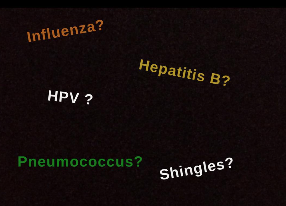
Vaccination should always be carefully reviewed with patients BEFORE starting immunosuppressive therapies. A number of vaccinations should be considered in patients with alopecia areata or scarring alopecias who are considering starting immunosuppressive agents. In general, one should speak to their physician about whether or not vaccination is needed for five infectious conditions: herpes zoster (Iive vaccine), HPV (recombinant vaccine), influenza (killed vaccine), hepatitis B (killed vaccine) and pneumococcus (killed vaccine). If so, these vaccines should be administered at least 4 weeks prior to starting the immunosuppressive agent.

1. Influenza vaccine (killed) - recommended for most before starting

Influenza infection can be a serious disease and sometimes fatal. The influenza vaccine can reduce morbidity and mortality from this infection. When we speak of the flu shot - we are typically referring to the inactive killed vaccine. It is important however to remember that there are two influenza vaccines: an Intranasal live attenuated influenza vaccine (LAIV) and inactive influenza vaccine (IIV)/trivalent inactivated vaccine (TIV). Patients on immunosuppressive medications should not receive live vaccine. 

The intramuscular attenuated vaccine is the vaccination is the vaccination that should be administered on a yearly basis. For patients who have demonstrated previous anaphylactic reactions to eggs in the past, there are recombinant flu vaccines available that lack ovalbumin.

These vaccinations can be re-adminsted yearly even for hair loss patients on tofacitinib, ruxolitinib and other immunosuppressive agents.

2. Pneumococcal vaccine (killed) - recommended for most before starting

The pneumococcal vaccine protected against infections with the bacteria S. pneumonia. The polysaccharide vaccine in adults protects against 23 serotypes. This is the common vaccine used in adults (i.e. Pneumovax). Conjugate vaccines are also available for adults. Normally these vaccines are given in childhood (4 vaccinations) and then repeated 5 years after the first vaccination. A lifetime revaccination dose at age 65 years or above.

3. Herpes zoster vaccine (live) - administered on a case by case basis

The zoster vaccine is a live attenuated vaccines to prevent shingles (herpes zoster) and may be appropriate for some individuals before starting immunosuppressive therapies. It is approved for individuals 50 years or older regardless of whether they have chickpox or shingles in the past. The zoster vaccine should be administered AT LEAST one month prior to receiving immunosuppressive therapy to reduce the chances of viral activation. Other live vaccines are shown in the list below.

4. Human papilloma virus HPV vaccine (recombinant) - administered on a case by case basis

HPV is the most common sexually transmitted disease in the world. Cervical cancer contributes to 8 % of all cancers in women worldwide. HPV is a clear risk for for cervical cancer. One in four individuals in the United States are infected with HPV. The HPV vaccines may be appropriate for some individuals before starting immunosuppressive therapies on a case by case basis. Current recommendations in Western countries include women through age 26 and men through age 21. In general, protocols are in place to vaccinate adolescents age 11 or 12 (two vaccination 6-12 months apart). Teens older than 14 years need three vaccinations over 6 months. Also, three doses are still recommended for people with certain immunocompromising conditions aged 9 through 26 years. Other patient subgroups (men who have sex with men, transgender adults, and young patients with immocompromsing medical condition) may slo benefit.

How best to vaccinate patients on immunosuppressants is not known and is therefore done on a case by case basis. Patients who do not fall in the above risk categories should speak to their physician.

5. Hepatitis B Vaccine - administered on a case by case basis

Recommendations for Hepatitis vaccination differ from country to country according to the prevalence of Hepatitis B in the country. In general, vaccination with Hepatitis B is appropriate for those at high risk. This includes health care workers, IV drug users, and those with multiple sexual partners in the last 6 months.

Patients who May be Considered for Hepatitis B Vaccination

1) Polygamous relationship (those with more than 1 sex partner during the last 6 months)

2) Persons seek evaluation or treatment for a sexual transmitted disease

3) Current or past IV drug users

4) Men who have sex with men

5) Health care workers who are exposed to blood or potentially infectious body fluids

6) All diabetics younger than 60

7) Diabetics over 60 years (at discretion of doctor)

8) End stage kidney disease

9) Chronic liver disease in those with HIV

10) Household contract of those with BSAG positivity

11) Clients and staff members of institutions for those with developmental delay

12) International travellers to counties with high Hepatitis B infection

5. Tetanus and diphtheria - administered on a case by case basis

Tetanus and diphtheria (Td) is recommended as part of the childhood DTaP 5 series injection. Revaccination is recommended every 10 years. If a patient has not had a tetanus shot in the last 10 years, it’s a good idea to have it before starting immunosuppression.

A note on Live vaccines

The following is a helpful list of live vaccines which must not be administrated to patients on immunosuppressants. If vaccination is needed, it should be done well ahead of starting immunosuppressants.

  • MMR

  • Varicella/Zostavax

  • Oral Polio

  • Flu mist (nasal vaccine)

  • Yellow fever

  • Typhoid vaccine (1 of 3 is live)

  • Small pox

  • BCG – bladder irrigation

Part 2. Vaccines AFTER starting Immunosuppressive Agents

For patients already using immunosuppressive agents, non-live vaccines can be administered on a routine schedule.

A few helpful points:

1. THE ANNUAL FLU SHOT IS OKAY. In general, the routine administration of the seasonal influenza vaccine is recommended for hair loss patents using immunosuppressive agents.

2. MOST INACTIVE VACCINES ARE OKAY. Most inactive vaccines (recombinant, subunit, toxoid, polysaccharride, conjugated polysaccharide vaccines), conjugated pneumococcal (CPV), tetanus-diphteria-acellular pertussis- hemophilus influenza type B (Hib) -inactive polio (DTaB-Hib-IPV), inactive influenza, tetanus-diphteria-acellular pertussis-inactive polio (DTaB-IPV), Hepatitis B, Hepatitis A)] can be administered in accordance with routine vaccine schedules.

3. LIVE VACCINES SHOULD NOT BE GIVEN. Live vaccines (see list above) MUST be avoided in patients already using immunosuppressive agents. Live viruses can replicate in patients who have received immunosuppressants and this can bring about potential harm to the patient. The CDC states that oral polio virus OPV) should not be administered to any household contact of a severely immunocompromised person. Measles-mumps-rubella (MMR) vaccine is not contraindicated for the close contacts (including health-care providers) of immunocompromised persons.

Summary

Discussion about previous and future vaccinations is important for all patients about to start immunosuppressive agents. The pneumococcal and injectable (killed) influenza are the only two vaccines universally recommended in all cases of immunosuppression. The remainder are dealt with on a case by case basis. Another important principle is that live vaccines must never be given to patients who are receiving immunosuppressive medications.


REFERENCES

https://www.cdc.gov/mmwr/preview/mmwrhtml/00023141.htm. Accessed Jan 4 2019.

Lopez et al. Vaccination recommendations for the adult immunosuppressed patient: A systematic review and comprehensive field synopsis. Journal of Autoimmunity 2017.


This article was written by Dr. Jeff Donovan, a Canadian and US board certified dermatologist specializing exclusively in hair loss.
Share This
No Comments


Post Partum Hair Loss

Hair Loss After Delivery

Post partum hair loss (hair loss after delivery) is common and nearly all women develop some degree of shedding after delivery. I was recently interview for the Huffington Post on this subject.

Postpartum Hair Loss Can Be Severe And Devastating For New Moms

Post-partum hair loss is a normal physiological process and not a sign of disease. During pregnancy, the rate of daily hair shedding decreases to extremely low levels and the result is an increased density on the scalp for most women. The result is that hair appears quite full for many women while pregnant. After delivery the changes in estrogen and progrestone trigger a resetting of the shedding rates back to what they once were - and this comes about only by increased shedding of hair initially.

Post partum hair shedding typically starts 2-3 months post partum and can last 6 months or more. By the time of the baby’s first birthday, hair shedding should be returning back to normal for the new mother. If not, careful review should be done by a dermatologist.

Practical Advice for Women with Hair Shedding

1. Wash and shampoo as often needed.  More hair will come out on the days that the hair is shampooed but this will not affect the long term density of hair.  The use of a volumizing or thickening shampoo may help the hair look fuller and feel thicker.

2. Use a conditioner formulated for fine hair. I recommend that women with shedding avoid heavy conditioners as these tend to weigh down the hair. A conditioner formulated specifically for "fine hair" tends not to weigh the hair down as much.  The conditioner should be applied only to the ends of the hair.   If it is applied to the scalp and the entire hair it tends to weigh the hair down.

3. Avoid hair styles that puts stress on the hair.  This includes tight braids, pigtails, cornrows, or a tight pony tail. These hair styling practices can lead to more hair being pulled out.

4. Avoid excessive combing of hair when it is wet.  This can lead to more hair breakage. The use of a large tooth comb can be helpful.

5. Eat as healthy as possible.

6. Talk openly about hair loss concerns. With so much focus on the new baby, there is often little attention given to the concerns of the new mom. It is normal to be worried about hair loss. Talking with others, especially other mothers who experienced hair loss, can be helpful.

7. Wear a wig or hairpiece for a short time if it helps cope with hair loss. Very rarely, a new mom with extensive hair shedding will ask whether wigs or hair pieces are safe or whether they weigh down the hair and prevent it from breathing. Wearing a wig or hairpiece is completely safe. This can be a helpful camouflaging option for women whose scalp can be seen.

8. Consider cutting the hair shorter. This will give more lift to the hair and weigh it down less. This can help camouflage hair loss to some degree. However, cutting hair won’t make the shedding stop faster or hair grow back quicker.  Shorter hair can also be much easier to manage.


This article was written by Dr. Jeff Donovan, a Canadian and US board certified dermatologist specializing exclusively in hair loss.
Share This
No Comments

On the Chasing of Dreams

On the Chasing of Dreams…


The dreams and ambitions of humans do not live inside of hair follicles. Rather, dreams live and are nurtured inside the human mind before they are finally translated by the individual into reality.

The loss of one’s hair should never lead to loss or modifications to one’s original dreams and ambitions - unless of course one has lost their mind somewhere along the way. The chances of the latter are, of course, rather unlikely. Dreams reside entirely in the mind.

Yes, it is true that the chasing one’s dreams in the face of hair loss may take more work for some individuals and perhaps even take more time for others...but the exact same dreams are right there for the taking.

Small dreams or big dreams. Let nothing stand in the way of your ambitions. Your mind is powerfully strong.


This article was written by Dr. Jeff Donovan, a Canadian and US board certified dermatologist specializing exclusively in hair loss.
Share This
1 Comment

Happy Holidays

On behalf of the Donovan Hair Clinic, I would like to wish everyone a wonderful holiday season and the very best in 2019.

whistler

This article was written by Dr. Jeff Donovan, a Canadian and US board certified dermatologist specializing exclusively in hair loss.
Share This
1 Comment

Hair, Health and Differences Between Men and Women

Multiple Health Parameters Differ in Men and Women

Many health conditions announce their presence differently in men and women. Heart disease, stroke and dementias are just a few examples. Women arrive at the clinic with different stories than men. Signs and symptoms often differ. Treatments work differently in men and women.

Hair loss also presents differently in men and women. Women arrive at the clinic with different stories about their hair loss than men. Signs and symptoms may differ. Treatments work differently in men and women. Modern medicine is just starting to recognize all these important differences.


This article was written by Dr. Jeff Donovan, a Canadian and US board certified dermatologist specializing exclusively in hair loss.
Share This
No Comments

Miniaturization in Alopecia Areata: Why is it reversible?

Association with Arrector Pili May Determine Reversibility

Miniaturization of hairs can occur in many hair loss conditions including androgenetic alopecia as well as alopecia areata. For many years, physicians have wondered why miniaturized hairs in alopecia areata hold on to the potential to regrow back to thick hairs whereas miniaturized hairs in AGA only have the potential to thicken up slightly . and only with treatment.

In 2016, Professor Rod Sinclair’s group in Australia proposed that alot of this may be due to a muscle known as the arrector pili muscle. The arrector pili muscle (APM) is not as well known as other muscles like the biceps, triceps or quadriceps. The APM is a very small muscle that is attached to every hair. When the APM contracts, the result for the patient is “goose flesh.” In the past, it was thought that the APM did not have any active role in any type of hair loss mechanisms and more or less acted as a ‘bystander.’ However, in 2016, Sinclair and colleagues proposed a new model of balding. They proposed that the APM has a key role in the decision of a hair follicle to ultimately "miniaturize" during the course of AGA.

By observing how miniaturization occurs within follicular units – the authors proposed that by maintaining tight attachment to the APM, some hair follicles are prevented from proceeding down the pathway of permanent miniaturization. The association with the APM was thought to preserve stem cells.

APM


The authors proposed that in other conditions like alopecia areata, hair follicles maintain their close association with the APM and therefore are protected from the possibility of irreversible miniaturization.

The photo here shows strong attachment of the APM to hairs in alopecia areata (right panel) whereas some hairs in androgenetic alopecia lose their attachment and become miniaturized (left panel).

Reference


Sinclair R, Torkamani N and Jones L. Androgenetic alopecia: new insights into the pathogenesis and mechanisms of hair loss. F1000Res. 2015 Aug 19;4 (F1000 Faculty Rev):585 -


This article was written by Dr. Jeff Donovan, a Canadian and US board certified dermatologist specializing exclusively in hair loss.
Share This
No Comments

Miniaturization in Alopecia Areata vs Androgenetic Alopecia

Miniaturization: Not Only for Androgenetic Alopecia


When most people think about the term “miniaturization”, the hair loss condition known as androgenetic alopecia “AGA” (male balding and female thinning) comes to mind. This common hair loss condition is associated wirh the progressive “miniaturization” of hairs. A biopsy from a patient with AGA or an up close dermatoscopic examination of the scalp shows this variation in the size of follicles. Some follicles are thick and some are thin and some are very thin. The variation in the size of follicles is known as “anisotrichosis” and is very much a part of what AGA is all about.

AGA is not the only hair loss condition that is associated with miniaturization. Alopecia areata for example also shows miniaturization. However, in contrast to AGA, alopecia areata often does not show the same degree of “anisotrichosis” - meaning that there is not the same degree of variation in follicle size as seen in AGA. In alopecia areata, follicles in the biopsy are generally either thick ones or thin without the dramatic variation in caliber that is seen in AGA.

Hair follicle miniaturization can occur in AGA and AA. Hairs are similar caliber in AA when miniaturization is present.

Hair follicle miniaturization can occur in AGA and AA. Hairs are similar caliber in AA when miniaturization is present.


The schematic diagram here shows these key differences. One can see on the left side (of AGA) that miniaturized hairs are all different thickness. In contrast, in the diagram of alopecia areata on the right the miniaturized hairs are all the same size. Lymphocytes are seen in both conditions although in alopecia areata they are classically around the bulb and in fibrous tracts whereas in AGA they are higher up in the skin in the infundibulum and isthmus. Eosinophils and melanin pigment can frequently be seen in fibrous tracts in alopecia areata.


This article was written by Dr. Jeff Donovan, a Canadian and US board certified dermatologist specializing exclusively in hair loss.
Share This
No Comments

Spironolactone for FPHL: Are routine potassium blood tests needed?

Spironolactone (Aldactone) for Healthy Women with FPHL: Is Potassium Testing Needed?

Spironolactone is an oral antiandrogen that is sometimes used to treat female pattern hair loss. It is also used to treat acne and hirsutism.

In addition to being an antiandrogen, spironolactone is a a type of blood pressure pill and diuretic. Spironolactone acts on the kidney (in the distal convoluted renal tubule) to promote sodium and water excretion and to promote potassium retention.

Do patients using spironolactone for hair loss needed blood tests for potassium?

Do patients using spironolactone for hair loss needed blood tests for potassium?


Previous recommendations had suggested that routine monitoring of serum potassium levels by having the patient periodically have blood tests performed was important. Recent studies have suggested this is not necessary for healthy women.

Layton and colleagues evaluated 10 randomized controlled trials (RCTs) and 21 case series pertaining to acne. The authors did not find that serum potassium was more likely to be elevated in healthy women using spironolactone for acne.

Plavanich and colleagues performed a retrospective study of healthy young women taking spironolactone for acne. The authors analyzed rates of hyperkalemia (high potassium) in 974 healthy young women taking spironolactone and also analyzed 1165 healthy young women taking and not taking spironolactone to obtain a profile for the baseline rate of hyperkalemia in this population.

The findings of the study were that young women receiving spironolactone had a hyperkalemia rate of 0.72%, equivalent to the 0.76% baseline rate of hyperkalemia in the general population. The conclusion was that the rate of hyperkalemia in healthy young women taking spironolactone for acne is equivalent to the baseline rate of hyperkalemia in this population and that potassium monitoring is unnecessary for healthy women taking spironolactone for acne.

Studies of potassium levels in women using spironolactone for hair loss have not been done but there is no reasons to believe there is any difference. Routine potassium testing in healthy women is not usually necessary. Women with cardiovascular disease, kidney disease, diabetes and women taking certain medications that affect potassium levels (ie potassium sparing diuretics), may or may not be good candidates for spironolactone but if they are candidates they will require periodic potassium measurements.


Reference


Layton AM et al. Oral Spironolactone for Acne Vulgaris in Adult Females: A Hybrid Systematic Review. Am J Clin Dermatol. 2017.

Plavanich M et al. Low Usefulness of Potassium Monitoring Among Healthy Young Women Taking Spironolactone for Acne. JAMA Dermatol. 2015.


This article was written by Dr. Jeff Donovan, a Canadian and US board certified dermatologist specializing exclusively in hair loss.
Share This
No Comments

New Players Identified in Hair Loss Mechanisms

DKK2 Proposed to be Key Player of Blocking Growth.

It’s well known in the hair research world that a molecule known as WNT is important for hair growth. New research from the Perelman School of Medicine at the University of Pennsylvania in Philadelphia shows that a protein known as DKK2 is responsible for blocking WNT from doing it’s job.

WNT signals are important in embryonic development and also have an important role in how cells signal with each other in adult life. WNT signals, when they are present, cause hair to grow. When they are absent, they cause hair loss stop growing or not grow at all.


DKK2

In order to study why some parts of the body don’t grow hair and to better understand what signals hairs to not grow, researchers studied the plantar skin of mice. These areas are similar to the palms of human hands in some ways. They lack hair follicles. The researchers found high levels of Dickkopf 2 (DKK2) proteins in this area. Remarkably when mice were engineered to lack the DKK2 proteins, the hair grew in these areas.


Summary

The DKK2- WNT pathway is relevant to human hair growth - and that’s of course why this study is exciting. Previous research has suggested that genes like DKK2 are plausible genes that are implicated in the pathogenesis of male balding and female thinning. More understanding of this important area is likely to be highly relevant and likely to lead us in the direction of better treatments. A DKK2 drug inhibitor for example has potential to help hair loss.

REFERENCE

Song et al. Regional Control of Hairless versus Hair-Bearing Skin by Dkk2. Cell Reports Nov 2018.

Heilmann-Heimbach S, et al. Meta-analysis identifies novel risk loci and yields systematic insights into the biology of male-pattern baldness. Nat Commun. 2017.


This article was written by Dr. Jeff Donovan, a Canadian and US board certified dermatologist specializing exclusively in hair loss.
Share This
No Comments

White hypopigmented scarring with FUE

FUE is not scarless but scars can appear barely detectable

Hair transplantation involves moving hair from one area of the body to another. Most commonly, hair transplants involve moving hair from the back of the scalp to the more frontal areas of the scalp (i.e. hairline, midscalp or crown). There are two main ways that hair transplant surgery is done nowadways. One is known as follicular unit strip surgery (FUSS or FUT) and the second is known as follicular unit extraction (FUE).

FUSS/FUT involves the removal of a strip of skin and hair from the back of the scalp. Sutures are used to close the wound that was created. The individual hairs are then obtained by delicately cutting up the hairs using a microscope. The result of FUSS/FUT is a linear scar that runs along the back of the scalp. 

The second type of surgery is increasingly referred to as follicular unit “excision” surgery and involves the excision of follicular units one by one from the back of the scalp using a “punch” device. One of the common and unfortunate misconceptions is that FUE is “scarless.” In fact, many advertisements list “scarless” as one the benefits of FUE over FUSS/FUT. FUE is not scarless. Anytime the skin is injured down in the deeper layers a scar has the potential to form. FUE involves removal of complete hairs - even hairs that were once rooted deep down in the fat layer.

Circular whitish scars in a patient with a previous FUE transplant

Circular whitish scars in a patient with a previous FUE transplant

By definition, FUE is not a scarless procedure. A tiny scar forms during the procedure. Fortunately, however, the scars with FUE tend to be quite small and sometimes quite difficult to detect. That usually makes it difficult for anyone to see evidence that the patient had a procedure in the past. 

For some people, very small circular whitish scars can be seen post surgery as seen in this photo. These circular scars may be quite difficult to notice but an astute eye can sometimes make out the areas in the donor area where the FUE punch took the hairs during the surgery. Some people also develop small amounts of redness in the donor area too which makes the whitish hypopigmented circles a bit more noticeable. Over time, redness may fade to some degree leaving the whitish circles less noticeable. Some people maintain permanent faint redness


This article was written by Dr. Jeff Donovan, a Canadian and US board certified dermatologist specializing exclusively in hair loss.
Share This
No Comments

Peripilar casts (Perifollicular scale) in LPP

Scaling in Lichen Planopilaris (LPP)

Lichen planopilaris (LPP) is a scarring alopecia that has the potential to cause permanent hair loss. Many patients first develop symptoms such as itching or burning or tenderness un the affected area. Some patients have all three. 

Clinical examination can sometimes look fairly normal. However, in more active LPP, the scalp is red to varying degrees. There is redness around the follicles (called perifollicular erythema) and there is often whitish scale around follicles too. The whitish scale is known by many names including “perifollicular scale” and “peripilar casts” and “follicular hyperkeratoses.” All these terms mean the same thing for the most part. Perifollicular erythema and perifollicular scale are signs the disease is active. 

Perifollicular scale (white color around follicles) in a patient with LPP.

Perifollicular scale (white color around follicles) in a patient with LPP.

Treatments for LPP have been discussed in other posts but include agents such as topical steroids, steroid injections, topical calcineurin inhibitors, low level laser therapy, oral doxycycline, oral hydroxychloroquine, mycophenolate mofetil, methotrexate, cyclosporine, low dose naltrexone, oral tofacitinib. Successful treatment stops symptoms and more importantly stops hair loss.


This article was written by Dr. Jeff Donovan, a Canadian and US board certified dermatologist specializing exclusively in hair loss.
Share This
No Comments

Frontal Fibrosing Alopecia (FFA): Perifollicular Scale/Peripilar Casts

Perifollicular Scale/Peripilar Casts

IMG_3779.png

Frontal fibrosing alopecia (FFA) is a type of scarring alopecia. In some patients, eyebrow loss is one of the first signs of the disease whereas in others the frontal hairline is affected first. Many patients are asymptomatic although a bit of itching is not all that uncommon.

FFA can affect the frontal scalp as well as the sideburns and even around the back of the scalp (ie behind the ears). In fact, any body hair can be affected. The appearance of the scalp in various regions has been shown to appear differently by dermoscopy.

In 2018, Cevantes and colleagues compared the differences between the dermatoscopic appearance of the sideburns and the frontal scalp regions. The sideburn area in FFA was found to have less redness around hairs (perifollicular erythema) and less scaling (perifollicular scaling or peripilar casts) compared to the frontal scalp.

This information is helpful for physicians as we often rely on the presence or absence of perifollicular erythrma and scale as helping to decide if the FFA is active or not. This study reminds us that monitoring redness and scale makes sense when examining and monitoring the frontal scalp in patients with FFA but is of limited value when monitoring FFA of the sideburn area. 

The dermoscopy photo shows the typical appearance of FFA of the frontal hairline. Scaling around hairs is known by many names including "perifollicular scale" and "peripilar casts."


Reference

Cervantes J, et al. Distinct Trichoscopic Features of the Sideburns in Frontal Fibrosing Alopecia Compared to the Frontotemporal Scalp.

Skin Appendage Disord. 2018.


This article was written by Dr. Jeff Donovan, a Canadian and US board certified dermatologist specializing exclusively in hair loss.
Share This
No Comments

Scalp Dysesthesias

What treatments are available for scalp dysesthesias?

I just posted a new answer to our “Question of the Week.” I was asked to outline what treatments are possible for patients who are diagnosed with ‘scalp dysesthesia.’

The full answer to this week’s question can be read here:

What treatments are available for scalp dysesthesias?

To submit a new question for consideration of our Question of the Week, simply visit complete our online form


This article was written by Dr. Jeff Donovan, a Canadian and US board certified dermatologist specializing exclusively in hair loss.
Share This
No Comments

Breast feeding and post-partum hair shedding

Women who breast feed experience less shedding at month 4 post partum

Many women notice that during pregnancy their hair is fuller and by the third month post-partum they are shedding more and more hair every day. This is a normal physiological process that is experienced to some degree by nearly all women.

The hormonal changes in pregnancy, especially the changes in levels of estrogen and progesterone seem to support the growth of hair and limit the shedding of hair. Normally about 10 % of hairs in the human scalp are preparing to shed within the next month or two. During pregnancy, this number drops markedly to just a few percent. The result of having less hair falling out and more hair growing in the scalp is thicker hair.

Despite the 353,000 mothers that give birth every day, there is actually little research about post-partum telogen effluvium. In 2014, Gizlent and colleagues performed a study involving 116 women. 28 women were in the 24th week of pregnancy, 30 pregnant women were at term gestation, 29 women were in the 4th post-partum month, and 29 in the first post-partum year. The number of breastfeeding mothers in the post-partum period and the month in which they were examined were also recorded. The researchers evaluated the ratios of growing hairs (anagen hairs) and telogen hairs using a Trichoscan instrument.

MONTH 4 IS ASSOCIATED WITH MORE TELOGEN HAIRS

The authors found that the mean anagen rate in the 4th post-partum month was significantly lower than that in the 6th and 9th month of pregnancy. The average telogen rate was significantly higher. Interestingly, women who breastfed had a mean anagen rate in the 4th post-partum month that was significantly higher than that in women who did not breast feed. The mean telogen rate was significantly lower. By the end of the first year, women who chose to breast feed had similar hair cycle rations as women who did not.

Conclusion

This study was interesting as it suggested that breast feeding was among one of the only known modifiable factors that has the potential to affect the severity of post-partum telogen effluvium.

Reference

Gizlenti S, et al. The changes in the hair cycle during gestation and the post-partum period. J Eur Acad Dermatol Venereol. 2014.


This article was written by Dr. Jeff Donovan, a Canadian and US board certified dermatologist specializing exclusively in hair loss.
Share This
No Comments

What constitutes a normal appearing scalp?

normal scalp

Recognizing the Normal Scalp

In order to appreciate what is “abnormal” on the human scalp, one needs to fully understand what is normal and what are all the variations of normal that are possible.

The normal scalp is not red and not flaky. Hairs emerge in groups of 1,2 and 3 hairs. In some cases (especially younger individuals) hairs come out occassionally in groups of 4 hairs too. Most hair follicles in an area are the same diameter (caliber) but a bit of variation is normal and acceptable. The finding of an occassional thin hair carries no significance and does not mean the individual has androgenetic alopecia.


This article was written by Dr. Jeff Donovan, a Canadian and US board certified dermatologist specializing exclusively in hair loss.
Share This
No Comments

Diagnosis of Scarring Alopecia in the Early Stages

Early Diagnosis More Challenging that Late Stage Diagnosis

Diagnosing scarring alopecias in the earliest stages is more challenging because the appearance of the scalp may not look all that unusual.  The photo here is from a patient with lichen planopilaris. There are subtle features like scalp erythema, pili torti and single hairs that might trigger  one to think about scarring alopecia but the features are not specific. This photos could just as easily be from a patient with androgenetic alopecia and seborrheic dermatitis.

EARLY LICHEN PLANOPILARIS: FEATURES ARE NON SPECIFIC IN THIS PHOTO

EARLY LICHEN PLANOPILARIS: FEATURES ARE NON SPECIFIC IN THIS PHOTO


However, diagnosing scarring alopecia in the early stages is made a lot easier by carrying a simple rule: if there is even the slightest possibility that what a physician is looking at could be a scarring alopecia, then a biopsy should at least be considered.


Not everyone that has scalp itching has a scarring alopecia. There are dozens of causes of itching. Not everyone with itching needs a biopsy.

Not everyone that has excessive shedding and a red scalp has a scarring alopecia. In fact, most don’t. There are many causes of shedding and a red scalp. Not everyone with a red scalp and shedding needs a biopsy.


Patients with Multiple Symptoms May Need A Biopsy

Scarring alopecias (particularly the symptomatic ones like lichen planopilaris and folliculitis decalvans) are very often associated with symptoms and signs that signal to the patient and physician that something is not right.  More times than not, these two diseases shout out clues to the patient and physician that something is not right. (In contrast frontal fibrosing alopecia can often be silent when it first develops and can go on for years without detection). For patients with lichen planopilaris and folliculitis decalvans there are often multiple clues that this is not indeed the diagnosis. The scalp sometimes itches in unusual patterns. The scalp might burn. The scalp is sometimes sore. The scalp feels bruised for some patients. The scalp is warm for many. Shedding occurs when there is otherwise no good reason.

Patients who report several symptoms (“more than itching”) often benefit tremendously from having a scalp biopsy. For example, a patient with scalp itching PLUS burning or a patient with itching PLUS scalp tenderness should at least be given consideration for a scalp biopsy.


This article was written by Dr. Jeff Donovan, a Canadian and US board certified dermatologist specializing exclusively in hair loss.
Share This
1 Comment

Scarring Alopecia: Is there any chance of regrowth?

Regrowth in Early Treatment of Scarring Alopecias

Regrowth in a patient with scarring alopecia following aggressive treatment

Regrowth in a patient with scarring alopecia following aggressive treatment

The classic teaching that physicians often learn is that the hair loss that occurs in scarring alopecias is permanent. In other words, physicians often explain to patients that “what you have lost is lost for good and the goal of treatment is to stop it from getting any worse.” We know now that this statement is not quite accurate.

The very early stages of scarring alopecia are associated with inflammation which causes hairs to shed.  It’s not so easy for hairs to grow in a soup of inflammation. However, if the inflammation can be stopped some of these hairs can regrow because there are still stem cells left inside the original hair follicle tract. The early and aggressive treatment of lichen planopilaris, frontal fibrosing alopecia, central centrifugal cicatricial alopecia, discoid lupus and dissecting cellulitis can all be associated with some degree of regrowth. Of course the regrowth is not always 100 % but it can be quite signficant.


The above photo shows massive amounts of regrowing hair in a patient with lichen planopilaris who started hydroxychloroquine, topical steroids and steroid injections about 6 months prior. 


This article was written by Dr. Jeff Donovan, a Canadian and US board certified dermatologist specializing exclusively in hair loss.
Share This
No Comments

Androgenetic Alopecia: The Brown Peripilar Sign

BPPS: Brown peripilar sign in AGA

Dermatoscopic image showing the brown peripilar sign.

Dermatoscopic image showing the brown peripilar sign.

It is increasingly clear that androgenetic alopecia (ie male and female genetic hair loss or “balding) is an inflammatory condition. Even though the scalp typically looks non-inflamed, scalp biopsies show that there is inflammation present just a few millimeters beneath the scalp in an area known as the isthmus and infundibulum. Studies have shown that inflammation occurs quite early in the course of androgenetic alopecia. This inflammation is believed to facilitate the progressive miniaturization of hair follicles.


Although the scalp usually looks fairly normal and non-inflammatory in patients with androgenetic alopecia, evaluation of the scalp via dermoscopy techniques may also show features that suggest there is inflammation under the scalp.


The 2004 Deloche Study

In 2004, Deloche and colleagues from France performed a nice study of 40 patients with androgenetic alopecia. The researchers showed that the brown discolouration around hairs that is seen with dermoscopy correlated nicely with inflammation under the scalp when evaluated by biopsy. They called this the peripilar sign (PPS). “Peri” means around and pilar means hair. The peripilar sign is known by many names as well including the “brown peripilar sign (BPPS)” and “peripilar halo.” Included here is a patient with androgenetic alopecia whose scalp hairs show the BPPS (photo below).


Treating Inflammation in AGA: Yes or No?

It’s almost a certainty that the inflammation needs to be treated in order to best stop hair loss. Modern medicine does not quite know how to best do this yet. There are many different types of anti-inflammatory treatment including corticosteroids, doxycycline, tacrolimus, TNF-inhibitors, immunomodulatory and immunosuppressants. It’s a bit of a guess as to how best to address the inflammation in AGA and more research is needed. It’s extremely likely this will play a beneficial role, particularly the earlier such anti-inflammatory treatment is started. Whether current treatments like antiandrogens or laser actually help with reducing inflammation is not known yet.


Reference

Deloche C et al. Histological features of peripilar signs associated with androgenetic alopecia. Arch Dermatol Res. 2004.


This article was written by Dr. Jeff Donovan, a Canadian and US board certified dermatologist specializing exclusively in hair loss.
Share This
No Comments

Blogs by Topic


Share This
-->