h1.qusth1 { display: none !important; }

QUESTION OF THE WEEK

Dr. Donovan's Articles

QUESTION OF HAIR BLOGS


Gynecomastia from Finasteride or Dutasteride: What are my options?

5 alpha reductase inhibitor induced gynecomastia

5 alpha reductase inhibitors like finasteride and dutasteride are widely used to treat androgenetic alopecia.  Gynecomastia or breast enlargement is a side effect that one needs to be aware of. It occurs in 4 to 10 out of every 1,000 users.  


Key points about gynecomastia from 5 aha reductase inhibitors

Gynecomastia from these drugs is often one-sided but can be both sides.  Changes can start as early as a few weeks but is typically a few months delay (if it is going to occur). It can also be 1-2 years before the phenomenon is appreciated. An important sign to watch for is the presence of pain or tenderness. This can occur in many males prior to any actual enlargement. Lower doses are less likely to cause breast enlargement in men compared to higher doses. The concept of the dose response is important because it means than for some men, 0.5 mg daily (or every other day) could be assocated with a lower risk of gynecomastia (while still potentially benefitting their hair). Finasteride induced gynecomastia can reverse in many individuals provided the drug is stopped in the early stages when the gynecomastia is noted. If the drug is not stopped, it can enter a irreversible stage (where only surgery will provide treatment). Finasteride induced gynecomastia is more likely with advanced age and in obese individuals. Blood tests may be appropriate  for some men depending on their history. However, most of the time blood tests and various hormonal tests are normal.


Treatment of Gynecomastia from 5 alpha reductase inhibitors

I'm often asked how gynecomastia should be treated. This simplest and more consistently helpful answer is to immediately stop the drug as soon as gynecomastia is noticed. Most men (but not all) will notice resolution of the gynecomastia with stopping the drug. Of course, the benefits for the hair will be lost. 

One should discuss the issue in detail with their physician. One need to make sure that the gynecomastia is indeed occurring due to the use of the 5 alpha reductase inhibitor and not another cause.  For some males, a reduction in the overall dose of the drug may reduce the gynecomastia yet still benefit the hair. For many males with gynecomastia, especially if it has been present for a longer period of time, surgery is needed to remove the excess tissue. 


This article was written by Dr. Jeff Donovan, a Canadian and US board certified dermatologist specializing exclusively in hair loss.
Share This
No Comments

Skin Pigmentation with Hydroxychloroquine (Plaquenil)

Skin Hyperpigmentation with Hydroxychloroquine

Hydroxychloroquine (Plaquenil is a common trade name) is an anti-inflammatory oral medication that is commonly used to treat many autoimmune diseases. In the hair clinic, we use it for diseases such as lichen planopilaris, frontal fibrosing alopecia, pseudopelade and discoid lupus. A variety of side effects are possible and users need to understand the potential side effects before starting treatments. Today we'll focus on the side effects of skin pigmentation.


What does hydroxychlorqoune hyperpigmentation look like?

Skin pigmentation related to use of hydroxychloroquine starts out as a yellow brown to slate gray or black pigmentation on many areas of the body - especially the front of the shins but also the face, forearms, mouth mucosa (essentially hard palace and gingivae) and nail beds.


How common is it?

It is estimated to occur in 7-10 % of patients who use these medication. Unlike the hydroxychloroquine-related pigmentation changes that affect the eye (retinopathy), the skin pigmentation changes are not directly related to duration of use and can even develop in the first year. In one study of lupus patients, 17% developed pigmentation changes after the first year of use and 40 % had developed them by year 5.


Why does it occur?

Hydroxychloroquine (Plaquenil) stimulates the pigment producing cells in the skin called melanocytes. As a result, there is both hemosiderin (iron) and melanin accumulation in the dermis. The exact reasons why this phenomenon happens is not clear. However, trauma and bruising may faciliate the developement of pigmentation.  This may be the reason why patients on blood thinners and anticoagulants appear to be at highest risk.   In fact, most patients who go on to develop these pigment changes first notice that the appearance of pigmented lesions was preceded by the occurrence of a bruised like appearance. This has often been likened to a “bruise that did not disappear.”


How are the abnormal pigmented areas treated?


After stopping the drug, (if indeed possible to do so), some will improve and the pigment will fade to some degree over time in a large percentage. Most, however, do not completely improve. Lasers are currently being studied as a therapeutic option for a variety of pigmentation issues.


REFERENCE

Coulombs et al. CMAJ. 2017 Feb 6; 189(5): E212.
 
Jallouli et al.  Hydroxychloroquine-Induced Pigmentation in Patients With Systemic Lupus Erythematous. A Case-Control Study. JAMA Dermatol. 2013;149(8):935-940. doi:10.1001/j


This article was written by Dr. Jeff Donovan, a Canadian and US board certified dermatologist specializing exclusively in hair loss.
Share This
2 Comments

Lichen Planopilaris

What is Lichen Planopilaris?

LPP pc.png

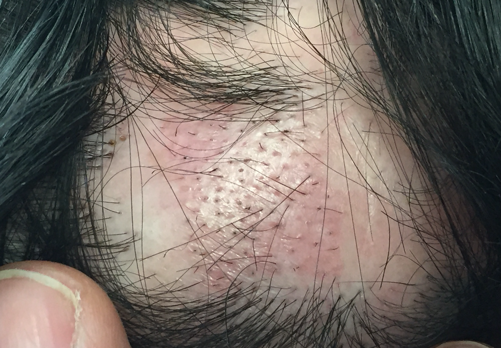
Lichen planopilaris (LPP) also known as lichen planus of the hair follicle is an immune based scalp condition that causes scarring alopecia. The early signs of LPP can be subtle but often include itching, burning, scalp tenderness or pain.

This up close “dermatoscopic” image of the scalp shows typical features of LPP including scalp redness, white scale around hair follicles and broken hairs. The blue dot is my marker and denotes the sight at which I will obtain a biopsy.

Treatments for LPP include topical steroids, steroid injections, topical calcineurin inhibitors, oral doxycycline, hydroxychloroquine, methotrexate, cyclosporine, isotretinoin, mycophenolate and others.


This article was written by Dr. Jeff Donovan, a Canadian and US board certified dermatologist specializing exclusively in hair loss.
Share This
No Comments

Vellus Hairs in Alopecia Areata: Sensitivity, Activity, Severity

AA-vellus-sensitivity.png

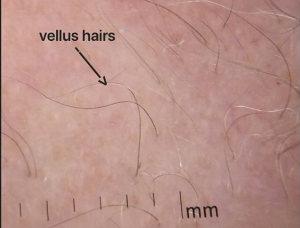
Vellus hairs are short, thin hairs are commonly seen in patients with alopecia areata. These hairs tend to be seen in patients with more severe and active disease. 
This photo shows vellus hairs in a patient with advanced alopecia areata involving 85 % of the scalp.


This article was written by Dr. Jeff Donovan, a Canadian and US board certified dermatologist specializing exclusively in hair loss.
Share This
3 Comments

Alopecia Areata: Broken Hairs

Hair Breakage

hair breakage.png

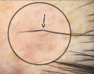
Alopecia areata is an autoimmune disease that affects hairs and nails. Inflammation deep under the skin in a region of the hair follicle known as the bulb leads to the production of weak hair follicles that break easily. Hair breakage is commonly seen in active alopecia areata. The photograph here shows a hair follicle that is about to break. Within hours the hair will likely break off at the site demarcated by the arrow. Treatment of alopecia areata can reduce inflammation and lead to the production of stronger hairs that do not break. 


This article was written by Dr. Jeff Donovan, a Canadian and US board certified dermatologist specializing exclusively in hair loss.
Share This
No Comments

Use of Isotretinoin for Facial papules in FFA

Facial papules in FFA

Facial papules occur in a subset of patients with frontal fibrosing alopecia. Its’ been difficult to ascertain what exactly these facial papules represent. Some of the difficulty comes from the limited number of biopsy specimens that have been obtained from such pappules. Some investigators have found small vellus hairs in the biopsies of facial papules, whereas others have only found hypertrophic sebaceous glands. 

Dr. Aline Donati and her colleagues were among the first to rigorously study facial papules in patients with FFA. She proposed that these papules contained vellus hairs and these vellus hairs showed typical LPP findings with perifollicular inflammation and fibrosis. 

In 2017, Pedrosa and colleagues from Portugal set out to further examine the features of these facial papules. The researchers showed that papules were present in 62 of 108 patients. 10 patients with facial papules underwent biopsy.  All 10 of these patients had similar histological findings, namely hypertrophic sebaceous glands but no evidence of a hair follicle in the biopsy and no evidence of lichenoid inflammation. Interestingly the skin was soft and thin which allowed for easy visualization of the sebaceous glands. 

Oral isotretinoin was reported helpful for these patients. The dose was 10 mg every other day and this was typically added to standard therapies that the patients was already on (such as anti-androgen therapies). Improvement was rapid – most patients saw changes with 2-4 months. 


Conclusion:

This study is interesting for two reasons.

1) It confirms that some biopsies for facial papules in patients with FFA will not contain hairs nor inflammation. Whether these sampled areas once contained hairs is unknown but presumably they did. The hypothesis then is that the vellus hairs were destroyed by the inflammation.

SEE: CURRENT HYPOTHESIS FOR FACIAL PAPULES IN FFA


2) The study is also interesting because it draws attention to the fact that low dose isotretinoin may in fact be helpful as a treatment for these facial papules.


Reference

Pedros et al. Yellow facial papules associated with frontal fibrosing alopecia: A distinct histologic pattern and response to isotretinoin. Journal of the American Academy of Dermatology 2017; 77:754-765

Donati et al. Facial papules in frontal fibrosing alopecia: evidence of vellus follicle involvement. Arch Dermatol 2011; 147: 424-1427.


This article was written by Dr. Jeff Donovan, a Canadian and US board certified dermatologist specializing exclusively in hair loss.
Share This
No Comments

Vitiligo & Alopecia Areata: Can they occur together?

Alopecia areata and Vitiligo are Closely Related

Alopecia areata is an autoimmune disease that causes hair loss. Vitiligo is an autoimmune disease that causes loss of pigment in the skin. The prevalence of vitiligo is estimate to be between 0.5 and 2 %. The prevalence of alopecia areata is estimate between 0.1 and 0.2 % of the population. 


Is the risk of vitiligo increased in alopecia areata?

There are studies that have reported both outcomes. For example, a 1994 study by Schallreuter and colleagues found no increased coincidence of vitiligo and alopecia areata. Nevertheless, three studies did should that patients with alopecia areata have a higher risk of developing vitiligo  however studies by Chu et al, Narita et al and Huang et al (see references below) showed that patients with alopecia areata probably do have a higher risk for developing vitiligo compared to the general population.  Overall, about 3 to 8% of alopecia areata patients have vitiligo (compared to the general prevalence of vitiligo mentioned above of 0.5 and 2 %).


Similarities of AA and Vitiligo

We have come to learn that alopecia areata and vitiligo share many similarities. Both conditions are common in children and adults. In fact, about one half of patients with vitiligo develop their condition before age 20. About one-half of those with alopecia areata develop their condition before age 20. 

Both diseases are relatively asymptomatic meaning that most patients do not have itching, burning or pain. There is inflammation in both conditions, but the amount of inflammation tends to be on the lower side. Most of the inflammation in alopecia areata and vitiligo consists of T cells: CD8+ T cells are present in the epidermis of the skin in vitiligo and in the hair follicle sheath in alopecia areata; CD4+ T cells are in the dermis.   

Both are associated with other autoimmune conditions, especially thyroid disease. The prevalence of thyroid disease has been estimated to be as high as 19.4% in those with vitiligo and 28% in those with alopecia areata.


Vitiligo and AA are TH1-diseases

Vitiligo and AA are driven more by interferon gamma (IFN-γ) signalling than TNF-α which makes one consider vitiligo and AA as so called "TH1 mediated diseases". The predominant Th1 cytokine is IFN-γ.  CD8+ T cells play a key role in alopecia areata and vitiligo and interferon is abundant in affected skin of both diseases. Vitiligo and alopecia areata appear to depend primarily on IFN-γ


Role of TNF in AA and Vitiligo

TNF-α is inflammatory cytokine. Its levels are elevated in TH17-mediated diseases and appears to be required for the diseases to occur. Psoriasis is an example of a TH17 disease and other examples include inflammatory bowel disease, and rheumatoid arthritis. These conditions require TNF-α as well as IL-17, IL-23, and IL-22.  

TNF appears less directly relevant in alopecia areata. However, some studies have suggested that TNF-α can be elevated in vitiligo and alopecia areata. Surprisingly though, treatment of patients with vitiligo and alopecia areata with TNF blocking drugs have been largely unsuccessful and sometimes treatment even triggers, flares or worsens the conditions. 


Conclusion

There are many recognized similarities between alopecia areata and vitiligo and the diseases are closer related than one might otherwise have thought. Both diseases are strongly driven by IFN-γ. Treatments that reduce IFN-γ, including JAK inhibitors, are proving useful for both diseases. Further studies of alopecia areata will likely yield some benefit for how vitiligo is ultimately treatment and vice versa. 


REFERENCE
Schallreuter KU, Lemke R, Brandt O, et al. Vitiligo and other diseases: coexistence or true association? Hamburg study on 321 patients. Dermatology. 1994;188:269–275.

Chu SY, Chen YJ, Tseng WC, et al. Comorbidity profiles among patients with alopecia areata: the importance of onset age, a nationwide population-based study. J Am Acad Dermatol. 2011;65:949–956. 

Narita T, Oiso N, Fukai K, Kabashima K, Kawada A, Suzuki T. Generalized vitiligo and associated autoimmune diseases in Japanese patients and their families. Allergology international: official journal of the Japanese Society of Allergology. 2011;60:505–508.

Huang KP, Mullangi S, Guo Y, Qureshi AA. Autoimmune, atopic, and mental health comorbid conditions associated with alopecia areata in the United States. JAMA Dermatol. 2013;149:789–794.  


This article was written by Dr. Jeff Donovan, a Canadian and US board certified dermatologist specializing exclusively in hair loss.
Share This
1 Comment

Hydroxychloroquine: 10% stop from side effects

hydroq.png

10% Stop from Side Effects

How often do patients stop hydroxychloroquine treatment because of side effects?

Hydroxychloroquine (also known by the name Plaquenil and generics) is an oral anti-inflammatory medication frequently used in the treatment of a variety of autoimmune diseases. For autoimmune hair loss, hydroxychloroquine is used in the treatment of lichen planopilaris, frontal fibrosing alopecia, discoid lupus, and pseudopelade of Brocq.

Side effects include irritation of the liver, pigment changes on the skin, reduced blood counts and retinopathy. The eye side effects are among the more worrisome side effects.

It’s helpful when prescribing a medication to have a sense of how common a side effect might be an how commonly a patient will discontinue a given medication.

Tetu and colleagues performed a retrospective study between January 2013 and June 2014 of patients receiving hydroxychloroquine for a variety of skin issues (not limited to hair). The study included 102 patients (93 of whom were women, with a median age of 44.5; range: 22-90 years). At least one adverse event was reported for 55 patients (ie 54%). 11 patients (10.75%) discontinued hydroxychloroquine due to a side effect that was thought to be directly attributable to the use of hydroxychloroquine.
 

Conclusion

It’s nice to have this kind of information when prescribing medications. Although the study did not solely focus on the use of hydroxychloroquine for hair loss, it’s reasonable that a similar proportion of hair loss patients would be expected to stop their hydroxychloroquine due to a side effect. Other oral options include doxycycline and tetracyclines, mycophenolate, cyclosporine, methotrexate and other anti-lymphocytic agents.
 

Reference

Tétu P, et al. Ann Dermatol Venereol. 2018.


This article was written by Dr. Jeff Donovan, a Canadian and US board certified dermatologist specializing exclusively in hair loss.
Share This
No Comments

Doxycycline and Gastrointesintal Side effects: What have we learned ?

Doxycycline for Scarring alopecia

Doxycycline is an antibiotic that has a unique property in that it is also anti-inflammatory. Doxycycline, at doses 100-200 mg is frequently used for treating scarring alopecias such as lichen planopilaris. The side effects of doxycycline must be carefully weighed against the benefits. Typical risks include gastrointestinal upset (including a risk of irritable bowel syndrome, gastritis, nausea, reflux, peptic ulcer disease and small intestine bacterial overgrowth) sun sensitivity, headaches, and weight gain.

I have increasingly been using subantimicrobial (40 mg) dosing of doxycycline in my clinic for patients with scarring alopecias that are entering a stable phase. Subantimicrobial doses of doxycycline are frequently used for treating rosacea. The hope is that by using the subantimicrobial dosing we can maintain benefits on inflammation without the gastointestinal side effects that accompany standard doses. 

I was very interested in a recently study by Hester Gail Lim and colleauges looking at the side effects on the gastrointestinal system with conventional dosing and subantimicrobial dosing of doxycycline.

Compared to conventional dosing, the use of subantimicrobial dosing was associated with a lower risk of gastrointestinal disease including lower irritable bowel syndrome, H. pylori infection, reflux disease, gastritis and peptic ulcer disease.

Importantly, patients using subantimicrobial dosing of doxycycline had similar levels of gastrointestinal disease to those who weren’t using these drugs at all.  Specifically, the incidence of celiac disease, irritable bowel disease, bacterial overgrowth, reflux disease and gastritis were not increased in those using subantimicrobial dosing. 


Conclusion

The goal of any treatment should be to keep the patient on the medication for as short of duration as needed. For many patients using doxycycline who can not quite quickly withouth risk of flare, the use of subantimicrobial dosing presents a treatment option. This study is encouraging that low dose doxycycline may not increase the risk of gastrointestinal disease. 


This article was written by Dr. Jeff Donovan, a Canadian and US board certified dermatologist specializing exclusively in hair loss.
Share This
1 Comment

Alopecia areata and the Bowel

Increased Risk of IBS in Patients with AA

Alopecia areata is an autoimmune disease. It occurs in about 2 % of the population and in all age groups and races.  An increasing amount of data is emerging looking at the role of “gut health” in many autoimmune disease. The fact that fecal transplants was propsed to benefit AA in limited studies further highlights the role of the bowel. 

In 2017, Conic and colleagues set out to retrospectively review the clinical features of patients with alopecia areata that were seen at the Cleveland Clinic over the period 2005 to 2014.  In total, data from 504 patients was tabulated and as a comparison group 172 patients with seborrheic dermatitis were also reviewed. 

The key bowel diseases that were studied were celiac disease, inflammatory bowel disease and irritable bowel syndrome. Interestingly, the incidence of irritable bowel syndrome was increased in patients with AA but there was no increase in inflammatory bowel disease and no increase in celiac disease. 

This study is interesting as it highlights the need to inquire about bowel symptoms in those with with AA, particularly those that related to irritable bowel syndrome (IBS). Extensive testing for celiac disease in patients with normal ferritin and hemoglobin levels is probably not advisable nor cost effective in adults. 


REFERENCE

Conic et al. Comorbidities in patients with alopecia areata. Journal American Academy Dermatology; 754-756.


This article was written by Dr. Jeff Donovan, a Canadian and US board certified dermatologist specializing exclusively in hair loss.
Share This
No Comments

Facial Papules in FFA: What is our current understanding?

What is our current understanding?

Facial papules are present in a significant proportion of patients with FFA. The papules appear as small yellowish colored bumps that may cause the patient’s face, forehead and chin to feel “rough.” For years, it’s been confusing as to what these papules really are. Early studies by Dr. Aline Donati et al showed that these papules contain inflamed vellus hair follicles.  More recent studies, including those by Pedros and colleagues showed that biopsies of facial papules contained no inflammation … and no hairs!

The following diagram is a diagram that I use when teaching about the facial papules in FFA. It’s a schematic cartoon of the current hypothesis about what these hairs represent and why they disappear.

facial papuels.png


It appears that early in the course of the facial papules, inflammation is present in the vellus hairs. Over time, the hairs disappear and what is left is a dome shaped papule containing hypertrophic sebaceous (SG) glands.

Over time, some papules do flatten and some even disappear. This can take a long time. Studies by Pedros and colleagues have shown that use of oral isotretinoin can help reduce the appearance of these facial papules.
 

REFERENCE

Pedros et al. Yellow facial papules associated with frontal fibrosing alopecia: A distinct histologic pattern and response to isotretinoin. Journal of the American Academy of Dermatology 2017; 77:754-765.


This article was written by Dr. Jeff Donovan, a Canadian and US board certified dermatologist specializing exclusively in hair loss.
Share This
2 Comments

Alopecia areata: Is it a medical disease?

Alopecia areata has widespread health implications

aa-medical

There are several expressions that we have in the English language that point to the fact that we as humans don't always feel comfortable giving things the name that they should be called. If you have even heard someone comment how they 'call a spade a spade a  spade" or if you've ever been told to "stop beating around the bush" you know exactly what is meant by the idiom. In some ways the expression points to the need for effective communicators to "tell it as it is"  and "call it as you see it."


Alopecia areata

A recent article, published by Korta and colleagues in the Journal of the American Academy of Dermatology titled “Alopecia is a Medical Disease” points to the position of alopecia areata as a true medical disease. 

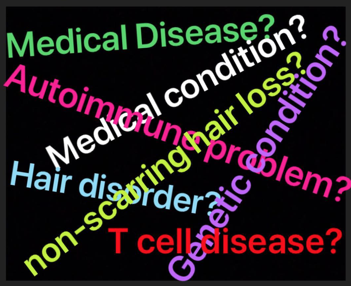
There is no doubt that alopecia areata fits the definition of a medical disease.  However, after many years of being involved in support groups for alopecia areata throughout the world, I can tell you that many individuals don't consider their alopecia as disease. 


"It's not a disease, Dr. Donovan. It's a medical condition of the hair"

"It's an autoimmune condition. It's like a disease, but not quite"

"It’s hard to describe, Dr. Donovan. I don’t have a disease like a disease disease”

"Dr. Donovan, alopecia areata is NOT a disease and even my dermatologist said so”


These comments are frequently similar among the 8 million people world wide who are directly affected by the condition today and the 140 million people that will become affected at some point in their lifetime.

I can tell you that many individuals with alopecia areata around the world don't like the use of the term disease. Some dislike it because they don't feel sick. Some don't like the label, stigma and stereotypes that come with having a disease.  I can feel how the room changes when I breach the subject with my support groups. There is sometime shock that I, given the privilege to be welcomed in a group of alopecia patients, would consider discussing this condition as a disease.


What is a disease anyways?

It's surprisingly difficult to get everyone in a room to agree on the definition of disease. Yet, everyone knows intuitively what's meant by a disease. I remember the definition that I memorized in my first week of medical school. It was a definition from the World Health Organization ( WHO) which defined a health as "a state of complete physical, mental and social well-being, not merely the absence of disease or infirmity." Others define a disease as some type of disorder of the structure or function that produces specific signs or symptoms.

For alopecia areata, there is no doubt that it impacts physical, mental and social wellbeing for affected individuals. From a pathophysiological perspective, there is little doubt that it is a disorder of the immune system that affects that structure and function of the hair.  So, from these criteria, alopecia areata is a disease.


So, why do some patients not consider it a disease?

Well, I’ve learned that defining a disease is not always so easy as the basic definitions allow give us permission to call something a disease.  Whether or not a person or society considers they have a disease depends on various factors including societal and cultural factors.  There has been a rapid expansion of what society calls diseases nowadays. (Ask someone 100 years ago what ‘texting thumb’ or ‘internet addiction’ was and I’m sure you’d be given some blank stares). In contrast, some conditions were viewed as diseases in the past and now have been ‘declassified’ from the list of diseases.

So classifying something as a disease is always open to some degree of interpretation. Many patients with alopecia view their hair loss as a disease. Yet, others do not.  I think we as physicians and society need to fully understand these views. 


The Importance of Calling Alopecia Areata a Disease. 

From the perspective of a physician, it’s clear that alopecia areata is a medical disease. In fact, it’s a complex medical disease that can affect the patient from head to toe. As a specialist who cares for patients with alopecia, one needs to not only focus on the hair, but on a range of medical issues that might be present. These need to be evaluated during a consultation. 

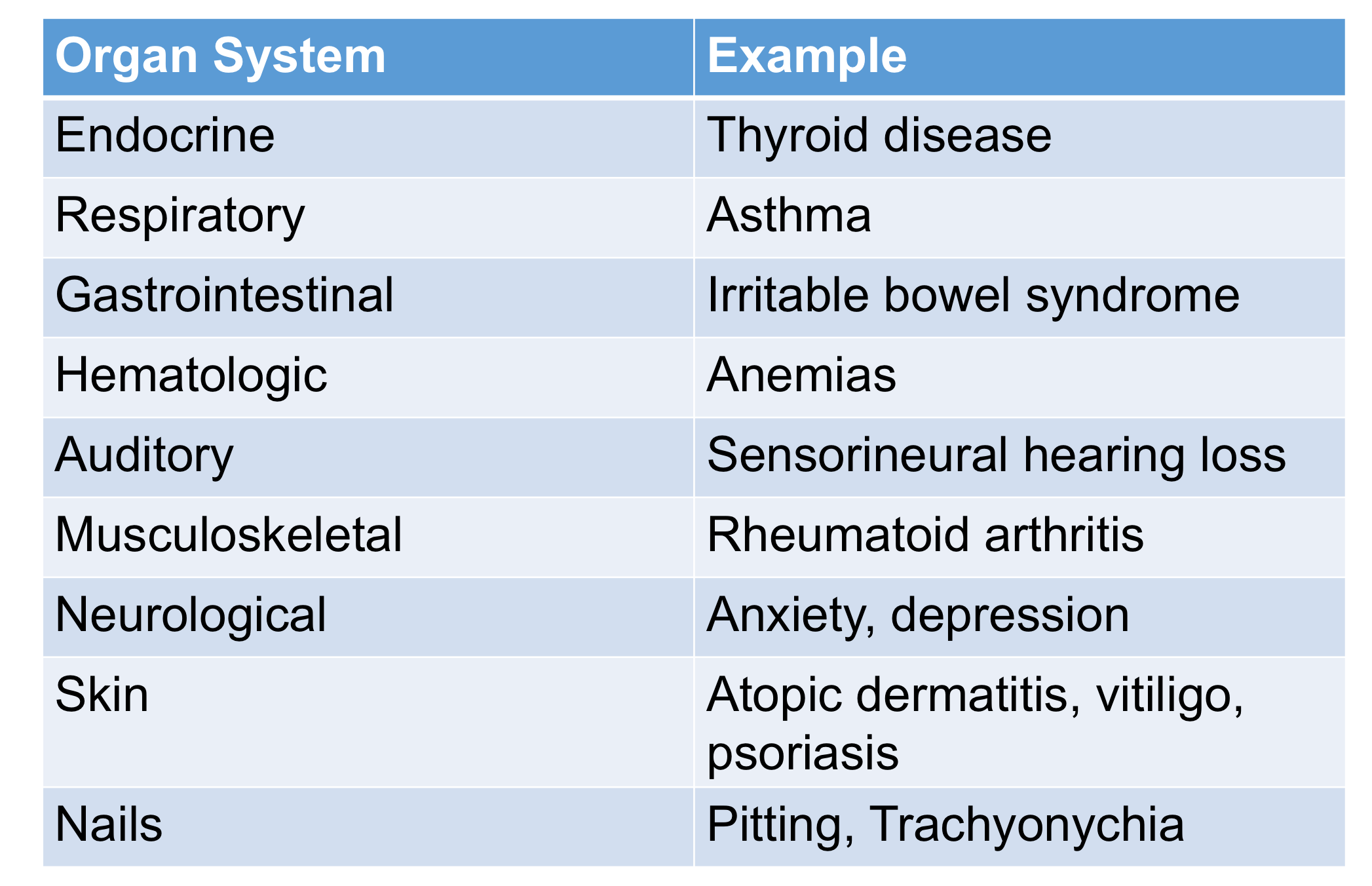
The following table summarizes the range of the more common medical conditions that need to be evaluated. Atopic dermatitis and thyroid disease are among the most common being present in 40 % and 20 % of patients respectively.  In addition to the medical issues, quality of life is adversely affected by alopecia areata. These include anxiety, depression and sleep disturbances.

Partial list of disease associations in alopecia areata

Partial list of disease associations in alopecia areata


Why we need to stop beating around the bush? 

Alopecia areata can sometimes have a significant financial impact on patients and their families as well as insurance groups that pay for various services. There is a need to make sure that alopecia maintains its position among the medical diseases and gets the appropriate recognition it deserves.  According to a recent Global Burden of Disease study which ranks the disability from having a specific disease, alopecia ranked higher than other common diseases such as psoriasis, melanoma and non melanoma skin cancer.   

Disappointedly, Korta and colleagues remind us that funding for alopecia areata remains low compared to its disease burden. It’s time to change that and perhaps the best way that one can change that is by stop beating around the bush, call a spade a spade and say it like it is: alopecia areata is a medical disease. 

It's important to position alopecia areata as a medical disease. It is important to have it recognized as medical disease in order to ensure that healthcare dollars and research resources are appropriated allocated to support this condition. Clearly, modern society has agreed that if one can't determine if something is a disease or not, it might not be worth allocating money to the area. Alopecia areata is a medical disease.

The hope is that by giving alopecia the title of a disease that it will get the recognition that it so deserves. Despite having over 25 treatments for alopecia areata on my list, it comes as a surprise to patients when I tell them that none are formally FDA approved. Making sure alopecia areata gets recognized as a disease is the first step to making sure insurance companies, drug companies, hospital, health agencies and research groups recognized alopecia areata in the way that it should be.   

Alopecia areata is a medical disease.


Reference

Korta DZ, et al. Alopecia areata is a medical disease.  J Am Acad Dermatol. 2018.

WHO (1946) Preamble to the Constitution of the World Health Organization. WHO, New York, USA

Karimkhani et al. JAMA Dermatol 2014. 


This article was written by Dr. Jeff Donovan, a Canadian and US board certified dermatologist specializing exclusively in hair loss.
Share This
No Comments

Pathology of Lichen Planopilaris: Lichenoid Change

Lichenoid Change

lichenoid change.png

There’s a few key things about scalp biopsies of lichen planopilaris (LPP) that are really is helpful to evaluate when looking under the microscope. These include the cell death of hair follicle keratinocytes (so called “lichenoid change” in the earliest stages as well as loss of sebaceous glands (oil glands) over time.

There are, of course, many other changes that can be seen and that a pathologist or dermatopathologist may offer comments. These include reductions in hair density, perifollicular fibrosis, and inflammation in the upper parts of the hair follicle. Changes in the skin layer (lichen planus-like changes) and dilated eccrine glands can also be a part of the pathology.

Unfortunately, there tends to be an extreme focus at times on documentation of perifollicular fibrosis and perifollicular inflammation when evaluating LPP. Certainly these are important and present in LPP. The problem is that they are not diagnostic of LPP as these findings are common in androgenetic alopecia too. In fact, up to 3/4 of patients with AGA have some degree of perifollicular fibrosis and about 1/3 or more have significant perifollicular inflammation.

The photo here shows typical features of the “lichenoid” change that accompanies LPP. There is inflammation in the root sheath and some hair follicle keratinocytes are showing vacuolar change and cell death. 


This article was written by Dr. Jeff Donovan, a Canadian and US board certified dermatologist specializing exclusively in hair loss.
Share This
7 Comments

On the media's reporting of "cures for baldness" - is there a cure for this?

On the Misrepresentation of Hair Research Findings by the Media

Several week ago, I wrote a article about the unusual practices on the media when reporting new hair research findings. The basic tenant of the article was that most articles that are written in the media have a exaggerated hope and unrealistic bias towards convincing the public that the new research finding bring us fairly close to a cure.  Interested readers can read the article here

On the Reporting of Infinitely Amazing Discoveries


A new osteoporosis drug as a model of the media's bias

As an example of the misrepresentation of the media, we'll focus on a recent study from Professor Ralf Paus's lab in Manchester. The results were published in the May 8 edition of PLOS BIOLOGY. Paus' group re-examined the molecular mechanisms of an old immunosuppressive drug, Cyclosporine A (CsA) which is known to promote hair growth.  Prof Paus' team ultimately uncovered a completely new understanding of how cyclosporine affects hair follicles.  The researchers carried out a full gene expression analysis of isolated human scalp hair follicles treated with CsA and found that CsA reduces the expression of SFRP1, a protein that inhibits the development and growth of many tissues, including hair follicles.  After some further work, the group found that a drug called WAY-316606 also antagonizes SFRP1. Surprisingly, WAY-316606 was originally developed to treat osteoporosis.


The Misrepresentation of the Media : 15 Examples

Even though WAY-316606 could impact the SFRP1 pathway, it's completely unknown whether the drug could have any benefit on human hair disorders. Yet the media's spin on the findings were that it was fairly close to a 'cure'. Here are some headlines from articles about the preliminary research of WAY-316606 which shows this misrepresented and skewed view:

WAy2


Way3


way6
 
WAy7
 
Way8
 
cure
 
way11
 
way12
 
way13
 
way13
 
way14
 
way15
 
way16
 
 
way16
 
way111
 

Conclusion

The media has come down with a serious case of excitement. The only problem is that many of these stories are not accurate, not realistic and manipulate the public away from the truth.  Whatever condition it is the media has, it's contagious and spreads quickly - I'm not even sure it's curable. 


This article was written by Dr. Jeff Donovan, a Canadian and US board certified dermatologist specializing exclusively in hair loss.
Share This
No Comments

L-lysine: It's role in iron and zinc absorption

It's Role in Iron and Zinc Absorption

LYSINE.png

L-lysine is an amino acid and is found in eggs, meat, fish and eggs. Generally speaking, lysine is one of the more challenging amino acids to get through normal food intake. This is especially true for those who do not consume these products.

There are very few studies looking at the role of L-lysine and hair. However, there is one in particular that one should be aware of. These are studies focusing on the role of L-lysine in iron and zinc absorption. In 2002, D.H. Rushton showed benefits of l-lysine in increasing iron and zinc levels and in reducing hair shedding.

Among 14 zinc deficient women, L-lysine at doses 1000-1500 mg daily led to an increase in zinc levels from 9.7 to 14.6 umol/L even without these women consuming actual zinc pills.

Similarly, Rushton showed L-lysine may help iron absorption. In his study, iron pills (100 mg per day) in 7 women with chronic telogen effluvium did not change ferritin levels at all. However, when combined with L-lysine (again at 1000-1500 mg per day), ferritin levels increased from 27.4 to 58.6 ug/L. This reduced hair shedding causing the proportion of hairs in the telogen phase to decrease from 19.5 to 11.3.

L-lysine is a key amino acid and I often add it to the overall plan for patients with chronic shedding abnormalities and those with deficiencies of iron and zinc that don't respond to standard treatments. If I do ultimately recommend patients use L-lysine, the dosing in our clinic is typically 500 mg twice daily, and rarely three times daily for short periods.
 

Reference

DH Rushton. Nutritional factors in hair loss. Clin Exp Dermatol 2002


This article was written by Dr. Jeff Donovan, a Canadian and US board certified dermatologist specializing exclusively in hair loss.
Share This
No Comments

Is topical clobetasol safe for the scalp?

On the Safety of Topical Clobetasol 

I've often asked if applying topical clobetsol steroid cream, lotion, foam or shampoo to the scalp is safe. Before we answer that, it's important to understand what clobetasol is and why it's used. 


Clobetasol is a class I steroid. Hydrocortisone is Class 7.

There are seven classes of steroid strengths. Class 1 steroids are the strongest and class 7 are the weakest. Clobetasol is a class 1 steroid and requires a prescription (in most countries). Hydrocortisone is a class 7 steroid that can often be bought 'over the counter' at the local drug store.  In simple terms, clobetasol is about 600 times stronger than hydrocortisone.  That does not simply equate to dangerous. It simply equates to stronger. A common steroid potency chart is found in the list below

TOPICAL STEROID POTENCY COMPARISON


Frequency, Duration, Amount

When someone tells me they are are using clobetasol, the first thing I want to know is how much are they using and how often are they using it? It comes as a surprise to some that how much steroid a patient is using is usually more important to me that the how often.  A patient who used clobetasol every day but it takes them 5 months to use up their bottle has a very different safety risk profile than someone who is using clobeetasol every day but goes through a bottle every two weeks.  Similarly, a patient who uses clobetsol twice per week could be using more than a patient using it everyday. The amount matters!


On the Fear of Topical steroids

There is quite a bit of inappropriate and misguided fear about topical steroids. I'm not saying topical steroids don't deserve respect, because they do. However, the fear that permeates society mainly comes form poor knowledge and also from the misuse of these products among the general population. Sadly, sometimes this misdirected fear comes from unethical practice and misguided motives. It's tough to change that but I can give at least 1000 examples from my own practice over the years of these situations:

A clinic wanting to sell product A for a child advising a parent "Oh you wouldn't want to put a steroid on your child would you?"

A clinic wanting to sell treatment B to there patient saying "Steroids are not safe. This treatment I am recommending is drug-free and natural."

A clinic wanting to establish 'trust' with a client and advises them "You need to stay away from that other clinic recommending you that steroid treatment. What was recommended is very unsafe. I can't believe they wanted to give you that. They should be reported."


It's difficult in the short term to change how hair medicine gets practiced throughout the world and it's difficult to regulate clinics and practitioners that prey on the vulnerability of their clients and patients. However, we can first and foremost recognize these patterns and spread accurate information as a starting point. I can assure you it's not always a popular view. Topical steroids can be quite safe when used appropriately.  Of course, they are unsafe when used inappropriately.


Logic, Practicality and other Forgotten Issues

When it comes to topical steroids, we need to be logical and practical.  Practical thinking does us good as humans, and we should not forget these principles:

A. It's safe to walk to your across the parking lot to your car on a blazing hot summer day, but it would not be appropriate to walk for hours across the entire city on the same hot day.

B. It's safe to add a bit of hot chili pepper to dish that one is preparing for dinner, but adding the entire chili pepper bottle would just not make sense.

C. It's safe to add a dab of toothpaste to one's toothbrush, but squeezing out the whole tube onto the brush would just be bizarre. 

D. It's generally safe to use a bit of topical steroid for short periods of time to calm down an inflammatory scalp disorder that is causing a patient extreme discomfort, itching and burning. 

E. It's generally safe to use a bit of topical steroid for short periods of time to reduce scalp inflammation that is preventing hair growth.


Safety Monitoring

Anyone using topical steroids needs to be monitored by an expert who knows how to use these prescriptions and what side effects they carry.  

First, patients using the steroid must understand how much to use and for how long. They should carefully record the amount of steroid they are using on a monthly basis and carefully record how long it takes them to go through their tube or bottle. 

Excessive use of topical steroids does lead to thinning of the skin, and even side effects from absorption into the body. These side effects are relatively uncommon with proper doses. 


Use of Topical Clobetasol in Hair Loss

In hair dermatology, we use topical steroids for many reasons. Topical clobetsol is commonly used to treat alopecia areata, and scarring alopecias such as lichen planopilaris (LPP). When used, these should be used for a short of time as needed and always under supervision. Frequent breaks from the steroid use ("steroid holidays")  are frequently helpful. For children with inflammatory scalp conditions that require topical steroids, we often prescribe topical steroids for 4-6 weeks straight and then give a 2-4 week steroid-free holiday period. This cycle is often repeated.


Summary 

Topical steroids can be both safe and effective when used appropriately. Of course, the don't help everyone and may not be enough of an immunosuppressive type treatment for certain kinds of hair loss. For example, some patients with alopecia areata and some patients with lichen planopilaris find that topical steroids help but not enough and hair loss still occurs despite using them.  In such as case the physician needs to decide whether to continue the topical steroid and add other immunosuppressive treatments or whether to stop the topical steroids altogether in place of the new immunosuppressive treatments. 


This article was written by Dr. Jeff Donovan, a Canadian and US board certified dermatologist specializing exclusively in hair loss.
Share This
1 Comment

Scalp Biopsies For LPP: Wonderful Tool When Used Properly

LPP

Scalp biopsies are wonderful tools but they must be interpreted properly. Getting a scalp biopsy “just because” it sounds like a good idea is rarely every never a good idea. One needs to have a purpose of doing a biopsy - to rule in a disease or rule out a disease.

There is much confusion when it comes to diagnosing LPP and AGA. Every year I see at least 30 patients who come into through my office with a diagnosis of LPP and leave my office with a diagnosis of AGA. It's not some treatment I did that changed the diagnosis, it's the diagnosis that changed. It’s a pretty remarkable and sometimes emotional consult.


How’s this even possible? How can a diagnosis be wrong?

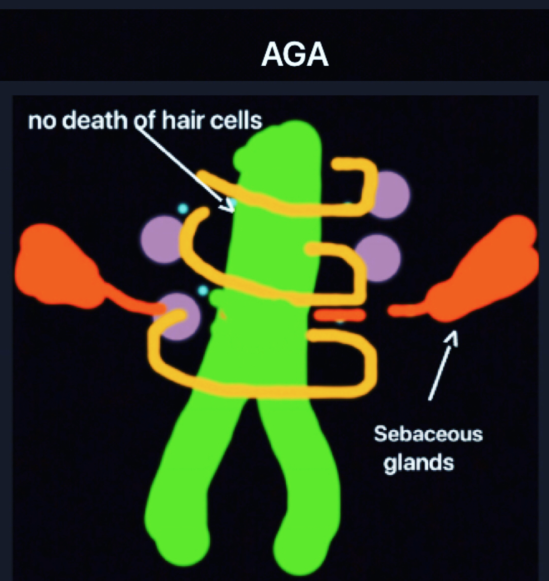
AGA

First off, let me say that most people who come into the office with a diagnosis of LPP actually have LPP. So what we are talking about here is something specific.

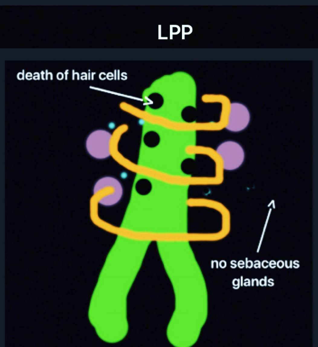
There is, however, tendency to overcall or overdiagnose LPP on account of a failure to recognize a few points. First, perifollicular inflammation and fibrosis is common in AGA. In fact, nearly 75 % of patients with AGA have perifollicular fibrosis and 30-40 % have perifollicular inflammation. So these alone are certainly not criteria for LPP! What needs to be properly recognized is that LPP is associated with “lichenoid change” in the outer root sheath and death of hair follicles keratinocytes.

LPP2

The other cardinal feature of scarring alopecia is loss of the sebaceous glands. These latter two features need to be the focus of the pathologist’s attention and not solely the perifollicular fibrosis and inflammation. As simple as it sounds, many lives can be altered be understanding these principles.


REFERENCES

Evaluation of Perifollicular Inflammation of Donor Area during Hair Transplantation in Androgenetic Alopecia and its Comparison with Controls.
Nirmal B, et al. Int J Trichology. 2013.

Diagnostic and predictive value of horizontal sections of scalp biopsy specimens in male pattern androgenetic alopecia.
Whiting DA. J Am Acad Dermatol. 1993.


This article was written by Dr. Jeff Donovan, a Canadian and US board certified dermatologist specializing exclusively in hair loss.
Share This
2 Comments

Hair Transplant Test Sessions for Scarring Alopecia

Common Errors in Performing 'Test Sesssions'  

Hair transplants can be an option for patients with scarring alopecia but only if certain criteria are met. First and foremost, the disease should ideally be completely 'inactive' for at least 2 years and the patient should be off all types of immunosuppressive medications. Unless these minimal criteria are met, we can't be sure the scarring alopecia is truly quiet (inactive).

test-sessions

Sometimes a hair transplant “test session” is performed 8-12 month prior to proceeding to the ultimate full hair transplant session. The purpose of the “test session” is to better estimate the chances of success of a larger transplant session. Patients with a successful test session are given the green light to proceed to a full session. Patients with a poor outcome on the tests session may be advised not to proceed with hair restoration in the near future. 

I think there is a lot of misunderstanding about how to perform a test grafting session for a patient with scarring alopecia. Below I outline some common errors. But first, let me begin with an analogy. 


Hair Transplant Test Sessions: An Analogy

I often use the following analogy when explaining the concepts I believe are important in  a hair transplant tes session. As an analogy. imagine yourself offered an exciting job position. 9 months of the year you’ll be travelling to the world's most exotic destinations. The only catch is that 12 weeks of the year, you'll need to be in the arctic circle during the middle of winter. Each day, you’ll need to walk 20 miles through ice and snow collecting various samples. You’ll sleep in a tent each night and you need to carry all your belongings with you. 

You are really not sure if you’ll be able to withstand the cold and all your friends think you’ll crazy for thinking about accepting the position as they think you are far to soft of a person to withstand the cold and extremes of the artic circles. To test whether you can really survive an article winter,  you decide to make a “test visit” for 1 month to see if you’ll really like living there or not. You figure if you can survive 4 weeks, 12 weeks should not be so bad. 

As you are deciding when to go, a friend advises you to go in the summer since she’s heard it’s lovely in the summer. Another friend advises you to go in the winter but stay at some luxury accommodations he’s heard good things about.   

In my opinion, neither pieces of advise from these friends is really ideal for a ‘test visit.’  To challenge yourself to see if you are really going to withstand the arctic winter, you need to visit in the winter - and you need to stay in lodging that best represents how you’ll live if you do decide to move. That lodging is, of course, a tent. It’s reasonable to bring two blankets with you and a few more warm belongings that most people who live in the area normally use – because let’s face it – the whole experience is completely new to you. 

The same is true with a transplant test session. The whole experience of moving from a warm, richly vascularized area into a scarred, poorly vascularized area is a big challenge for a little hair follicle. During a 'test session, I believe one needs to challenge these little hairs and see if they really can make it. One needs to make the scalp suitable to growing but not too perfect and luxurious that it falsely misrepresents the challenges that will ultimately be present if a real transplant is performed. Newly transplanted hairs are not really used to growing in scar tissue and not used to growing in between the little bits of inflammation that are often there – so it’s reasonable to help them out a bit. But my opinion is that a test session should truly be a test session. The hairs need to be tested !


COMMON ERRORS IN "TEST SESSIONS"

1.     Excessive topical corticosteroids and immunosuppressives are used

A test session is really all about testing whether hairs can withstand the challenges of growing in less than ideal conditions. The new grafts need to grow in scarred scalp tissue that may lack ideal blood supply. The skin itself may be too thick or too thin.

As part of the test session, I believe on should use minimal corticosteroids (and minimal other immunosuppressives if at all possible). It's still okay to use them but just not excessively.  I typically recommend steroids a few times per week before the test transplant with steroids stopped one month before. Steroids are started again 1 month after the test session but only twice weekly for two months and then once weekly thereafter.  A mid potency topical steroid should be sufficient.

One needs to challenge these little hairs and see if they really can make it. One needs to make the scalp suitable to growing but not too perfect and luxurious that it falsely misrepresents the challenges that will ultimately be present if a real transplant is performed. The analogy is similar to the arctic circle analogy I used above. 


2.     The grafts are spaced too far apart

Ideally, 100-200 grafts should be put in an area at a density of 25-30 follicular units per cm2 and this area carefully followed over time. The problem with placing grafts at a lesser density is that it does not adequately ‘stress’ the hair follicles during the test session. When it comes to growing in scar tissue, it’s easier for hairs to grow when they are far apart rather than close together. My view is that during a ‘test session’we need to understand how the hair follicles survive under realistic situations. If the patient ultimately proceed to a full hair transplant session, follicle are going to be transplanted at a density of 20-25 follicular units per square cm AT MINIMUM and so a test session should slightly exceed this. We need to test the follicles!


3.     Too many grafts are put in

I don’t recommend that test sessions be done with more than 200 grafts and 150 is often ideal. The problem with doing more than 200 is that it become difficult to count the grafts. 

I have seen patients who come to see me after have 500-800 grafts ‘peppered’ into the front of the scalp or ‘peppered’ into the crown. Instead of putting 100-150 follicles in to an area the size of a golf ball, 500-800 grafts get put into an area the size of a large melon or small dinner plate. It is easy to count 100-200 grafts (1, 2, 3, 4…) but more difficult to count 500. When it comes to hari transplant test session, I believe we need accurate survival numbers. Knowing that 90% survived is very different than knowing 54 % survived. Knowing that the survival 'seemed ok' is very different than knowing that the survival was 'excellent.'


4.     The grafts are put into an area already containing hair follicles

In my opinion, a test session should be performed in an area with as few hairs as possible, ideally with no hair. Sometimes this is not possible, but if it is –this should be followed. This makes it easier to document “before and after”photographs, count hairs and document clearly the survival of grafts. 

The problem with putting in 500-1000 grafts in area area that already has hair, is that it becomes impossible to really get a sense of the proportion of grafts surviving at a time 9-12 months post op. I saw a patient recently who had 500 grafts placed into his crown. The test session was performed in an area that was thinning but not bald. It was impossible to really get a sense of how many grafts survived, despite the fact that the surgeon estimated survival waspretty good. Photos suggested there were no changes to the density and the patient felt it had worsened! Objective measurements are what is needed in these test session.  


5.     The grafts are put in an area that is not representative of the actual scalp to be transplanted

Hair follicles need to be placed in an area that is representative of the average quality of the skin of the scalp, and possibly even in an area that represents slightly poorer than average quality. If much of the scalp contain thicker areas of scar tissue, this is not unreasonable to perform the test session in there. If most of the scalp is thick pale, poorly vascularized tissue and only a small portion of the scalp is pink normal appearing skin, it is misrepresentative to perform the test tession in the pink, normal appearing area.  


Returning to the analogy above, it is unreasonable (in my opinion) for a person undergoing a test visit to the article circle to stay in a luxury accommodation during the visit when the whole purpose of the visit is to see if one can really withstand the extremes of living in the article circle. In the same way, it is unreasonable to make the test session a wonderful opportunity for hair growth.


A test session should represent a true... test!


This article was written by Dr. Jeff Donovan, a Canadian and US board certified dermatologist specializing exclusively in hair loss.
Share This
No Comments

Donovan Hair Clinic rated a top authority on Hair Loss

Agilience.com rates Donovan Hair Clinic among top 10 

We're honoured that Agelience.com recently rated donovanmedical among the top 10 authorities on hair loss. The Paris-based Agelience measures the overall relevancy of social media and web content. The Agilience Authority Index provides recognition  among social media groups in similar categories.  


This article was written by Dr. Jeff Donovan, a Canadian and US board certified dermatologist specializing exclusively in hair loss.
Share This
No Comments

Lichen Planopilaris and the Eye

Lichen Planopilaris and the Eye

LPP and the eye.png

In the pathways that lead to scarring alopecia, there is a close (but poorly understood) relationship between hair follicles and oil glands. In fact, inflammation and reduction in oil glands in the scalp appear to be one of the various first steps in lichen planopilaris.

Although eyelashes can be lost in lichen planopilaris and closely related frontal fibrosing alopecia, there has been little study of how the oil glands in eyelashes are altered in patients with lichen planopilaris.

The oil glands of the eyelid are known as “meibomian glands” and they have a key role in secreting an oily substance meibum which helps prevent evaporation of tear film. Without meibomian glands, our tears would evaporate quickly or constantly run onto the face. There are about 50 such glands in the upper eyelid and 25 in the lower eyelid.

Problems within the Meibomian glands are increasingly recognized by eye doctors under the term “meibomian gland dysfunction” (MGD) and are a major cause of dry eye.

In a new study, researchers set out to study whether meibomian gland dysfunction is present in patients with lichen planopilaris. They performed a case-control study involving 23 patients with histologically confirmed LPP and 23 healthy controls. The researchers used a specific test known as “tear breakup time” as a measure of meibomian gland function.

Interestingly, patients with LPP had different results than controls. Specifically, patients with LPP had lower tear breakup time meaning that evaporation of tears occurred more readily in patients with LPP.

Other findings of the eye examinations were normal in patients with LPP including conjunctiva, lid margin shape, eye pressures, and fundoscopy.

In summary, patients with LPP scored worse in ocular surface tests. This study draws attention to the importance of monitoring for dry eye and meibomian gland dysfunction in patients with LPP.
 

Reference

Gheisari M et al. Ocular Surface Findings in Patients With Lichen Planopilaris. Cornea. 2018.


This article was written by Dr. Jeff Donovan, a Canadian and US board certified dermatologist specializing exclusively in hair loss.
Share This
2 Comments

Blogs by Topic


Share This
-->