QUESTION OF THE WEEK

Dr. Donovan's Articles

QUESTION OF HAIR BLOGS

Filtering by Category: Biopsy


Hair Loss: What's causing my hair loss?

What’s causing my patch of hair loss?


I’ve selected this question below for this week’s question of the week. It allows us to review some concepts regarding clinical and trichoscopic examination of acute hair loss.


Question

Hi Dr. Donovan.

I’m a 31 year old female. While giving birth I almost died, went in to septic shock and lost a massive amount of blood. 2.5 months later I lost a lot of hair, I had thinning all over but more obvious around my ears, sides of my head and on the nape of my neck. My dermatologist (and biopsy) said it was TE and gave me steroid shots and my hair is growing back normally with no thinning.

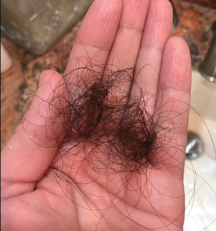
I developed seborrheic dermatitis, my head is a little itchy and I’m on ketoconazole shampoo. 5 months after birth, I had to have major surgery on my kidney, the surgery itself lasted 8 hours. On the 2nd or 3rd day at the hospital I noticed a painful bump on my parietal lobe. On the 19th day after surgery, I washed my hair and then noticed the hair loss on the same area as the swelling. Ive attached photos of the first time noticing it.

My dermatologist injected it with steroids and it isn’t growing back. It’s been 3.5 months since it fell out. There are tiny hairs in the area so my dermatologist is sure it isn’t scarred.

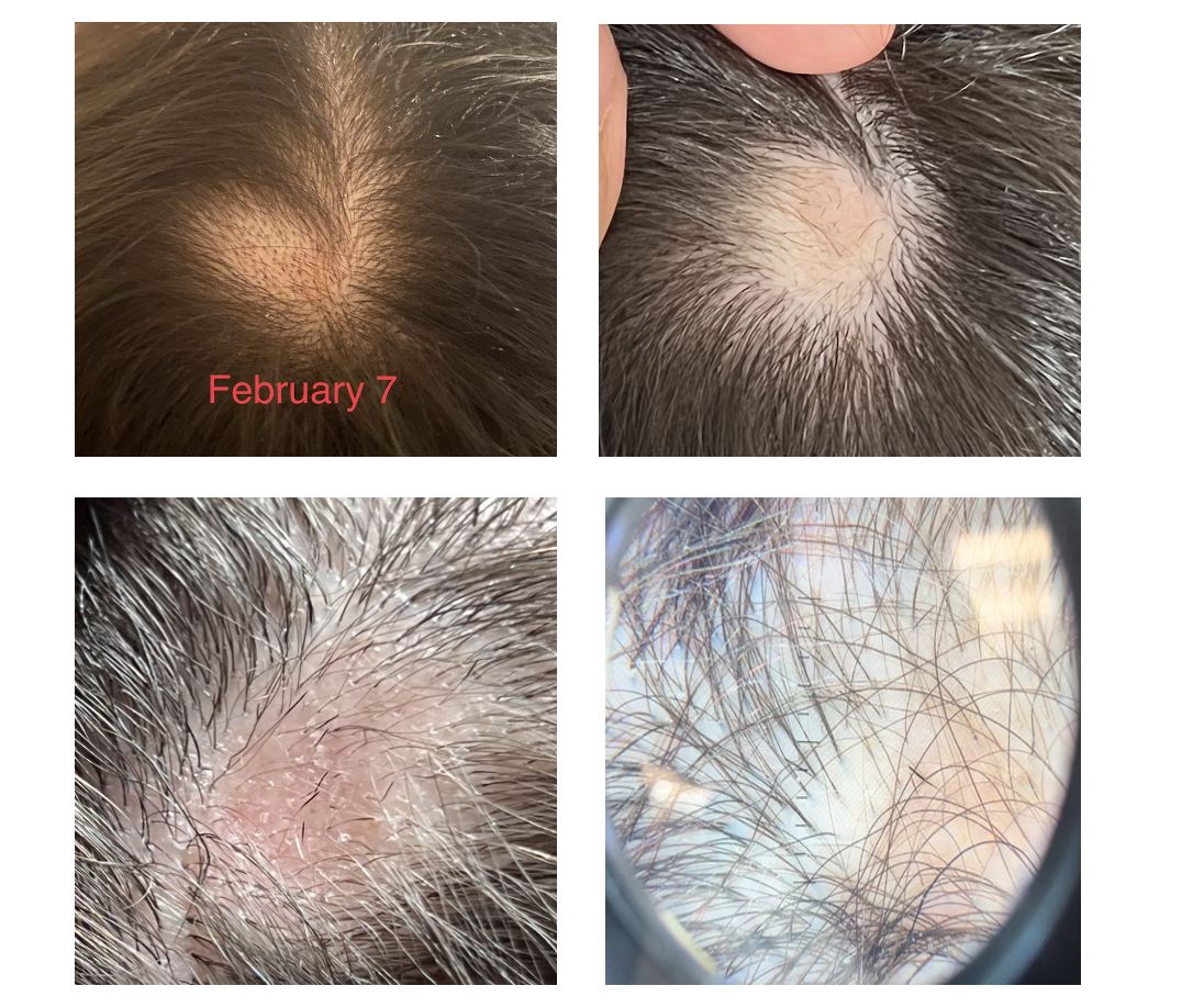
But there are also exclamation looking hairs so we are not sure. The area is reddish. I have no hair loss anywhere else. It hasn’t gotten bigger and I don’t have any patches elsewhere. The picture labeled February 7 is the day I noticed it.

Answer

Thanks for submitting this question. I hope that you are feeling well. At first overview, it certainly would appear that the diagnosis is alopecia areata with some overlapping findings of seborrheic dermatitis. In addition, it appears that you first had a telogen effluvium (of alopecia areata again) that settled after delivery. I still favour alopecia areata as the diagnosis in the current photos but there are a few things in your story and some of your images that cause me to pause and ask “is it possible there is anything else going on here?”

The reason I’ve chosen this question is that it allows us to review some of these features today.

There are several scalp conditions that can cause localized hair loss in this manner with possible ‘exclamation mark like hairs”. The top 4 include:

  1. alopecia areata ** most likely **

  2. dissecting cellulitis

  3. pseudocysts (alopecic and aseptic nodules of the scalp

  4. infections (syphilitic alopecia)

Other diagnoses to consider here but do not have good evidence include:

  1. tinea capitis

  2. pressure alopecia

  3. infiltrative conditions.

  4. trichotillomania

Let’s take a look first at some of the images supplied in this question and then we’ll go into these possibilities a little further and come to some conclusions.
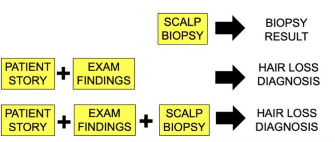
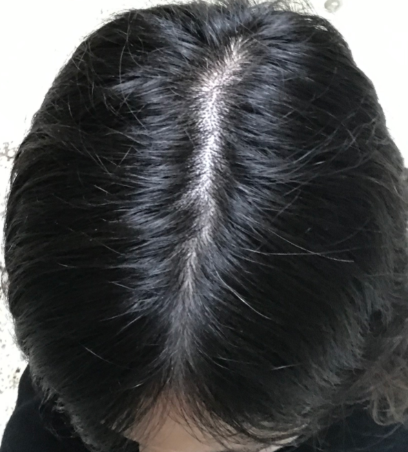
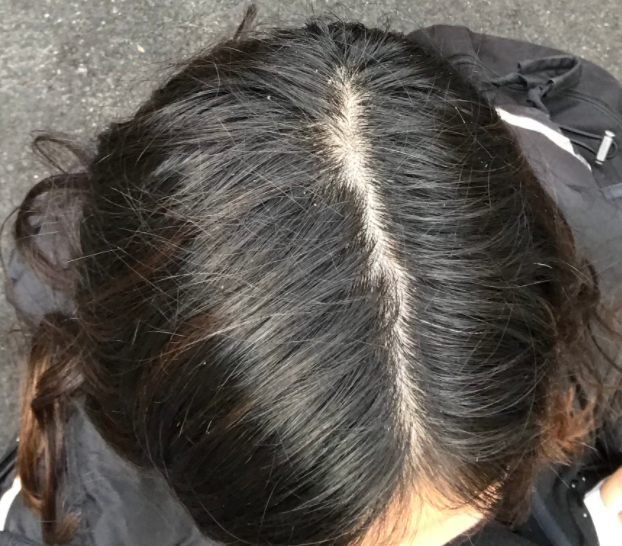
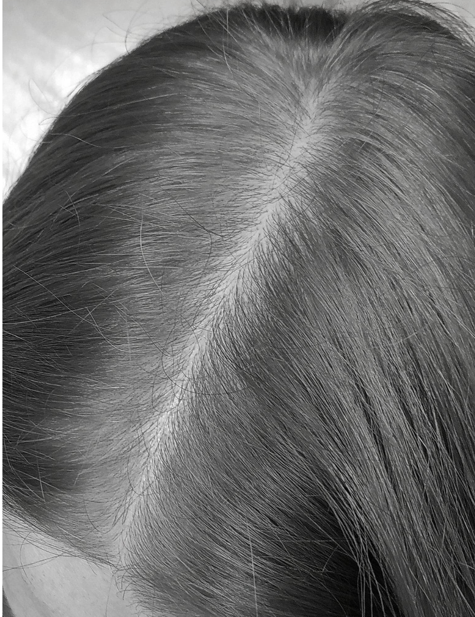
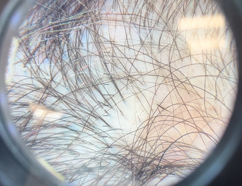
Submitted Image 1

This image shows patchy hair loss. There are broken hairs. Inflammation is mild. The top diagnosis at this magnification would be alopecia areata. The differential diagnosis from this image might include trichotillomania, tinea capitis, and pressure induced alopecia. Alopecia areata would be the top diagnosis. Exclamation mark hairs are not clearly seen in this image but are seen in other images. There is no evidence for a scarring alopecia. Density may be reduced in the more anterior portion of the scalp (top of the photo) suggesting ongoing TE or another hair loss diagnosis happening in this area.
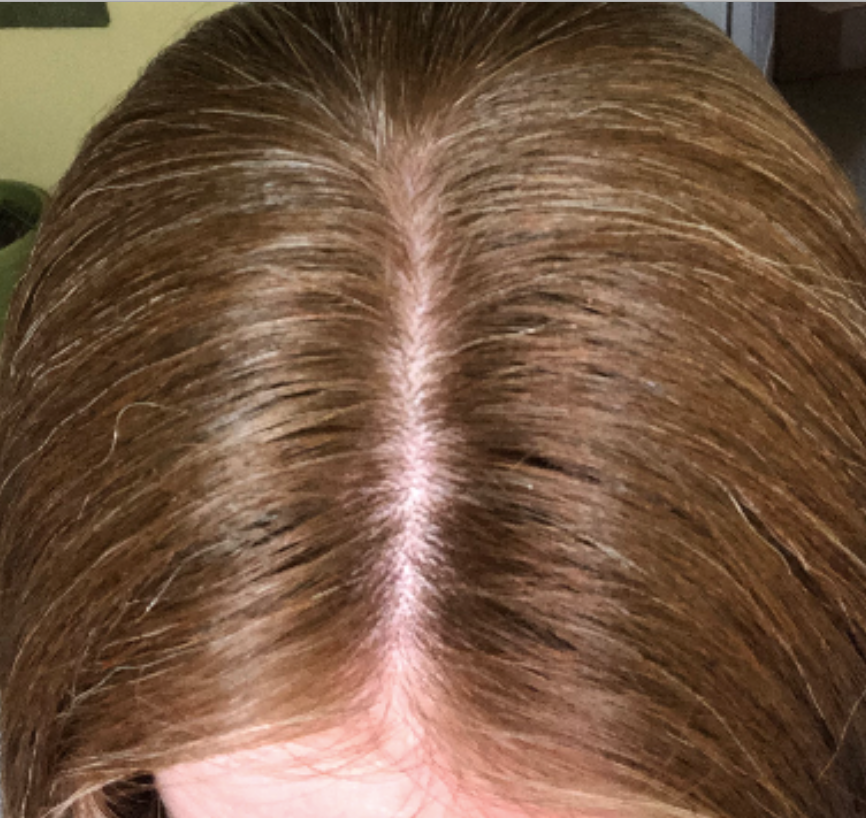
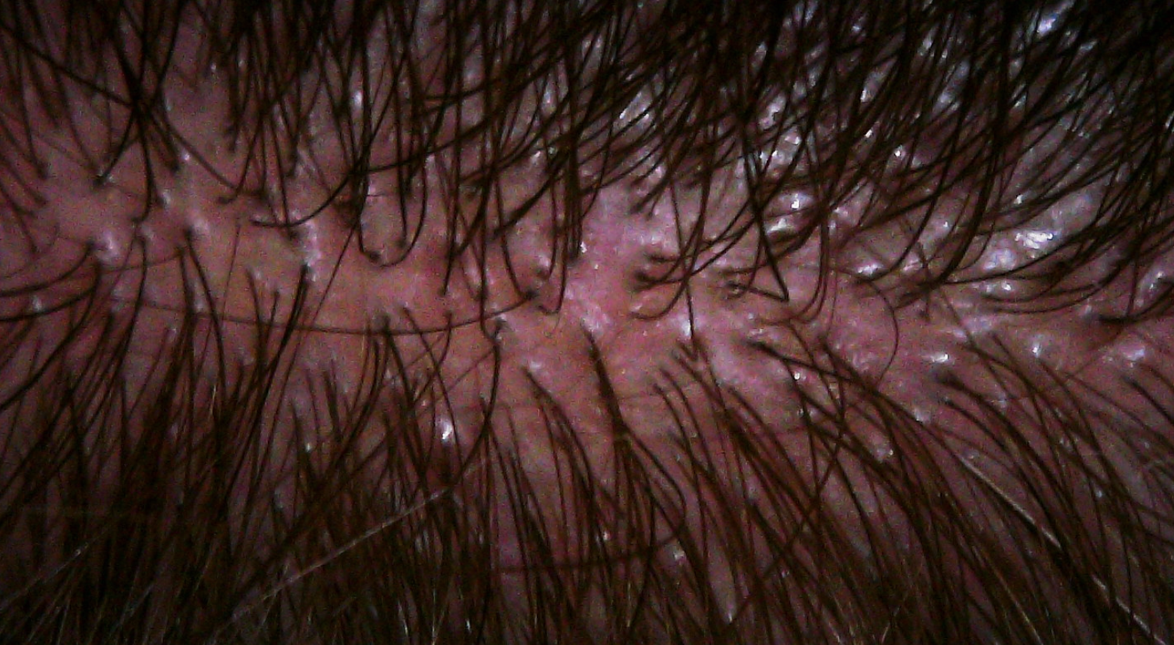
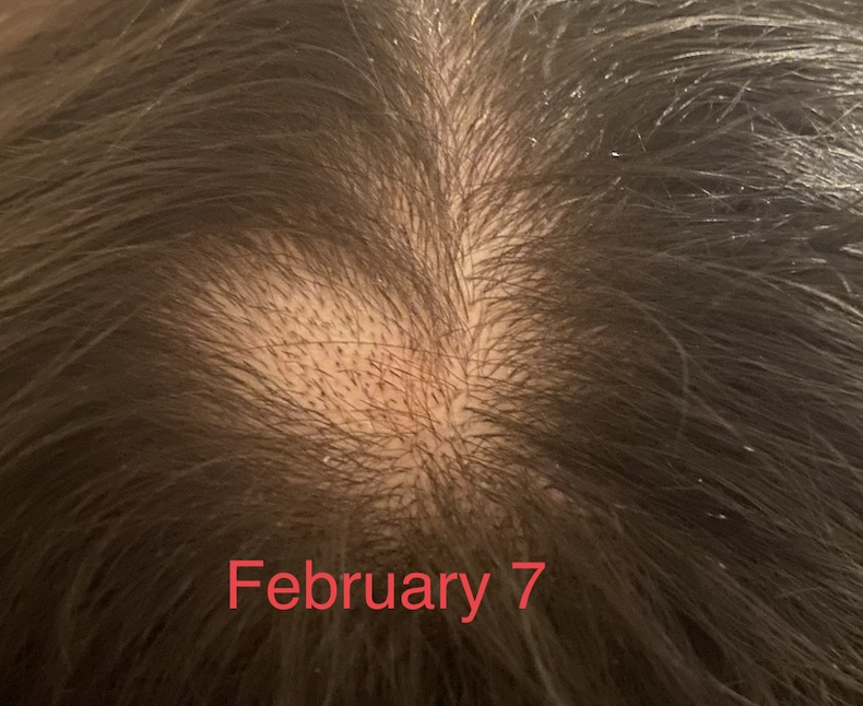
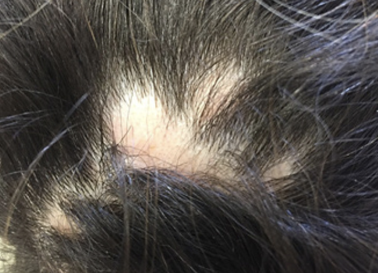
Submitted Image 2

This image shows a well cicumscribed area with minimal inflammation. There are vellus hairs and broken hairs. Some hairs have hair shaft changes suggestive of a pseudo-monilthrix like change (Pohl Pinkus constrictions). Exclamation mark hairs are not clearly seen in this image but are seen in other images. There is no good evidence for a scarring alopecia. Alopecia areata remains a favoured diagnosis.
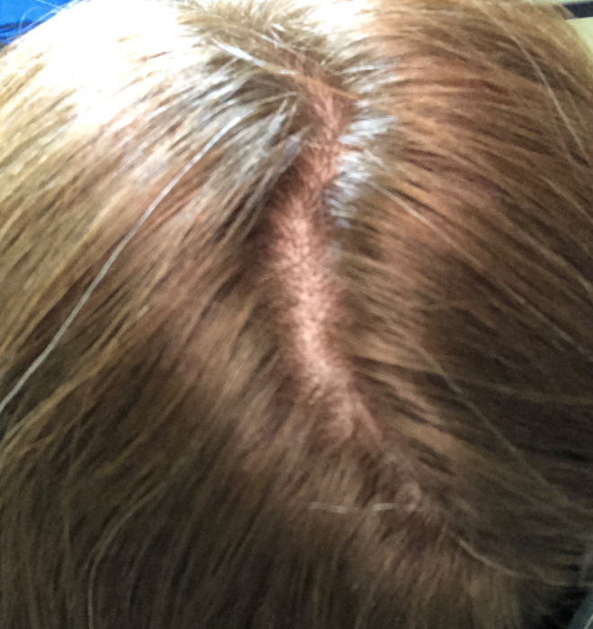
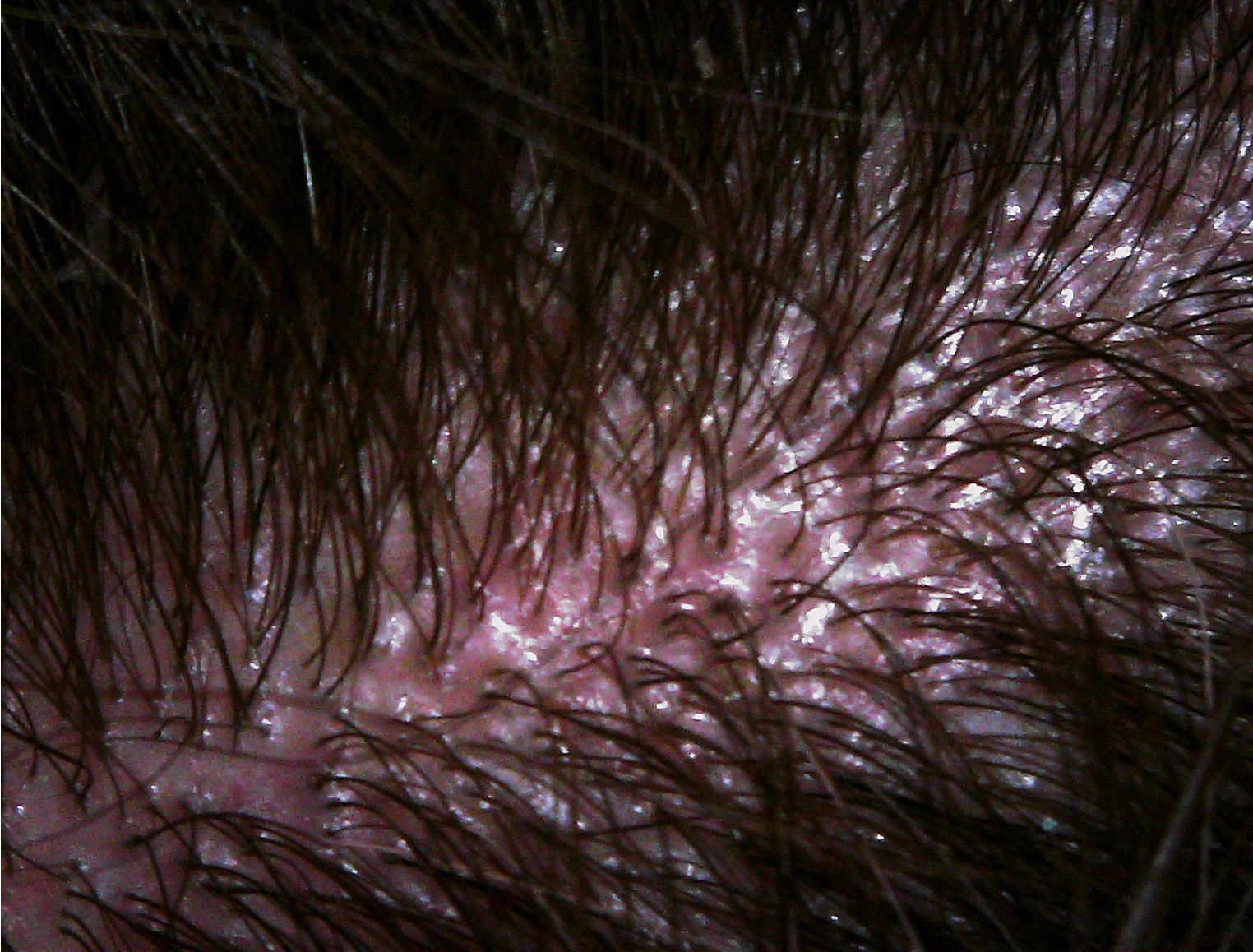
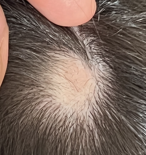
Submitted Image 3

Numerous exclamation mark hairs are seen in this image. Elbow hairs are seen. Yellow dots are seen. There is an inflammatory type change with whitish scale. There is a mild pigmentation alteration which is somewhat non specific. The exclamation mark hairs make other diagnoses quite unlikely as exclamation mark hairs of this kind do not occur in pressure alopecia nor in inflammatory connective issues issues. The appearance of the scalp in this image differs quite a bit from the appearance seen in other images.
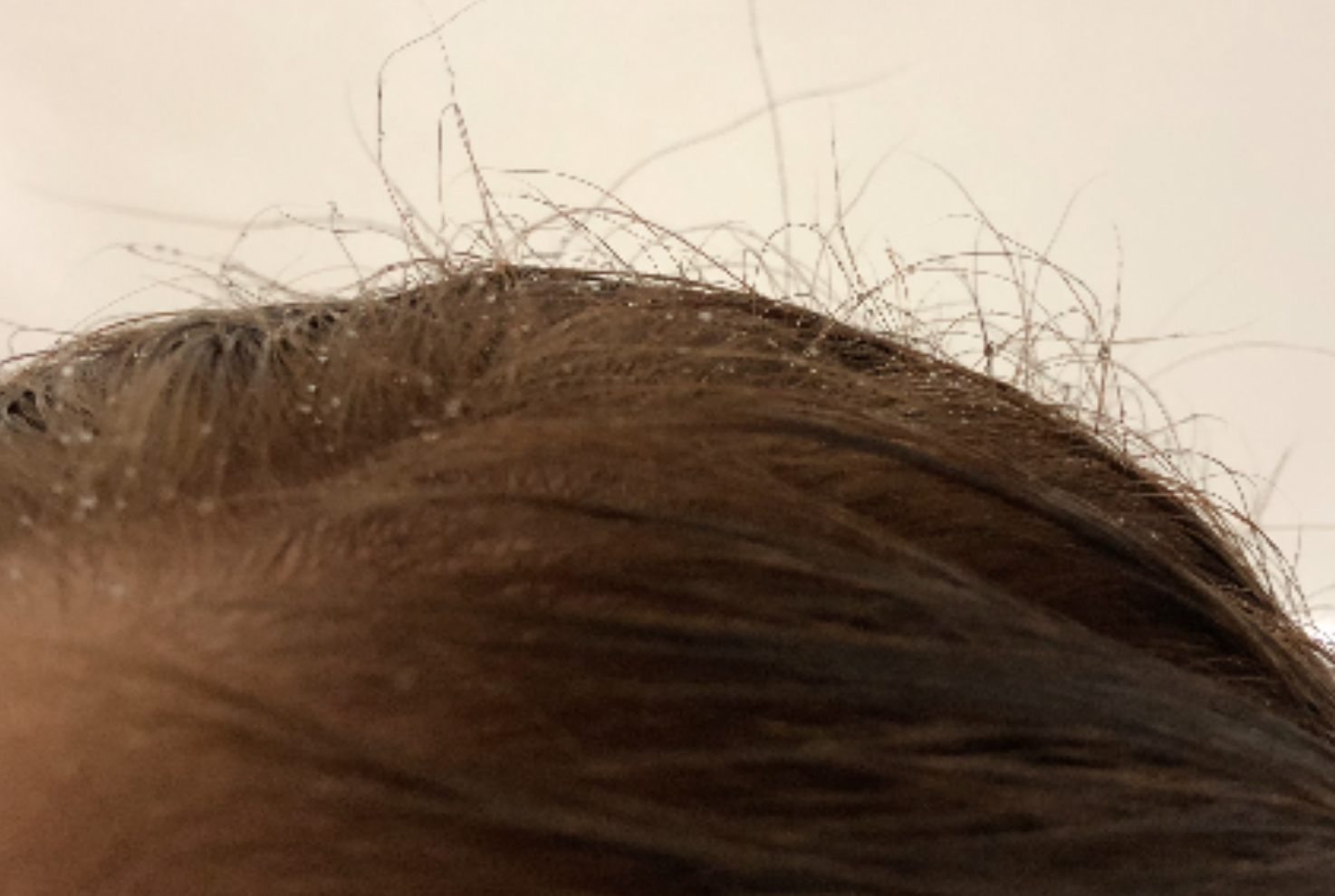
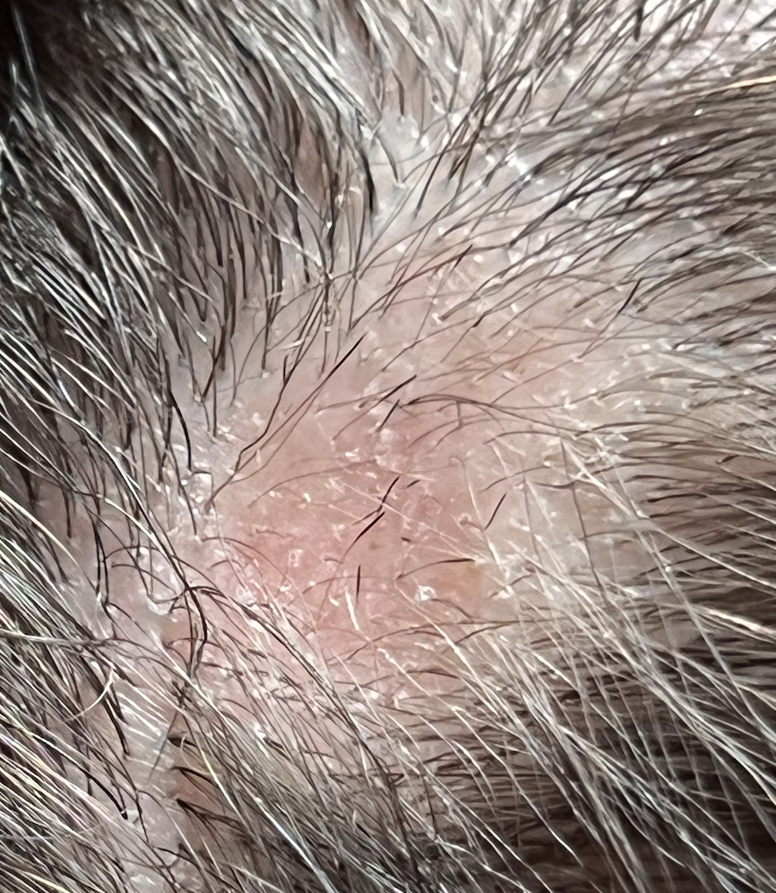
Submitted Image 4

This image shows several exclamation mark hairs with regrowing vellus hairs. There is mild yellow scale which may be in keeping with seborrheic dermatitis (of psoriasis) or an artefact of the photo itself. There is no evidence for a scarring alopecia.


Further Discussion

Thanks again for submitting this case. I favour alopecia areata but of course it’s nice to have more information and see the entire scalp eyebrows eyelashes, and nails. The most accurate way to diagnosis hair loss is to collect all the information about the patient and then examine all the scalp.

The features that support alopecia areata are the exclamation mark hairs, vellus hairs, regrowing hairs and localized nature of the hair loss.


What other conditions cause exclamation mark hairs?

As we think about this question, it’s helpful to think about all that conditions that cause exclamation mark hairs. After all, one of the key features in the submitted images are the exclamation mark hairs.

Exclamation mark hairs are seen in alopecia areata, trichotillomania, thallium poisoning, dissecting cellulitis. Syphilitic alopecia has been rarely described to have a type of tapered hair closely resembling a true exclamation mark hair. This is very rare.

Trichotillomania

There does not appear to be good evidence here for trichotillomania. The story does not fit. It’s one of the famous causes of exclamation mark like hairs. Certainly extensive broken hairs can be a feature but other findings like black dots, V hairs, coiled hairs, hook hairs, hair powder just don’t appear to be a feature of this patient’s hair loss. I don’t think we’re dealing with trichotillomania.

Thallium poisoning

Of course, thallium poisoning is rare.

Dissecting Cellulitis and Alopecic and Aseptic Nodules of the Scalp

The description of the ‘painful bump’ is a bit unusual in the submitted question. It’s not typical of alopecia areata. It may be a ‘red herring’ and unrelated to the case here or it may truly be a valuable clue. Also, it would be helpful to know more about what is meant by a painful bump and how big of a bump is the individual referring to.

As we think about painful bumps, we need to think about small bumps and things like a folliculitis. As we get into larger and larger bumps we need to consider more significant inflammatory conditions of the scalp. Dissecting cellulitis can cause a larger dome shaped bump when it occurs and is famous for mimicking alopecia. Another closely related entity is “alopecic and aspetic nodules of the scalp” (AANS). AANS can resemble alopecia areata. The condition was first called “pseudocyst” but the name AANS was proposed in 2009 by Abdennader and Reygagne when it became clear that not all of these lesions show a pseudocyst morphology under the microscope.

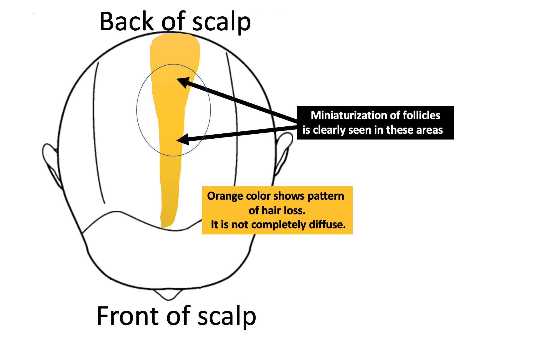
The back of the scalp is a common area for AANS. Often patients present with just a single painful bump. Some authors feel that AANS is closely related to dissecting cellulitis.

Exclamation mark hairs have not been described in AANS but have been described in dissecting cellulitis.

Dome shaped area of hair loss on the vertex scalp, consistent with a diagnosis of alopecic and aseptic nodules of the scalp. Image from Anna Isabel Lázaro-Simó et al. Alopecic and Aseptic Nodules of the Scalp with Trichoscopic and Ultrasonographic Findings. Indian J Dermatol. Sep-Oct 2017;62(5):515-518. Image used with creative commons license.


Trichoscopic image from alopecic and aseptic nodules of the scalp (AANS), also known as pseudocysts. There are black dots, yellow dots, vellus hairs and broken hairs. Image from Anna Isabel Lázaro-Simó et al. Alopecic and Aseptic Nodules of the Scalp with Trichoscopic and Ultrasonographic Findings. Indian J Dermatol. Sep-Oct 2017;62(5):515-518. Used with creative commons license.

Trichoscopy of alopecic and aseptic nodules of the scalp. Image from Khalil I. Al-Hamdi and Anwar Qais Saadoon. Alopecic and Aseptic Nodules of the Scalp with a Chronic Relapsing Course. Int J Trichology. 2019 Nov-Dec; 11(6): 244–246. Used with creative commons license.

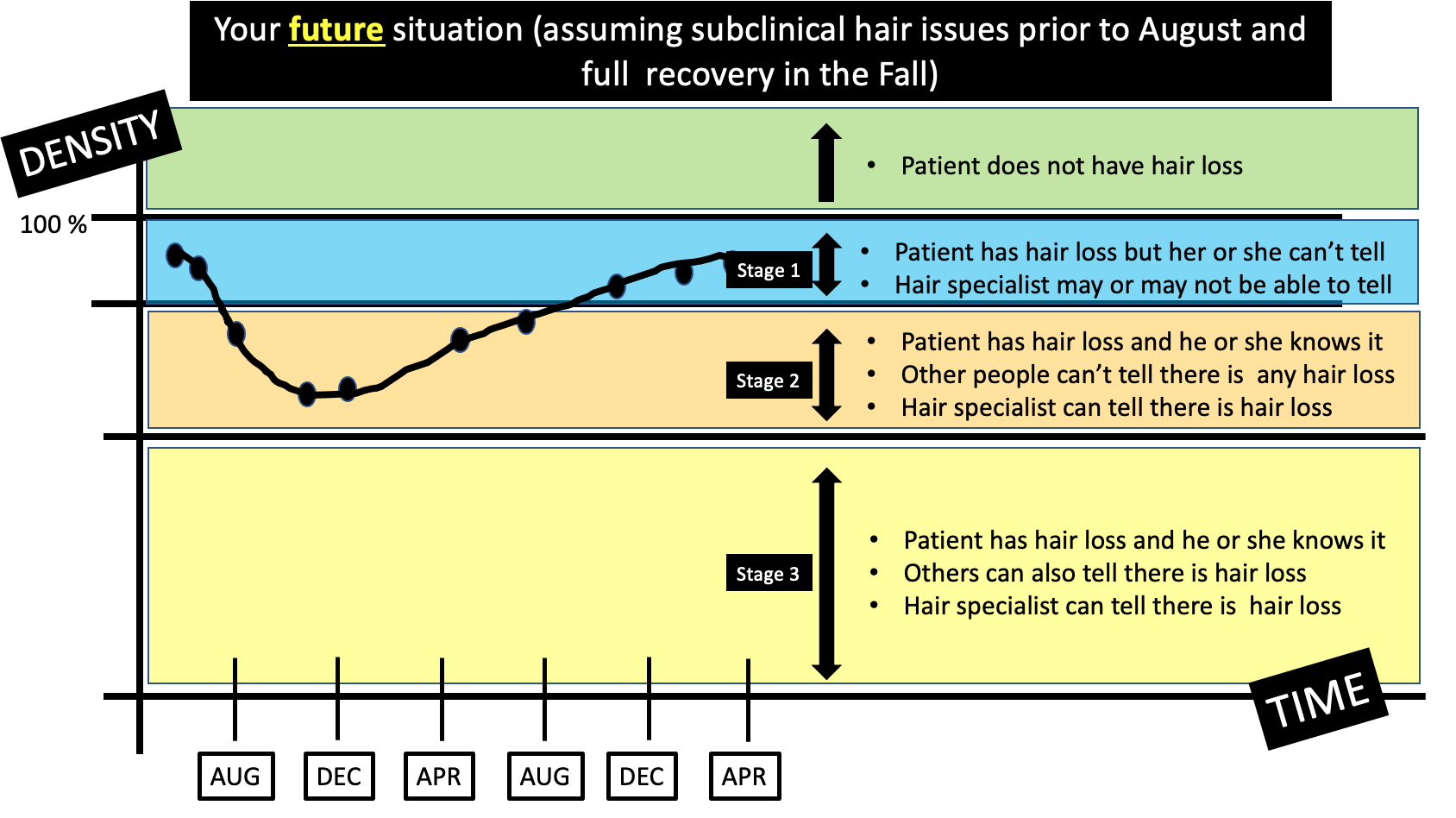
There are three stages of appearance to AANS lesions as described by Al-Hamdi and colleagues. It’s important to understand this - especially in this case.

Stage 1: Firm nodule. A firm and often tender nodule is present and the nodule lasts 1-3 weeks. There may be lymphadenopathy. If the nodule is punctured, it does not usually express any fluid. But if it does, the fluid is sterile and does not grow bacteria

Stage 2: Fluctuant Nodule with Hair Loss. In this stage, the nodule becomes less tender and hair loss is clearly seen. If the lesion is punctured in this stage a yellow fluid is expressed. This stage lasts 3-7 days.

Stage 3: Patchy Hair Loss Stage. In this stage, the nodule is no longer present as it has flattened either spontaneously or by puncture. This stage may last 2-3 month at which point hair growth normally occurs. It’s common in this stage for the patchy hair loss to be given a diagnosis of alopecia areata.

Was the bump described by the patient in this case actually stage 1 or stage 2 of AANS? Clearly, more information is needed. I would say it’s still quite unlikely.

Infections (Syphilitic Alopecia)

In a case like the one presented, one must never lose sight of alopecia areata as the most likely diagnosis. Most things fit well and it could be simply that this patch is more refractory and needs further steroid injections. However, we do need to consider rare mimickers (like AANS) - and another rare mimicker of syphilitic alopecia.

I don’t think that there is much in this case that makes a diagnosis of syphilitic alopecia high on the list. However, it can be a cause of patchy hair loss - especially with tapered exclamation mark like hairs, scale and redness like we see in the photos sent in by the patient.

Atypical trichoscopy of a patient with syphilitic alopecia in a 32 year old male. Exclamation mark like hairs are seen. Tapered bended hairs, erythematous background, diffuse scaling and perifollicular hyperkeratosis were present. Testing revealed a positive Venereal Disease Research Laboratory (VDRL) at a titer of 1:256 and a reactive Treponema pallidum particle hemoagglutination assay. Image from Linda Tognetti et al. Syphilitic alopecia: uncommon trichoscopic findings. Dermatol Pract Concept. 2017 Jul; 7(3): 55–59.


Other Diagnoses to Consider


There are several other diagnoses to consider here but they do not really have good evidence. These include:

  1. tinea capitis

  2. pressure alopecia

  3. infiltrative conditions.


Tinea capitis

Tinea capitis can be a mimicker and the appearance can be altered by steroid injections. I don’t know the patient’s history well enough to know if there are predisposing factors that might make tinea capitis more likely. (In fact, I don’t have enough information in this patient’s history including information about the kidney surgery at 5 months post partum). It’s always possible that an inflammatory tinea developed and was flattened by steroid injections and persists in some manner. Of course, that’s unlikely and there do not really appear to be any trichoscopic features of tinea. There are no corkscrew hairs, comma hairs, bent hairs, i hairs, morse code hairs and no zig zag hairs. I don’t think this is tinea.


Pressure alopecia

In anyone with patchy hair loss after surgery, we need to consider pressure alopecia. It’s thought that hypoxia and altered blood flow predisposes to hair loss. Studies of patients with pressure alopecia have not suggested that exclamation mark hairs are part of the pressure alopecia diagnosis. Therefore, a diagnosis of pressure alopecia would not be likely in this case. According to Neema et al, trichoscopic findings of pressure alopecia include comedone- like black dots, black dots and area of scarring. In 2016, Francine Papaiordanou et al proposed that black dots, broken and dystrophic hairs were main features of pressure alopecia. In 2020, Tortelly et al proposed that black dots and vellus hairs were key features.

It’s not impossible that pressure from surgery facilitated the development of alopecia areata. in fact, R L Zuehlke et al in 1981 suggested that pressure may be a risk for alopecia areata too. So it’s going to be important to review if this area on the scalp shown in the photos had pressure during surgery. I don’t think it’s likely that what we’re seeing is related to pressure alopecia.

Trichoscopy of pressure alopecia showing black dots, broken and dystrophic hairs. In Image from Papaiordanou F et al. Trichoscopy of Noncicatricial Pressure-induced Alopecia Resembling Alopecia Areata. Int J Trichology. Apr-Jun 2016;8(2):89-90. Used with creative commons license.

Infiltrative conditions.

The term “infiltrative conditions” refers to a massively long list of cells that can enter into an area of the scalp (infiltrate) and cause localized hair loss. A variety of inflammatory and neoplastic cells can trigger patchy hair loss so one needs to always keep these in mind. They don’t usually cause exclamation mark hairs. Alopecia neoplastica refers to hair loss from metastatic cancer and can mimic alopecia areata in some cases.

This would not be expected in this case but this is added to the list and discussed here for completeness as we review patchy hair loss and considerations in the setting of refractory patchy hair loss. It does seem that hair is growing back in your case which makes infiltrative type causes quite unlikely. I don’t have a good sense of the time course of the photos you’ve submitted so that too would need to be carefully reviewed.

Alopecia neoplastica due to breast cancer. Image from Efthymia Skafida et al. Secondary Alopecia Neoplastica Mimicking Alopecia Areata following Breast Cancer.Case Rep Oncol. 2020 Jun 11;13(2):627-632. Image used with creative commons license.

Alopecia neoplastica due to breast cancer. Image from Efthymia Skafida et al. Secondary Alopecia Neoplastica Mimicking Alopecia Areata following Breast Cancer.Case Rep Oncol. 2020 Jun 11;13(2):627-632. Image used with creative commons license.

Conclusion and Summary

Thank you for this question. There are a few important points here in this case as we conclude. The first is that a full history and full examination are needed. This area is the area that you have photographed but one needs to always examine the entire scalp. A full history is needed of health and medical conditions over the past 31 years. The reason for the kidney surgery is completely unknown and would need to be included in the full story. I need to know everything about patients to confirm diagnoses with absolute certainty.

That said, the photos and clinical case still fit with undertreated alopecia areata as a top diagnosis. The exclamation mark hairs here and vellus hairs and regrowing hairs support this diagnosis. There are mimickers of course and these need to be considered.

For my own patients with similar stories I would first take a full history and do a full examination of the scalp, eyebrows, eyelashes and body hair. Then I might inject with 2.5 mg per mL triamcinolone acetonide (steroid) with 2 to 3 mL injected into the area. I would not be too concerned if hair does not immediately grow back as it might take 2-3 sessions one month apart. I would not do more frequent than this. There is an option to add topical minoxidil to the plan but you’d want to review side effects with your supervising doctor.

If the area was slow to regrow I might add periodic use of clobetasol and minoxidil at home while doing these steroid injections.

Your photos would suggest you are already growing back significant hair.

If the area did not respond, I might do a biopsy. I don’t see this as necessary right now. The area needs to be properly treated and then if it does not respond to proper treatment, one can move on to step 2.

I don’t see AANS as a likely diagnosis (alopecic and aseptic nodules of the scalp), or pressure alopecia as being likely. We don’t see exclamation mark hairs in most cases of pressure alopecia. It would be helpful to know just how lumpy or raised this area was when you noticed it as this might lead one to at least consider AANS. The reality is that even if it is AANS and it’s some unusual pattern of exclamation mark hairs, it should respond to steroid injections at this point. I don’t think it’s likely we’re dealing with AANS.

Finally, anyone with this story should have blood tests for CBC, TSH, ferritin, vitamin D, creatinine. An antidandruff shampoo should continue to be used. Ketoconazole is reasonable. As mentioned, a biopsy will be needed if the area is not responding to appropriate doses of steroid injections (+/- minoxidil or clobetasol).

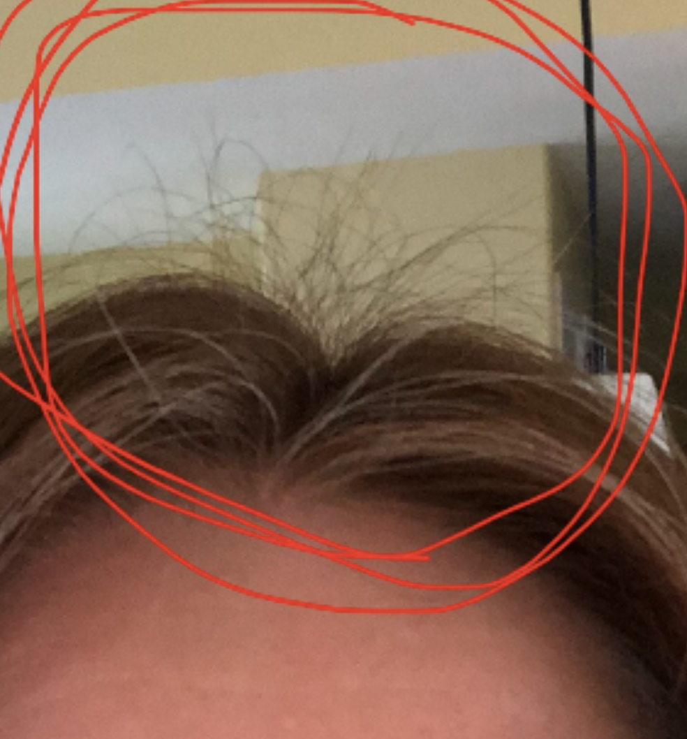
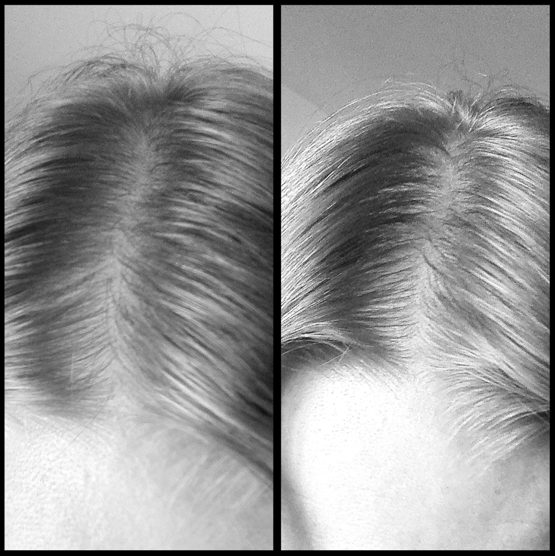
A full scalp examination is needed to determine if there are any other issues too. The area to the front may be thinner than prior years and that needs to be evaluated. It could be part of a resolving telogen effluvium or another diagnosis. A full examination of the scalp is needed in this case (as well as full examination of eyebrows, eyelashes and body hair as mentioned)

Thanks again

REFERENCE

Anna Isabel Lázaro-Simó et al. Alopecic and Aseptic Nodules of the Scalp with Trichoscopic and Ultrasonographic Findings. Indian J Dermatol. Sep-Oct 2017;62(5):515-518.

Khalil I. Al-Hamdi and Anwar Qais Saadoon. Alopecic and Aseptic Nodules of the Scalp with a Chronic Relapsing Course. Int J Trichology. 2019 Nov-Dec; 11(6): 244–246.

Abdennader S, Reygagne P. Alopecic and aseptic nodules of the scalp. Dermatology. 2009;218:86.

Linda Tognetti et al. Syphilitic alopecia: uncommon trichoscopic findings. Dermatol Pract Concept. 2017 Jul; 7(3): 55–59.

Neema S et al. Trichoscopy of Pressure-Induced Alopecia and Alopecia Areata: A Comparative Study. Int J Trichology. Jan-Feb 2022;14(1):17-20.

Papaiordanou F et al. Trichoscopy of Noncicatricial Pressure-induced Alopecia Resembling Alopecia Areata. Int J Trichology. Apr-Jun 2016;8(2):89-90.

Tortelly et al,Pressure-Induced Alopecia: Presence of Thin Hairs as a Trichoscopic Clue for the Diagnosis Skin Appendage Disord. 2020 Jan; 6(1): 48–51.

R L Zuehlke et al. Pressure-potential alopecia areata. Am J Orthod . 1981 Apr;79(4):437-8.

Efthymia Skafida et al. Secondary Alopecia Neoplastica Mimicking Alopecia Areata following Breast Cancer.Case Rep Oncol. 2020 Jun 11;13(2):627-632.


Share This
No Comments

Should I get another biopsy?

Should I get another biopsy?


I’ve selected this question below for this week’s question of the week. It allows us to review some concepts in interpreting biopsy results.


Question

I’m a 34 year old female with previously beautiful hair. My biopsy came back showing “non-scarring alopecia with features consistent with androgenetic alopecia.” I’m wondering if I should get another biopsy? Could it be wrong?


Answer

Thanks for the question.

Without knowing more about your medical history, and seeing your scalp (or photos), it’s difficult for me to say.

There are a number of possibilities here:

a) Your story and examination confirm you have androgenetic alopecia and doing a biopsy was not needed but simply served to confirm the diagnosis. Doing another biopsy would not be advised.

b) Your story and examination confirm you have androgenetic alopecia but there is a suggestion in the story or examination that a second diagnosis is also present. In this case, doing another biopsy could be a good idea if one is not 100% confident about this second diagnosis. If one is 100% confident in the second diagnosis, then another biopsy is not needed.

c) Your story and examination do not suggest androgenetic alopecia and there is a suggestion in the story or examination that a different diagnosis is present. In this case, doing another biopsy could be a good idea if one is not 100% confident about this diagnosis. If one is 100% confident in the diagnosis, then another biopsy is not needed.


I hope this helps. I don’t know your diagnosis because I haven’t seen your scalp and I don’t know your story. Certainly, if your practitioner is highly experienced in diagnosing women’s hair loss and says “This can’t be androgenetic alopecia” then I’m more likely to feel something is strange here and more likely to feel that a re-evlauation of things is needed. Early androgenetic alopecia is tricky to diagnose so many women have AGA even though it might not look like it.

If the practitioner is less experienced in diagnosing women’s hair loss, I’m less bothered by him or her saying “This can’t be AGA.” it’s tricky to diagnose some of the early forms of AGA. The reality is that if the terminal to vellus hair ratio on biopsy is dipping down below 4:1 and sebaceous glands are present in the biopsy there’s a good chance we’re dealing with AGA. The key question is whether this is the only diagnosis or the correct diagnosis.

Finally, I’d like to point out that we don’t diagnose hair loss just with biopsy results alone. One needs to take the biopsy finding and see if it ties in with the history of hair loss and the clinical examination findings. I would never every commit to saying a person has a certain diagnosis without the chance to see their hair and know their story. Even with the biopsy sitting in front of me, I need to know the story and see the scalp.


Share This
No Comments

Do I just need patience or is my hair density not going to fully return?

Is my hair density going to return?

I’ve selected this question below for this week’s question of the week. It allows us to the review some key concepts in the diagnosis of hair loss in the early stages.


QUESTION


I am a 40 year old female. I have always had a lot of hair, and coarse hair. I have always been a shedder, but it never made a difference on how dense my hair was. Until now. I had my "normal to me" hair up until August/September 2020. But, in September/October, I started to see a lot more hair coming out in the shower/brushing afterwards/when blowdrying.

Handfuls would come out in the shower when in the shower. It was definitely the worst/at it's peak in November 2020.

I remember after one shower the entire wall was covered with the hair I collected. By this time I started to freak out a bit. It was definitely making a difference on my head now as far as density. I went to my family doc, and he did blood work. My ferritin came back at 21, but my hemoglobin was ok. I started taking iron supplements at the end of November. At the end of Jan 2021 I went to see a dermatologist. She only had a physical look at my scalp, did not do a biopsy and did not look at my scalp with any sort of magnifying tool or anything. She said based on my story she thought it was either Androgenetic Alopecia or Telogen Effluvium. She had my vitamin D tested. It was a bit low, so I started taking 2000IU of vitamin D daily. The hair shedding continued like this until end of February-ish/beginning of March. (The lost hair was mostly long hairs, some medium length, barely any short hairs)


In March/April 2021 the hair fall slowed down a lot, and now I would say it is back to a normal amount with each wash.

But, I can definitely see a difference on my head. It is most noticeable on the top/sides of my head, and down the back of my head (I have weird parts all along the back of my head). It also sort of looks like I lost hair at the nape of my neck. My part has not gotten wider at all, but sparser. I do have a lot of new growth at the top and back of my head, but it doesn't seem like enough to make a difference in terms of overall thickness, even when it grows longer.

My scalp hurts often, as if it has been in a super tight ponytail, even though it has not. Sort of hurts to move it around. My scalp can be quite dry/itchy at time (always has been like this, even before hair loss)

I am still taking iron supplements, as when I was re-tested in February my ferritin had only gone up to 30. I am also still taking the vitamin D daily. I should mention I take 2.5 or Ramipril daily.

My question is … would Androgenetic Alopecia happen that quickly and then taper off that quickly? And, if it is Telogen Effluvium would I expect to have more re-growth by now? Or, is there any chance I could have some sort of diffuse Alopecia Areata, based on what is happening at the nape of my neck and the weird parts down the back of my head? I have attached some photos. I am trying to be patient, as I know hair takes a long time to grow.

Thank you for your input!!

Image 1. Hair density in the central part.

Image 1. Hair density in the central part.

Image 2: Hair density in the crown.

Image 2: Hair density in the crown.


Image 3: Hair regrowth.

Image 3: Hair regrowth.

Image 4: Hair regrowth.

Image 4: Hair regrowth.

ANSWER

Thanks for the great question. The short answer is that many diagnoses are possible for you. I’ll get into these in just a moment.

I would need to see your scalp and know more about your full story to tell you which diagnosis (or diagnoses) you actually have…. but the 6 possibilities are outlined below. Each of these possibilities has different probabilities for being your actual diagnosis. If I was to see your scalp, these ‘estimated’ probabilities would change. However, with the information provided, we have six scenarios. The most likely is scenario 1 and 2 followed by scenario 3.


Six Possible Scenarios for Your Hair Loss


There are six possible scenarios for your hair loss. The most likely is scenario 1 and 2 followed by scenario 3.

Scenario 1) You have a telogen effluvium due to low iron or low vitamin D. This has now been fixed and you need to give it until October/November in order for your density is going to come back.

Scenario 2) You have a telogen effluvium for some other reason (other than simply low iron and vitamin D) and it has now somewhat resolved and you need to give it until Oct/November in order for your density is going to come back. Causes of telogen effluvium that could be relevant for you would include stress last summer 2020, low iron (which you might have), thyroid problems, medications started last summer, weight loss last summer, COVID infection last summer. Other causes are possible too.

Scenario 3) You have actually had a hint of subtle “subclinical” androgenetic alopecia for a while and this recent telogen effluvium has “unmasked” the subtle androgenetic alopecia. Your density is going to improve by the Fall 2021 now that your telogen effluvium is resolving but you might or might not get back all your density - but you may come pretty close.

Scenario 4) You have an inflammatory scalp condition that has been present for a while and is now acting up to give periods of hair shedding. The iron and vitamin D are unrelated in this particular scenario and are simply a true red herrings. Your inflammatory scalp condition has now settled again but you need to give it until November/December to see if things will fully settle. Such inflammatory condition could include seborrheic dermatitis, psoriasis, scarring alopecia or contact allergy (ie to some ingredient in a shampoo, conditioner, hairstyling product or dye). This scenario number 4 carries a risk of flare again.

Scenario 5) You have an inflammatory scalp condition that has been present for a while but it’s not enough to give hair loss. A new telogen effluvium has come along that will resolve and time will tell whether the inflammatory scalp condition also settles fully. If the inflammatory scalp condition is a low grade scarring alopecia, density won’t come back fully but still will improve to some degree when the current telogen effluvium resolves.


Scenario 6) You have an inflammatory scalp condition that has been present for a while but it’s not enough to give hair loss. You also have a subtle amount of androgenetic alopecia that has now been unmasked by the new telogen effluvium. If the inflammatory scalp condition or androgenetic alopecia is active enough it may prevent density from coming back to your full normal by Fall 2021. 


Detailed Review of the INITIAL Situation (August 2020 to Dec 2020)


Let’s go further into the situation that you describe in your question. Before we do, let me point out that there are three stages of hair loss for most people. At least that’s a helpful way that I view hair loss. These stages are important to appreciate because it impacts how I approach your question.

In “stage 1” of hair loss, the patient has hair loss but doesn’t really know it. For all practical purposes, the patient feels the hair looks the same as it always did and feels the same as it always did. Perhaps when they look at a photo from years gone by they might say something like “Wow, I can’t believe how much hair I had back then!” Otherwise stage 1 of hair loss is unrecognizable by anyone - patient, doctor, specialist or hairstylist.

In “stage 2” of hair loss, patients themselves realize they have hair loss - but others around them don’t believe it or don’t realize it. The patient feels the pony tail is smaller or the scalp is more see through or something is just not the same. A spouse, sister, parent, daughter, son, barber, hairstylist or friend usually say the same thing - “You’re exaggerating ! Everything looks fine to me! Sometimes that sentence is delivered by the doctor or other hair expert that has been asked to help.

Stage 2 is sometimes frustrating and lonely and anxiety provoking. Patients feel something is wrong but the world around them says repeatedly that everything is just fine.

Now, some patients in stage 2 resolve their hair loss and go back into stage 1 and so they do end up feeling they were exaggerating because everything resolves itself. Some patients stay in stage 2 and eventually find an answer to their hair loss issues. If specialist A does not believe them, they move on to specialist B. If specialist B does not believe them, they move on to specialist C.

Some patients in stage 2 do progress on to stage 3 of hair loss where hair loss becomes more noticeable to others. With hairstyling and camouflage a patient in stage 3 might still be able to hide their hair loss. With treatment of course, a patient may be able to return to stage 2 or even stage 1.

3 stages of hair loss


With these stages in mind, let’s delve a little further into this situation you have mentioned in your question.

There are two main scenarios that may have been present before you noticed hair loss in August. The first is that your hair density was completely normal and the same as it was when you were 20. You then lost hair in the August - December period and the density went down. In other words, you went from no hair loss to stage 2. This is shown below.

scenario  1


The second scenario is that you felt that your hair density was completely normal but it was not, in fact, completely the same as it was 20 years ago. You then lost hair in the August - December period and the density went down. In other words, you went from stage 1 of hair loss into stage 2. This is shown below

scenario 2


Both of these situations above would appear identical to you. In the first situation, you had normal hair to start and then you lost density. In the second situation, you had (what you thought was) normal hair to start and then you lost hair. The only difference is that in the second sitatution you actually didn’t have quite normal hair - it just seemed that way to you (and everyone else).


Detailed Review of the RECOVERY (April 2021 to Dec 2021).

Your hair loss is now in a recovery phase. Your shedding has stopped. You are sprouting hair everywhere!

Let’s spend some time looking at the recovery of your hair loss and how the hair might respond over the next few months. The most likely are the following 2 scenarios.

If you don’t have any underlying issues that are affecting how hair grows, then it’s likely that this telogen effluvium will continue to settle and a you’ll get a return to full growth by the end of the year. In other words, you’ll go from stage 2, into stage 1 and back to full hair. The chapter on hair loss will be closed

scenario three


Even if you do have some kind of “subclinical” hair loss situation happening in your scalp, there is still a good chance that you’ll recover your density by the end of the year and you’ll return feeling that your hair feels ‘full’ to you. In other words, you’ll move from stage 2 into stage 1. Stage 1 of hair loss looks just as good of having no hair loss at all so for all practical purposes it does not matter.

scenario four


What happens if my density does not recover by the end of the year?

The final scenario is a bit trickier to explain. If you did in fact have some sort of subclinical hair loss situation going on in the scalp before August 2020 and this condition got a little bit worse from August 2020 through summer 2021, then you might not find that you have a full recovery by the time the Fall 2021 comes around. This could be due to several situations including

a) you had some subclinical androgenetic alopecia prior to August 2020 and the androgenetic alopecia got a bit worse from August 2020 to August 2021.

b) you had some subclinical scarring alopecia prior to August 2020 and the scarring alopecia got a bit worse from August 2020 to August 2021.

c) you had some subclinical psoriasis or contact dermatitis prior to August 2020 and the inflammatory issues got a bit worse from August 2020 to August 2021.

In these situations, it’s possible you stay in stage 2.

scenario five

This final scenario is the least likely but a proper scalp examination and full review of your story is going to help me decide just how likely it is in your specific situation. For now, I estimate it as unlikely (but not zero).

What you can see here in these examples above is that you really need some definite diagnoses. If you allow time to help you with a solid diagnosis then that’s one good strategy. For example, if your density comes back perfectly to normal by the end of the year, then there’s probably no real hair issues at all that need treating or need any kind of workup. In other words, if your density returns back to full by December 2020 (and you enter stage 1 or no hair loss), it’s pretty unlikely there’s any other hair loss issue going on.

But if density does not return, I strongly believe that you need to have some formal diagnoses put on paper for BOTH the hair loss and the scalp symptoms. The reason we need different diagnoses is because every hair loss condition is treated differently. Unless we have a diagnosis, we can’t formulate the right treatment plan.


My Final Comments

Thanks again for the great question.

I’m really glad you are seeing all this hair growth sprouting everywhere as it’s a really good sign. The hairs are about 5-6 cm so it seems that the telogen effluvium you had in Aug/Sept 2020 is settling down. It could be that the iron and/or vitamin D is helping or that could just be a coincidence. It’s difficult to prove.

I do feel your scalp symptoms (dry, itchy, hurts to move) needs a formal diagnosis. Your scalp symptoms need a name of some kind. Now, keep in mind that the diagnosis of that situation might not be anything concerning given how long you have had it, but it still needs a formal diagnosis. If nobody is sure of what to call your itching and soreness, then you need a scalp biopsy. That is pretty clear in my mind. There is flaking present in some of your photos so there is some kind of inflammatory issue present. I would need to see the scalp up close to give a diagnosis. Please be sure to follow up on that.

I am glad you are taking photos as that will be key over the next 6 months. If you feel by November/December 2021 that you are really happy with your hair and how the density has returned, then this chapter of your hair story is likely done: you had a telogen effluvium and it resolved. It went away. In this situation, it’s unlikely there is some subclinical androgenetic alopecia present but it does not really matter much. I wouldn’t treat hair loss if you go back to feeling good about your hair. I’d simply repeat photos in 1 year. Put the shedding episode behind you for now.

If your density does not return to normal by the end of the year, then there is a good chance that there is some androgenetic alopecia that has entered the picture. No, it’s not 100% but that becomes increasingly likely. There’s very small chance that another diagnosis besides androgenetic alopecia is responsible but that’s pretty rare. Of course, the itching and tender issues on your scalp need to be diagnosed properly. That may or may not have any role here. But someone needs to give it a name.

But if you are pleased with your density in November/December 2021 and your shedding is completely back to normal (and stays normal), and your scalp symptoms are not worrisome to you and your doctors …I would put it all to rest and simply take a photos again in 1 year. It’s helpful to have your doctors follow you closely but nobody really knows their hair better than you. If you feel your hair has not returned back to normal, then you have remained in stage 2 and need a solid convincing diagnosis.

I hope this helps.

Many thanks for the question.

Share This
No Comments

What is the reason for my hair loss?

Why am I shedding ?

I’ve selected this question below for this week’s question of the week. It allows us to discuss shedding issues in women with hair loss.

QUESTION

Hi!

I am a 42 year old women and have been shedding about 200 hairs a day since March, 2020. I have seen 4 dermatologists and my General Practitioner and they have different diagnoses from TE to FPHL or a combination of both. Prior to March, 2020 I was under extreme stress which started in November 2019. In March 2020 my hair loss was sudden and I have had diffuse shedding since then for the past 10 months. I have always had full, thick and healthy hair and no issues with my hair until the past 10 months. There is no family history of hair loss and my bloodwork came out normal.

Increased hair shedding.

Increased hair shedding.

I have been taking vitamins, biotin and Lysine (since June 2020) daily. I am washing my hair every other day, air dry my hair and do not use styling or heating products and eat healthy. I am also taking spirolactone since December 2020 (one month as of today). My dermatologist suggested I take spirolactone (50 mg twice a day) because I have irregular periods. My hair loss slowed down in September 2020 to about 100 hairs a day and went back up to 200 plus in November 2020. I am experiencing itchiness, pins and needles sensation on my scalp and my hair texture changed from straight to wavy for the past 10 months. My hair is also now flat, dull and I have some dandruff that comes and goes. My middle part is widening (compared to pre-March 2020 before the shedding) and with the ongoing shed the part has somewhat looked the same since March.

PHOTO 2
PHOTOS 3

I lost about 30% of my hair and cannot style it the way I used to because of the thinning in the front. The last two dermatologists I saw performed a pull test and scalp examination and one of them said it is CTE and that there is nothing I can do but wait it out. The other doctor said it's FPHL and that she can tell just by looking at the front of my hair because of the way its thinning. I do see hair growth and my hair is full of static with short hairs coming out but I am also losing a lot of hair in all different lengths including short ones every day. I am frustrated because it has been 10 months and my shedding is not stopping. I do not know which diagnosis is right and what treatment I should start. Also It would be great for the itching and "pins and needle" feeling on my scalp to go away...

Thank you for reading and I'm so happy to find this website.


ANSWER

Thanks for the question.

I’d like to discuss several important things in the question you ask and the information you have submitted.

Before we do go further, I’d like to point out that the ideal way to diagnose hair loss is using what I termed the ”Diagnostic S.E.T.” I refer to these as the diagnostic “set” because theses 3 aspects all go together. These 3 items include:

1) the patient’s story

2) the findings uncovered during the process of the scalp examination including trichoscopy

3) the results of relevant blood tests. 

The first letter of each of the three words 1) story, 2) examination and 3) tests spell out the word “S.E.T.” - again a helpful reminder of how the information obtained from reviewing each of these 3 aspects helps solidify a proper diagnosis.

There is lots more to your story that I need. I would want to know exactly what your lab tests showed and which ones were tested. In about 20 % of patients who tell me they had blood tests and all came out normal, the labs are either insufficient (more are needed based on their story and examination) or the labs are not in fact really normal. I always like to see the labs. I would want to know about other symptoms like joint pains, headaches, fatigue, weight loss, eyebrow changes, eyelashes changes, body hair changes, nail changes, and rashes.

I strongly suspect that androgenetic alopecia with seborrheic dermatitis are part of the diagnoses. The 2 key questions here in your case are:

  1. Do you have really have telogen effluvium as well ?

  2. What really is the reason for the ‘pins and needles’ sensation ?

Let’s look at a few key points.

POINT 1. Androgenetic alopecia (female pattern hair loss) appears to be at least one of the diagnoses.

I do think that at least one of the diagnoses here is androgenetic alopecia (also called female pattern hair loss, FPHL). The widening of the part does not itself mean the diagnosis is AGA. however, the pattern of the part widening is not the same front to back. There is a slight increase in thinning noted in the mid scalp and crown compared to the frontal one third of the scalp. This leads me to believe there is a patterned nature of the hair loss. I’m open to the possibility that some of the hair loss is diffuse in nature (ie all over) but some is likely not. In other words, I don’t think this is entirely a diffuse type of hair loss.

Also, when I look up close at the images, it’s clear that some follicles are thinner than others. This is a phenomenon called anisotrichosis and is a feature of AGA. Some hair follicles are miniaturization (getting thinner).

pattern of loss

Women with AGA often experience shedding of hair in the early stages and shedding can fluctuate in intensity. Other hair loss conditions can cause shedding as well so we’ll address that in just a moment. Women with AGA often notice that the texture of hair changes. There are many such patterns of texture change and a change from straighter to curlier is quite common as you too have described.

The fact that you note increasing numbers of short hairs is not confirmatory for a diagnosis of AGA but certainly is supportive of this diagnosis.

POINT 2: Seborrheic dermatitis/dandruff is likely another diagnosis.

I agree with you that dandruff (or its close cousin called seborrheic dermatitis) is likely present. Flakes are noted in some of the photos. I’d need to perform trichoscopy to confirm this diagnosis but it appears to be a component of the issues present. Mild dandruff is not usually a cause of hair loss but it certainly can cause all sorts of scalp symptoms. To eliminate the possibility that dandruff or seborrheic dermatitis is contributing to symptoms, I often encourage my own patients to aggressively treat their seborrheic dermatitis so we can remove this as a factor. Shampoos with zinc pyrithione, ketoconazole, selenium sulphide or ciclopirox can be used 2 times per week and left on 90 to 120 seconds before being rinsed off. The duration that these shampoos are left on the scalp can certainly be increased but I don’t recommend that to start with as many antidandruff shampoos can be drying and then the dryness starts causing itching and symptoms. I often recommend to my own patients that 5-10 drops of betamethasone valerate lotion 0.1 % can be applied in the scalp after their hair is shampooed and dried. This is a weak steroid and can be safely used for 2 week periods to help settle itching. If the use of shampoos settles the itching, tingling and pins and needles, then it’s not needed.

POINT 3: Telogen effluvium could be present.

Telogen effluvium is one of those conditions that can come and go. Sometimes it’s easy to prove a TE is present and sometimes it’s a bit more challenging. It could be that a TE was present when your AGA first started. You were under extreme stress in November 2019 and yes this could most definitively give shedding in March 2020. Telogen effluvium usually follows 2-3 month after some kind of trigger and can last 3-6 months or more. Other causes of telogen effluvium are low ferritin levels, thyroid issues, medications, diets, weight loss and internal illness. I don’t really have enough information to evaluate these other issues so I’ll go with your assessment that your blood tests were normal. Hopefully you had a reasonable set of tests including TSH, ferritin, CBC. With your irregular periods you describe it would make sense to have FSH, DHEAS, testosterone. One needs to consider whether you are entering a perimenopausal transition and how this could contribute to hair shedding and AGA. With any pins and needles sensation, it’s nice to know that liver enzymes (AST, ALT) are normal and that kidney function is normal (creatinine).

Telogen effluvium can sometimes precipitate or accelerate an underlying AGA. This is a well accepted phenomenon. it does not happen to all women with AGA. However, women with shedding who have AGA that is about to begin (ie very early onset AGA) often find that the AGA component of the hair loss gets sent into a more rapid speed of development if a TE is present. This could be a feature here.

With your normal blood tests, it’s unlikely that a TE is still driving hair loss all this time. Not impossible of course, but unlikely. What is more likely is that AGA is not fully being treated. Spironolactone helps but does not fully suppress AGA in all women at 50 mg twice daily. Sometimes higher doses are needed OR other treatments for AGA are needed (other anti androgens, laser, minoxidil, etc)

POINT 4: If you want to assess the degree to which AGA and TE are present, you could have a biopsy or 5 day modified hair wash test (or a proper trichoscopic examination). I don’t think these are really needed.

For your physicians/specialists who think that AGA is not a diagnosis here for you, a biopsy or 5 day modified hair wash test could help prove them wrong (… or prove them right!). This is a wonderful test but adds to the stress of collecting hairs so I’m not always in favour of it. Biopsies leave scars but if interpretted by a knowledgable dermatapathologist, they can be very helpful.

But, let’s be clear. A biopsy showing a terminal to vellus hair ratio of less than 4:1 taken from your mid scalp area puts to rest any argument about whether AGA its present of not. End of discussion. A 5 day modified hair wash test (done properly !) showing less than 100 hairs and more than 10% hairs being tiny 3 cm hairs also points to an underlying AGA.

Of course, simply examining the scalp with trichoscopy can also confirm this diagnosis but not all specialists are skilled with trichoscopy. If a specialist knows how to use a handheld dermatoscope, we don’t even need biopsies or hair collections to diagnose AGA. If they don’t then yes, we need to go to the extra effort to prove it.

POINT 5: The ‘pins and needles’ is a bit trickier given how many conditions can cause this.

There are a very large number of conditions that can cause pins and needles in the scalp. Stress can cause it. AGA can cause it. TE can cause it. Alopecia areata can cause it. Dandruff can cause it. Scarring alopecias cn cause it. The list is long and includes issues even outside the scalp like cervical spine disease.

I would need to know more about your story and carefully examine the scalp and eyebrows and eyelashes and nails to get a sense of what is causing this.

For pins and needles sensations, I usually advise treating any dandruff or seborrheic dermatitis and using a few drop of betamethasone lotion as outlined above. If it’s still there and the patient has AGA, I usually recommend treating the AGA more aggressively. This often help stop pins and needles. Low level laser, minoxidil and other antiandrogens can be considered.

Conclusion/Summary

Thanks for the question. I hope this helps you in your search for answers and helps with further discussion with your doctors. I think it’s really important for you and your doctors to feel confident with the diagnosis and not proceed with any sort of “maybe.” It would appear that AGA is a component of the issues here but if there is any doubt, a trichoscopic examination, biopsy or 5 day modified hair wash test can help confirm this.

Photos are really important moving forward to document changes - hopefully for the better.

if spironolactone is not fully helping then you and your dermatologists might discuss together whether or not to increase the dose or whether other treatments need to be considered. These include topical minoxidil, oral minoxidil other topical or oral antiandrogens and low level laser. PRP can be considered too. The important thing to note about minoxidil, laser and PRP is that if there is any amount of chronic shedding issue present these treatments can help promote more normal shedding patterns. This is assuming all your blood tests are normal. If any of your blood tests are abnormal and if, in fact, you have not had a proper work up then those issues need to be addressed first. it sounds like you’ve had a good set of blood tests through all the doctors you have seen.

Thank you.

Share This
No Comments

Sudden Loss of My Hair: What are possible reasons?

Why am I losing hair?

I’ve selected this question below for this week’s question of the week. It allows us to discuss several important features, including the different diagnosis of red flaky scalps with increased shedding, and the concept of “follicular prominence.”


QUESTION

I am in my early 40s, female and experiencing sudden hair loss that includes scalp & eyebrows (and even eyelashes, but to a lesser extent). I have lost about 25% overall density in the past 2 months. I’m not sure how many hairs I shed each day (too many to count!), but hair is constantly falling out.  I usually wake up with a several scalp & eyebrow hairs on my pillow.  Prior to then, my hair/scalp was healthy except for a mild episode of telogen effluvium about 10 years ago that resolved within a few months. I do not have any bald spots, the loss appears diffuse. My scalp is moderately itchy/tingly, flaky & slightly tender. I do not have any significant pain or burning.  The itchiness & tenderness tend to be worse on the top of the scalp, but overall it is quite itchy.  

Right around the same time my hair loss began, I developed small patches of dermatitis/eczema on a few areas of my face, underarms & body.  I’m not sure if it’s related, but I’ve never had skin rashes before.  I had blood tests (CBC, ferritin, hormones, ANA, ESR & TSH) and all came back within normal ranges (only testosterone at 20 and ferritin at 48 were slightly low).

I am healthy except for early perimenopause symptoms & high cholesterol. I started a new cholesterol medication about a month prior to the hair loss (which I discontinued in the case that was the cause) but the hair shedding has only worsened. My doctor was concerned about Frontal Fibrosing Alopecia due to the eyebrow loss and did two 4 mm punch scalp biopsies. One biopsy was non-diagnostic and the other showed female pattern loss:

bx results nov 1

I never noticed any hair loss prior to 2 months ago, but I suppose it makes sense to have some female pattern loss given my age. However I was told that probably was not the cause of the rapid shedding. After seeing the biopsy results and doing a scalp exam, the doctor thought the shedding of scalp/eyebrows must be telogen effluvium due to stress/hormones & the itching was seborrheic dermatitis. The only things mentioned during my scalp exam were inflammation and flaking (attached pictures of middle part) and some re-growth in my eyebrow area.

Image 1 of scalp from individual experiencing rapid hair loss.

Image 1 of scalp from individual experiencing rapid hair loss.

Image 2 from the scalp for individual experiencing hair loss.

Image 2 from the scalp for individual experiencing hair loss.

My concern is since one biopsy was non-diagnostic, could I have a more serious type of hair loss going on that I should be addressing? Does this sound like a telogen effluvium I should wait out or do you recommend any next steps if a biopsy doesn’t fit with all the symptoms or give a definitive diagnosis? Thank you!


ANSWER

Thanks for the great question. I’d like to discuss several important things in the question you ask and the information you have submitted. Let’s get to it.

Before we do go further, I’d like to point out that the ideal way to diagnose hair loss is using what I termed the ”Diagnostic S.E.T.” I refer to these as the diagnostic “set” because theses 3 aspects all go together. These 3 items include:

1) the patient’s story

2) the findings uncovered during the process of the scalp examination including trichoscopy

3) the results of relevant tests - blood tests and biopsy tests

The first letter of each of the three words 1) story, 2) examination and 3) tests spell out the word “S.E.T.” - again a helpful reminder of how the information obtained from reviewing each of these 3 aspects helps solidify a proper diagnosis.

Certainly a more complete story is going to be needed in your case. As I go about reviewing your story, several conditions pop up into the list of possibilities for what’s causing your hair loss. These include one (or MORE) of the following (and likely there are more than one issue):

  1. Androgenetic alopecia

  2. Lichen planopilaris and its variants (fibrosing alopecia pattern distribution & FFA)

  3. Telogen effluvium

  4. Seborrheic dermatitis

  5. Contact dermatitis

  6. Other types of scarring alopecia

I do think with your story that another biopsy is going to be important as I do think lichen planopilaris with androgenetic alopecia together with some degree of seborrheic dermatitis is at the top of the list. Another biopsy (or two) can help clarify this.

Let’s go deeper now into the case that has been submitted.

You present with sudden onset of hair loss and a great deal of shedding. It’s sure sounds like a telogen effluvium at first glance. It’s easy to feel perhaps it’s a TE from the cholesterol drug. The problem with that is that the biopsy does not support that. Second, that’s relatively uncommon. However, biopsies are not really how we nail down a diagnosis of telgoen effluvium so it would be mistake to jump to the conclusion that there is no TE here. However, there is not a great deal of evidence.

You have redness, and itching and tenderness in the scalp and the middles areas are particularly affected. What is the most likely diagnosis in a 42 year old female with new onset itchy, tender, and red scalp especially in middle accompanied by markedly increased shedding? Lichen planopilaris by far. Is it the only possibility? Of course not. But LPP is at the top of the list and we can’t rest until we decide whether it stays on the list of gets removed from the list.

Let’s take a look at some of your “up close” photos a bit more.

1. You have miniaturized hairs.

photos up close 1

Your photos show some degree of variation in the caiber of follicles. It’s not much but there is some. I suspect this is being captured to some degree on the biopsy in biopsy “B” where your terminal to vellus ratio was 4.5 to 1. This could suggest some mild female androgenetic alopecia. It’s mild and does not account for the shedding, itching, redness and tenderness you have.


2. Your images illustrate a phenomenon known as “follicular prominence”

photos up close 2

Your up close images show what is termed follicular prominence. This is a phenomenon whereby the follicles create little elevations above the surface of the scalp at the point at which they emerge from the scalp.

Put into a cartoon-like representation, here is what’s happening:

F-P


Now, let me say that follicular prominence is by no means conclusive of a diagnosis of LPP. But what it does mean is that LPP remains at the top of the list in this case until someone definitively proves it’s not LPP. We’ll go into that more in a moment. Follicular prominence is an early finding in LPP and is due to a reactive hyperplasia of the follicular epidermis.

3. Your images show some scaling as well as possible loss of the follicular openings.

photos up close 3

As you have pointed out in your submission to the “question of the week” program, you do indeed have scaling - or as you described a somewhat ‘flaky’ scalp. There are many reasons for people to having flaking, but I do note that some of your scale tends to hug the hair follicle quite closely. We call this “perifollcular” scale (a term that simply means scale that occurs around hairs). Perifollicular scaling can be seen in several conditions but certainly LPP is on that list.

Your scalp also shows areas where there is no hair. We call such areas of no hair “atrichia.” These areas of atrichia are shown in yellow in the diagram above. Focal atrichia can be seen in more advanced androgenetic alopecia, but your androgenetic alopecia is certainly not advanced so that can’t really apply. The tiny follicular openings or “pores” can not be clearly seen in these areas in yellow raising the possibility this could be an area of scarring. That’s by no means definite but it needs to be explored further by your doctors.

4. Your images show two types of possible scaling.

photos up close 5

There may be two reasons for your scaling or ‘flaking’. The redness and somewhat yellow color and somewhat whitish color scaling calls into question the possibility that there is some degree of seborrheic dermatitis present on the scalp. However, the marked shedding, the follicular prominence and close “perifollicular scale” indicates that seborrheic dermatitis alone as the diagnosis makes little sense. It’s likely a component.

Some of the scale, as shown in the image above, is strikingly tubular - like tubes around the hair. The scale rises up around the hair. This type of scale is unexpected in seborreheic dermatitis alone. It’s again by no means pathognomonic of a scarring alopecia but it’s an important finding.

5. Your images show redness around the hairs.

photos up close 4

Redness around the hairs is known as “perifollicular erythema”. It’s quite a non-specific finding but it does remind us that the immune systemic is likely causing some sort of issue in and around the hairs. It’s not a random inflammation anywhere in the scalp - it’s focused on the hairs. We see perifollicular inflammation in many conditions.


6. Your biopsy shows loss of sebaceous glands in biopsy A

Perhaps one of the most important findings in your story is the reduction of sebaceous glands that your pathologist reports in biopsy A. It’s for this reason that LPP is still very much on the list of possibilities.

Loss of sebaceous glands is very much a part of what scarring alopecia is all about. All of the scarring alopecias no matter the cause show loss os sebaceous glands at some point in their evolution. In LPP, reduction in sebaceous glands occurs relatively early in the disease course.

The following cartoon shows what we see under the microscope in LPP. We see loss of sebaceous glands along with inflammation in the upper part of the hair follicle. So what does your biopsy A report ? Loss of sebaceous glands and inflammation in the upper part of the hair follicle (known as the infundibulum).

SG - LPP - early

Does this prove it’s LPP? No, but is is certainly pointing in that direction. Other features we often see in early LPP are not reported in the biopsy report. These include a specific type of inflammation known as “lichenoid inflammation.”

So what’s left to confirm the actual diagnosis ?

I do think in your case another biopsy is going to be helpful. The diagnosis of lichen planopilaris is still at the top fo the list. The discussion around whether this is frontal fibrosing alopecia too depends on whether the frontal hairline is involved. But one thing is for certain - if you do have FFA, you also have LPP because of the involvement of the mid scalp and crown areas.

If you or your doctors are against rebiopsying, you certainly could ask the pathologists to perform a very special stain of your biopsy to look at elastic fibers. This elastin staining technique is very helpful in these challenging cases like you have. It the special test shows your elastic fibers are destroyed, this is likely a true scarring alopecia.

But most likely a repeat biopsy is going to prove really useful.

Where to biopsy?

I do think that if another one (or maybe two) 4 mm biopsies is going to be done, it should be done from an area that shows perifollicular scale, follicular prominence and perifolicular redness. Ideally, this should be from an area of the scalp that is itchy and tender and ideally from somewhere in the middle or crown of the scalp. If the biopsy can be taken from an area that shows all these features that’s even better. The biopsy must contain hairs and ideally if it can include some areas of no hair, that’s great too because that’s where the pathologist will very clearly see the sebaceous glands are lost (if they are indeed lost). Taking from the sides or back is less helpful in my opinion although is often done to ‘hide’ a scar. I don’t recommend this. Some examples of where to biopsy are shown here:

A possible biopsy site: the area on the right shows scale and follicular prominence. This would be preferred over the area on the left side.

A possible biopsy site: the area on the right shows scale and follicular prominence. This would be preferred over the area on the left side.

A second possible biopsy site: the area shows follicular prominence.

A second possible biopsy site: the area shows follicular prominence.

Conclusion

I hope this helps and good luck on the journey to find the precise diagnosis. I do think that one can not rest until LPP is ruled in or ruled out in this particular situation. I certainly favour this diagnosis (along with some androgenetic alopecia and mild seborrheic dermatitis). But it’s not 100% of course.

There are other considerations here too like an allergic contact dermatitis but your biopsy does not show this and the loss of sebaceous glands would not occur in a contact dermatitis. However, this too can be a great mimicker. I don’t really think this is an issue here but one needs to respect this great mimicker, especially when other rashes are present on the body.

Studies have not shown differences in cholesterol profiles between patients with LPP and those who do not have LPP. My own thought is that there is a connection - and I’m always a bit suspicious when young women show up with high cholesterol, itchy red scalps, follicular prominence and shedding. There may be more of a link to the metabolic dysfunction and lipid metabolism issues that we realize. Be sure to follow up on your cholesterol issues.

I do think that repeat biopsies are key here snd that the pathologist should include some elastin stains as well to get a sense of whether the delicate elastic network is destroyed or not. This too can point your doctors in the direction of LPP.

If your testosterone is low, you might consider estradiol, LH and FSH testing to see where in the perimenopasual spectrum you are (if you are at all). Perhaps you’ve already had these tests done. There is a link to earlier menopause in a small proportion of women with some types of scarring alopecia and so these tests should be done as they are quite easy to do. A high FSH and lower estrogen level would be suggestive of this.

If there is any uncertainty that still exists after all these results come back, a trial of topical steroid, steroid injections can be considered in combination with mild allergen free antidandruff type shampoos. If that fails to help other systemic agents can be considered as well.

I wish you the best and thank you for submitting your question.

Share This
No Comments

Excessive Shedding in the 30's: Why is my hair still shedding?

Excessive hair shedding in the early 30s: What are the reasons?

I’ve selected this question below for this week’s question of the week. It allows us to discuss diagnosis of hair loss in women 30-40 years of age with chronic shedding. Here is the question….


QUESTION

Can oral vitamin + iron supplementation increase shedding the way minoxidil does?

I am a 35 years old female. I have always been under a lot of stress, especially in 2016-2017. In spring 2018 I noticed my hair got thinner (I always had rather fine hair); my scalp could be seen under direct light. I used castor oil and took spiruline tablets hoping it would improve; got the impression it did so I stopped. I was vegetarian then, too. I got preoccupied with the fear of getting bald, did a lot of research on the Internet that frightenend me even more and finally I got the courage to get an appointment with a dermatologist this summer (2020). She didn't notice hair loss (pull test); said my density was normal and scalp looked ok. She said it didn't look like AGA at all. She prescribed my iron supplementation (low ferritin (24)) and advised me to change my diet. I lack vitamin B12 too. From the end of July onwards I've been taking iron, spiruline, biotin and B12 supplementation. Since I didn't agree with the diagnosis ("no visible hair loss") I began counting the hair I'm shedding each day. The amount is horrible: it's more than 200 hair/day! The supplementation I'm taking and the changes I made to my diet don't seem to decrease the shedding at all. I've booked a appointment with anonther dermatologist for a second opinion (I'm truly terrified: my scalp feels strange; a bit of itching and burning + "crawling" sensations; my hair keeps falling out and for my dermatologist there's no problem...!) I'm surprised I still have hair left on my scalp when I see the amount that's falling every day... 
I have read that those who use Minoxidil experience shedding in the first months which is a sign that new hair is on the way (I do see regrowth but it doesn't make my hair volume look any better). So I am wondering: can oral supplementation cause a similar shedding, which proves that the treatment is working? If not, what should I do? I got no "real" dagnosis; from what I read on the internet it seems to look like TE but how can I be sure?

photo 1
photo 2


I would like to add that from time to time I have small pimples on my scalp that come and go. Not a lot of them though, but they can be itchy. My skin (on face) is oily, I have the same sort of sores on my face from time to time too. I don't know if this information is important.

Thank you for reading and I hope you'll be able to answer my question since my own dermatologist doesn't seem to take my problem seriously...I think the thinning is all over, but mostly noticable on the top of my scalp and at the temples. My hair become very flat, no volume at all. I wash it daily because it greases very fast (eversince I was in my early teens).


ANSWER

Thanks for the question. There’s really two very good ways to determine the cause of your hair loss - and that is to share your story with a hair specialist and have him or her

1) Evaluate your scalp up close with “trichsocopy” (magnified imaging)

or

2) Perform a 4 mm scalp biopsy


So there is a way for you to get your answer.

I’d like to discuss several important things in the question you ask and the information you have submitted. Let’s get to it.

Before we do go further, I’d like to point out that the ideal way to diagnose hair loss is using what I termed the ”Diagnostic S.E.T.” I refer to these as the diagnostic “set” because theses 3 aspects all go together. These 3 items include:

1) the patient’s story

2) the findings uncovered during the process of the scalp examination including trichoscopy

3) the results of relevant blood tests. 

The first letter of each of the three words 1) story, 2) examination and 3) tests spell out the word “S.E.T.” - again a helpful reminder of how the information obtained from reviewing each of these 3 aspects helps solidify a proper diagnosis.


AGA must be the Default Diagnosis in Women 30-40 with Increased Hair Shedding

I would need to examine your scalp to determine if you have androgenetic alopecia or telogen effluvium or both …. or some other diagnosis.

However, I strongly believe that the first diagnosis that must be ruled in or ruled out in any female patient with hair loss in the 30s is androgenetic alopecia. One must not move on until this issue has been fully settled. Once that it settled one can determine if the patient has or does not have telogen effluvium (with AGA or by itself ) and whether or not the patient has some other hair loss condition.

How does AGA present or ‘announce itself in women’? With shedding ! .. and with thinning in the top or often also diffusely!

How does telogen effluvium TE present itself or announce itself? With shedding ! …and with thinning diffusely !

It’s important to be aware that TE and AGA can look identical - at least at first glance.

What’s the most likely cause of hair loss in a 30-35 year old female with hair loss for 3 years and shedding and thinning? Androgenetic alopecia by far.

Of course, I can’t say what you have as I have not examined your scalp. But these are the principles that guide the entire discussion.

Therefore, the key question that must be asked in your story is “Does this patient have androgenetic alopecia (AGA)?” That’s the key question. That’s the number one question. The key question should not be what supplement can this patient take? ….. or what shampoo should this patient use? The key question is “does this patient have androgenetic alopecia?”

What is needed now is proof that you do have AGA or proof that you don’t have AGA. One should not rest until this question has been solved. Once we solve that question, we can move on to figuring out if any other diagnosis is present.

For now, we need to determine if AGA is present. That is what is needed now. Your doctors might be able to solve this with trichoscopy or they might need to solve it with a biopsy.

We can not always solve it with simply looking at the scalp from afar.

Only you know what your hair looked like before and your doctors do not. If you hair looks thinner to you but just fine to another person - then guess what? You still have hair loss.

AGA as default diagnosis


The Three Stages of Hair Loss


There are 3 stages of hair loss that I describe for patient’s with androgenetic alopecia. What is so important in your case is to determine once and for all as to whether you are in stage 2 AGA or whether you don’t even have AGA at all. Here are the stages.

Stage 1 of Androgenetic Alopecia

In stage 1, hair density is slowly reducing but the patient is unaware. There may be a slight increase in hair being shed in the shower or coming out daily in the brush. However, this generally goes by unnoticed by the patient. A biopsy can sometimes (but not always) capture a T:V ratio below 4:1 and some degree of miniaturization (and anisotrichosis) may be present. Much of the time it's challenging to confidently diagnose AGA in this stage. Some stay in stage 1 for a very long time; others just a matter of months.


Stage 2 of Androgenetic Alopecia

In stage 2, the patient first becomes aware that something is not quite right. They may see a bit more scalp showing when they look in the mirror. They may feel the hair does not feels as thick when they run their fingers through the hair. Under bright lights they may feel a bit more aware of these changes. When the hair is wet, the thinning is evident.

Nevertheless, in this second stage everyone else tells the patient they look fine. Some patients are told they are "crazy". Even some physicians will tell the patient they "look fine" and need not worry. Patients often feel isolated in this stage because nobody believes them when they say they are losing hair! A biopsy definitely shows a T:V ratio less than 4:1 and miniaturization is clearly seen in more than 20 % of hairs. Many never progress to stage 3 especially those with onset of AGA later in life.


Stage 3 of Androgenetic Alopecia

In stage 3, the hair loss has progressed to a stage where hair loss may become evident not only to the patient but also to others. Of course with use of various hairstyles, products, camouflaging agents it may still be possible to hide one's hair loss from others. As stage 3 progresses it becomes more and more difficult to hide hair loss.


3 stages

Understanding the Patterns of Hair Loss

Both AGA and TE can cause hair to look thinner. With AGA is typically affects the middle of the scalp whereas with TE is affects all of the scalp fairly equally. We call this a ‘diffuse’ pattern. AGA can sometimes have a diffuse pattern too but very often than not it affects the middle more than other areas. In addition, AGA often affects some areas of the middle a bit more than others.

Your photos show the hair parted in the middle. These types of photos are great for evaluating the scalp. If your part width at the back of the scalp seems smaller than the front of the scalp, the chances start to increase that you might have AGA. By part width, we simply mean the amount of scalp showing when you part your hair in the middle.

In your photos, it’s difficult to get a sense of the exact patterns because I only have photos of the middle. But when I look at these photos I do wonder whether the density towards the crown is a bit less than the density up front. In other words, it seems that even in the mid scalp the density is not reduced equally.

TE vs AGA


aga  pattern

Summary: Putting it All Together

Thanks again for the question. Let’s review everything again.

1. You first asked if oral vitamins can increase shedding like minoxidil does. That answer is not usually. The mechanism is different.

2. You have high shedding rates so something is probably different with your hair cycles than it was 20 years ago.. One can shed 200 hairs daily in AGA and 200 hairs daily in TE so this information is not helpful to actually get to the diagnosis. You could have one, You could have both. You might have neither. Statistically speaking, a 30-35 year old female with shedding has either AGA or TE and with your history AGA is far more likely to be a diagnosis. Of course, we are not statistics and each person requires a proper examination.

3. You mention increased oiliness of the face so one needs to also consider whether you have a component of “seborrheic dermatitis”. This can increase these scalp sensations like you describe - and so can telogen effluvium. Your doctors can determine if you have SD by carefully examining your scalp.

4. Overall, it may be that you’ve had TE at some point in time - and perhaps you also have it now too. It may be that stress was a trigger before for a TE and perhaps maybe now you have different triggers that are causing a TE (such as lower iron). I suspect there was some component of TE back in 2016-2017 when your hair shedding stopped. Your doctors can evaluate these ‘triggers’ for shedding in greater detail. You may or may not need more blood tests but your doctors can review that in detail.

A full work up is needed at this point. You may need more blood tests. However, what you do need next is a thorough scalp examination with trichoscopy. If there is significant “anisotichosis” on trichoscopy then you may have AGA. I can’t tell these with your photos - it needs an up close examination. If it’s still difficult for your doctors to determine with trichsosopy, then a scalp biopsy (with use of horizontal sections) is going to be helpful. The pathologist can determine the number of large terminal hairs and tiny vellus hairs and the number of telogen hairs. A terminal to vellus hair ratio of less than 4:1 usually signals a diagnosis of AGA in women. You can review more about scalp biopsies here Scalp Biopsy Interpretation


I hope this helps and thank you again for the question.

Share This
No Comments

Central Centrifugal Cicatricial Alopecia: Does my Biopsy Show I have CCCA?

I Received My Scalp Biopsy Results. Does it suggest I have CCCA?

I’ve selected this question below for this week’s question of the week. It allows us to discuss the important points in evaluating scarring alopecia. Here is the question….


QUESTION

I'm a female of African descent in my early 30s. I'm on oral minoxidil and taking Nutrafol. I received the attached scalp biopsy result and I can't tell if it’s concluded that I do in fact have CCCA as it says that findings are compatible to it. To me, is not a definitive answer. 

I'm really not sure what to make of this in layman's terms. Can you help?

bx


ANSWER

Thanks for the question.

It’s important to understand that we don’t diagnosis central centrifugal cicatricial alopecia (CCCA) “just” with a biopsy. We diagnose it with two things - a biopsy PLUS the clinical examination. I only have the biopsy in front of me and I haven't examined your scalp - so I can’t say with 100 % certainly.

However, with that behind us….. it does seem like you have CCCA. I would be surprised if you did not have CCCA with the limited information you have provided to me in your question and with your submission of biopsy results.

In other words, even though I’m not 100 % sure, I’m about 95-99% sure that a patient with the information you have given would have CCCA.

But let’s delve a bit deeper and look at a few key points:

Point 1. Your biopsy shows loss of sebaceous glands

Sebaceous glands just mean the oil glands of the scalp. Loss of the sebaceous glands is a really important finding because only scarring alopecias have this finding on biopsy. If you did not have a scarring alopecia, the sebaceous glands would not be lost like they are in this biopsy. Your biopsy also shows scar tissue and the typical inflammation of lymphocytes and plasma cells we see in CCCA.

Taken together, these two points tell me you have a scarring alopecia with 100 % certainty.

Point 2. CCCA does not have have its own biopsy findings. Other scarring alopecias can look 100 % identical to CCCA.

It would be great if a doctor could biopsy CCCA and send it to the pathologist and the pathologist could just look under the microscope and say “yes, this is CCCA." Unfortunately, that’s not how it works.

The biopsy findings of CCCA can be 100 % identical to a few other scarring alopecias including lichen planopilaris (LPP) and frontal fibrosing alopecia (FFA). You might be interested to know about a 2005 study where worldclass pathologists were given biopsy slides from several patients who already had known diagnoses. Some had CCCA. Some had LPP. Some had FFA. The pathologists were not told what the patient had and could only look under the microscope to make a diagnosis. What was so interesting was that the expert pathologists could not distinguish CCCA from LPP and FFA. They all look the same under the microscope! These were not amateur or newbie pathologists. These were some of the very best pathologists in the world. Their answer was simple : CCCA looks just like LPP and FFA under the microscope. Of course, these scarring alopecias don’t look the same when the scalp is examined in the clinic - and that’s why ideally I’d need to examine your scalp myself to tell you with 100% certainty what you have.

But I’m 95-99 % sure you have CCCA. 95-99% is pretty close to 100 %.

The key point is that the best you can ever receive from a scalp biopsy of even the most classic clinical presentation of CCCA is ‘compatible’ with CCCA. If you are looking for the pathologist to commit to saying “Yes this is CCCA!” - it will never happen because a few conditions look 100 % identical. It’s up to the dermatologist to take the results from the biopsy and the results from the scalp examination and come up with the final diagnosis. The responsibility lies with the dermatologist not the pathologist.

3. You have hair loss in the crown (vertex).

Your biopsy tells me have a scarring alopecia. I looked carefully at your report. Given that your biopsy was taken from the crown, I know now that your concerns relate (at least in part) to hair loss in the crown.

What is the “most likely” type of scarring alopecia in a female of African descent in her early 30s with hair loss in the crown?

CCCA ….. by far.

It certainly does seem like CCCA is the diagnosis that relates to this biopsy and story.

Conclusion

Thanks again for the great question. It does appear that CCCA fits with your biopsy results. That’s what the pathologist is referring to by ‘compatible.’ You’ll want to review treatments for scarring alopecia and CCCA with your dermatologist. The treatments for CCCA include topical steroids, steroid injections, topical calcineurin inhibitors. Oral medications like doxycycline can be helpful for some. All patients with CCA should have blood tests. Vitamin D levels should be checked (test is called 25 hydroxy vitamin D) as well as iron levels ( test is called ferritin test) and thyroid tests (test is called TSH). If vitamin D or iron levels are found to be low they must be replenished and this typically involves taking supplements. Other blood tests might be important too depending on your full story.

You and your dermatologist can sit down and review which is best in your case. Some patients with early staged CCCA can get a bit of hair back but the main goal is to stop the hair loss from spreading and from getting worse over time.

Thank you again. I’ll include some references that may be of interest:

Use of Topical Metformin for CCCA

Reduction in Sebaceous Glands Might Not Be An Early Feature of CCCA

Uterine Fibroids and CCCA

Trichoscopy of CCCA: The Peripilar White-Grey Halo

Increased risk of fibroids in women with CCCA

Can CCCA occur in Children?

Hair Transplantation for Women with CCCA

An overview of CCCA

CCCA and Diabetes Risk

Reference.

Mirmirani et al. Primary cicatricial alopecia: histopathologic findings do not distinguish clinical variants. Journal American Academy Dermatology 2005 Apr;52(4):637-43.

Share This
No Comments

Still losing hair despite my biopsy showing I have end stage scarring alopecia: Why?

Why does my biopsy show I’m end stage yet my hair loss is continuing?

I’ve selected this question below for this week’s question of the week. It allows us to discuss the important concept of end stage scarring alopecia and how it differs from “burnt out”. Here is the question….


QUESTION

Dr. Donovan, I’m 63 years old and I was recently diagnosed with something called end stage scarring alopecia. My doctor says I won’t get back my hair but says the good news is that my disease is over now and that I don’t need any sort of treatment to stop it from getting worse. In other words, the feeling is that the disease came and went. I find this hard to believe because I’m still losing more and more hair! Can you please help us understand what this disease is all about and what you recommend in these cases?


ANSWER

Thanks for the question.

Your question is a good one. I’d need to know more about your story to figure out if you’re losing hair still on account of the scarring alopecia or something else. If you are losing hair on account of something else, then yes - your doctor is right. You might not need treatment for the scarring alopecia component. But you would need treatment for the “other diagnosis.” I’d need to see your scalp and review your whole story to figure out the “other component.” 

However, many times, patients with a diagnosis of end stage scarring alopecia are losing hair because of their scarring alopecia. 

Let’s take a look at what end stage scarring alopecia - is and more importantly what it is not. 

End stage scarring alopecia (ESSA) is term that’s often confused. It’s a term that pathologists use when describing biopsies of scarring alopecia that are so far advanced that one can no longer tell what kind of scarring alopecia that patient has. Inflammation is generally no longer present in the specimen so all the valuable diagnostic clues are gone. For example, the pathologist cannot tell if it’s lichen planopilaris or discoid lupus or folliculitis decalvans. 

They can however tell it’s a type of scarring alopecia - but that’s where their help ends. 

I dislike the term “end stage scarring alopecia” quite a bit because it generally causes more harm than good to many patients with hair loss. So many patients come to see me after losing hair simply because a biopsy told them their disease was end stage. The dermatologist figured that treatment was no longer needed and treatment was stopped. In some cases, treatment was not started because of this result.

essa3


A Common Mistake in the Management of End Stage Scarring Alopecia. 

Let’s look together at a very common scenario I see - it mimics your story almost perfectly. 

Step 1: A sample is taken from the scalp and submitted to the pathologist for processing. 

Step 2: The pathologist looks down the microscope and see massive amounts of scarring and little in the way of inflammation and concludes it’s ESSA. Clues are missing. The pathologist assumes inflammation was once present in that area of the skin but now the disease is so far advanced that all clues are gone. Usually the report goes on to say that the disease is end stage and burnt out.

Step 3: The dermatologist gets the report and says “Oh my patient’s disease is no longer active”

Step 4: The patient and dermatologist share in the excitement that treatment is no longer needed because the disease is thought to be burnt out. The patient leaves the clinic and stops medications.

Step 5: The patient returns back to clinic in 7 months with more hair loss.


essa+1


A Very Simple Way to Generate Better Outcomes in The Diagnosis of End Stage Scarring Alopecia (ESSA). 

Your question is so important. We need to put an end the days when end stage means an end to treatment. This common view is sometimes simply not correct. 

There is only one way to really know if a patient’s scarring alopecia is quiet and inactive and truly burnt out. It’s so simple too: a camera! If photos are taken 1 year apart (even better if two years ago) and show no changes in hair density and the patent has no symptoms and the patient’s shedding is low and there is minimal redness - the scarring alopecia is likely burnt out. 

So here’s how the scenario really show play out:

Step 1: A sample is taken from the scalp and submitted to the pathologist for processing. 

Step 2: The pathologist looks down the microscope and see massive amounts of scarring and little in the way of inflammation and concludes it’s ESSA. Clues are missing. The pathologist assumes inflammation was once present in that area of the skin but now the disease is so far advanced that all clues are gone. 

Step 3: The pathologist generates this report:


“The histological findings here support a diagnosis of advanced scarring alopecia. The pathological findings in the current specimen that was submitted are advanced to such a degree that it is not possible to differentiate this scarring alopecia into a specific subtype type (lymphocytic, neutrophilic, etc). This has traditionally been called end stage scarring alopecia in the past. Clinicopathological correlation is needed to accurately determine if these histological findings actually imply inactive disease (ie. so called burnt out scarring alopecia) or whether the patient may in fact have ongoing and progressive hair loss in this or other areas of the scalp. The clinician is advised to take note that histological findings of end stage disease may not always correlate with the patient’s disease truly being burnt out. Close monitoring is advised.”


Step 4: The dermatologist gets the report and says “Oh my patient’s disease seems end stage - I’m going to follow him or her closely and see if this is what’s really happening in the scalp or not”

Step 5: The patient and dermatologist review the unknowns of ESSA. Photos are taken before leaving with a plan for the patient to return in a defined period. The patient may or may not continue treatment. 

Step 6: The patient returns back to clinic in a defined period for review of photos, symptoms and signs (shedding, redness).

Step 7: A decision is made but the clinician as to whether the scarring alopecia really is “burnt out.” 


essa 2

Summary and Conclusion:

I appreciated your question. I can’t say whether your scarring alopecia is burnt out. This requires a clinical evaluation. If you are losing hair because you have a thyroid disorder then yes, it’s possible your scarring alopecia is burnt out and you now need to address the thyroid disorder. However, it’s certainly possible that the scarring alopecia is not truly burnt out despite a biopsy coming back showing end stage scarring alopecia. A biopsy is just a piece of the skin. It could be that a sample take from another area shows disease activity. Or it could be that the biopsy truly is lacking inflammation but other disease processes that go along with the scarring alopecia are causing the hair to be progressively lost. 

End stage scarring alopecia is a pathological term. It is best made in the pathology department. Burnt out is a clinical term - it is best made in the clinic with the patient in front of the doctor.


Share This
No Comments

What features do you look for in a good biopsy report?

QUESTION

biopsy-reports

QUESTION:

What kind of things do you look for in a biopsy report that tells you it was a good biopsy report?

ANSWER

Thanks for the great question. This is a topic we don’t touch upon all that often and so I’m glad you’ve brought it to attention. Let me begin by saying that biopsies are important for some complex cases of hair loss. They are not needed in every patient. Biopsies can be extremely useful if done properly. They can also be extremely misleading if certain pieces of information are left out. Here are some of the top 10 things I look for when reading a report.


TOP 10 FEATURES OF A BIOPSY REPORT


1. SIZE OF THE BIOPSY SHOULD 4 mm

A punch biopsy is deal and should be 4 mm in size. Too often smaller punches (3 or 2 mm) are used in attempt to limit scars for the patient. While this is a good thought, smaller biopsied provided limited information.


2. THE PATHOLOGIST SHOULD COMMENT ON THE DEPTH OF THE BIOPSY

Ideally, it’s nice to see that the biopsy goes deep enough into the scalp so that the pathologist has a good chance to see what’s happening at the very bottom of the hair follicles and even into the fat. Conditions like alopecia areata and many autoimmune and inflammatory conditions and scarring alopecias (dissecting cellulitis) go quite deep. A biopsy must be deep enough to capture. this.

Many biopsies are not deep enough. Sometimes, there’s just too much bleeding during the biopsy and a physician is afraid to go deeper. Sometimes the punch technique the physician is using is not adequate and the punch is simply not pushed deep enough when taking the sample.


3. HORIZONTAL SECTIONING OF THE BIOPSY SAMPLE WAS CONSIDERED.

A biopsy specimen can be cut side to side (horizontal sections) or up and down (vertical sections). Many labs nowadays will do both. It’s nice to have horizontal sections as this gives the pathologist a lot of information on 12-30 hair follicles. Vertical sections give information on 3-7 hair follicles. I prefer to work with a pathologist who reads horizontal sections as it gives a great deal more information. Most labs perform horizontal sections.


4. THE PATHOLOGIST COMMENTS ON THE PROPORTION OF TERMINAL HAIRS, VELLUS HAIRS, ANAGEN HAIRS AND TELOGEN HAIRS IS GIVEN.

If horizontal sections are used to process the sample, it’s important to know exactly what the pathologist sees. In this regard it’s nice to have information about the proportion of terminal hairs, vellus hairs, anagen hairs and telogen hairs in the biopsy.

1) A high proportion of vellus hairs relative to terminal hairs given a clue to possible androgenetic alopecia. Horizontal sections allow the pathologist to comment on the ratio of terminal hairs to vellus hairs - which is a wonderful clue for diagnosing andrognetic alopecia and in some cases also chronic telogen effluvium. A terminal to vellus ratio less than 4:1 means androgneetic alopecia in most cases and a T:V ratio above 8:1 signifies chronic TE.

2) A high proportion of telogen hairs may also offer information about possible underlying telogen effluvium. For example, normally there are less than 12 % telogen hairs in a biopsy. As the percent of telogen hairs rises above 12-15 % one must also wonder if a telogen effluvium is present. The diagnosis of telogen effluvium is more of a clinical diagnosis than a pathology diagnosis. So, even if the percentage of telogen hairs is less than 12 %, it’s still posisble that a patient has a telogen effluvium.


5. THE PATHOLOGIST COMMENTS ON THE SEBACEOUS GLAND (OIL GLAND) DENSITY

When I read a report, I want to know what’s happening to the sebaceous glands (oil glands). This is often surprising to hear as one would normally imagine one would like to know what’s happening to the hair follicles themselves. I certainly do want to know what’s happening to the hair follicles (see below), but I’d like to know if the sebaceous glands are present, if they appear bigger in the biopsy or if they are reduced. Reduction in the density of sebaceous glands is very much a feature of scarring alopecias. A relative increase in the appearance of sebaceous glands is a feature of many non scarring alopecias such as androgenetic alopecia. This information is sometimes left out of reports and it’s so incredibly helpful to have.


6. THE PATHOLOGIST COMMENTS ON THE PRESENCE OR ABSENCE OF SCAR TISSUE (FIBROSIS)

It’s important to know if there is scar tissue present in the skin. Scar tissue is often referred to as fibrosis, and it’s nice to know if there is scarring around the hairs (perifollicular fibrosis) or more widespread in the skin (interfollicular fibrosis).

This is perhaps the most easily confused part of interpreting biopsies since perifolliclar fibrosis can be seen in both non scarring as well as scarring alopecias. It’s easy to over interpret the presence of perifollicular fibrosis as indicating a scarring alopecia.

For example, perifollicular fibrosis is seen in many biopsies from the non scarring hair loss condition known as androgneetic alopecia (male balding). However, the tip off that a biopsy from a patient with male balding is not a scarring alopecia is the fact that the sebaceous glands are still present and there is no lichenoid inflammation (see below). In lichen planopilaris (one of the scarring alopecias), there is perifollicular fibrosis, lichenoid change and reduction in sebaceous glands.


7. THE PATHOLOGIST COMMENTS ON THE PRESENCE OR ABSENCE OF INFLAMMATION.

Most biopsies have bits of inflammation here and there (sparse inflammation) and some have more Inflammation. When inflammation is present, it’s nice to know whether the pathologist feels it’s mild, moderate or severe. In addition, it’s important to know where the inflammation is found. Is it up high in the skin… or is it in the middle or is it down low at the level of the bulb. In scarring alopecias and androgenetic alopecia, the inflammation is high up in the level of the so called isthmus (fairly close to the skin level). In alopecia areata, the inflammation is quite deep in the skin around the hair follicle bulb.

I also look for the type of inflammation. Most inflammation in biopsies is comprised of a type of white blood cell called lymphocytes. But the presence of other inflammatory cells like lymphocytes, neutrophils or plasma cells might mean different things in different situations.


8. THE PATHOLOGIST COMMENTS ON CELL DEATH

If hair follicle cells in the biopsy are dying this is important. There are two mains ways that cells die - apoptosis and necrosis. These is a specific pattern of cell death in scarring alopecias called “lichenoid inflammation” that gives death of hair follicle keratinocytes. The presence of lichenoid inflammation in a biopsy really points towards a diagnosis of certain scarring alopecias such as lichen planopilaris or frontal fibrosing alopecia. . This information on cell death is desperately needed but too often left out of reports.

9. THE PATHOLOGIST COMMENTS ON WHAT’S HAPPENING ELSEWHERE IN THE BIOPSY

It’s nice to know the biopsy was looked at carefully. Ideally a report should comment on the presence of inflammation in other parts of the biopsy - not only around the hair follicle. This would include the presence of inflammation around blood vessels (called perivascular inflammation) and the presence of inflammation around other gland structures (like eccrine glands). In addition, some biopsies show that the body has produced and left behind extra material in the skin (like mucin for example). All this information is very helpful to know about.


10. THE PATHOLOGIST DECIDES IF ANY SPECIAL STAINS WERE NEEDED AND COMMENTS ON THESE FINDINGS

A variety of special ‘stains’ are available in the world of pathology. These stains are used to help identify specific substances in the skin or specific markers on cells. Special stains are commonly used to identify fungi (PAS stain). Other stains like gram stain may be used for identifying the presence of bacteria. An Alcian blue stain is commonly used to identify a substance in the biopsy known as mucin which may point to come autoimmune processes. Elastic stains are used to identify patterns of scarring.


Conclusion

I don’t usually need a biopsy to make a diagnosis. But if I’m going to do a biopsy, I want the pathology report that comes back to be a good one. I want it to be useful to my patient. I want the report to contain all the features that allows be to be as close to 100 % confident as to what’s going on in the scalp of the patient.

Share This
1 Comment

Seborrheic Dermatitis vs Lichen Planopilaris: Which do I have?

QUESTION

Screen Shot 2018-10-15 at 5.54.06 AM.png

QUESTION:

My doctors can’t decide if I have seborrheic dermatitis or lichen planopilaris. My scalp does feel less itchy and becomes less red with anti dandruff shampoos. However, it also becomes less red and itchy with topical steroids. Overall my shedding has improved after 4 weeks of treatment. Does this information suggest one diagnosis over the other?


ANSWER:

Thanks for the great question. The short answer is that the information provided here does not actually suggest one diagnosis over another. You may have scarring alopecia and you may have seborrheic dermatitis. The key point I would like to make is that you may have both! Up to 50 % of patients with lichen planopilaris have seborrheic dermatitis too. A scalp biopsy can fully answer your question.

Let’s take a closer look at both of these conditions and we’ll see why some patients with lichen planopilaris will benefit from anti-dandruff shampoos and we’ll see why some patients with seborrheic dermatitis benefit from topical steroids (the same ones used to treat lichen planopilaris.)

SEBORRHEIC DERMATITIS
First, seborrheic dermatitis is closely related to dandruff. The exact cause is still being worked out but yeast such as Malassezia may have an important role. Patients with seborrheic dermatitis have many similar (and sometimes identical) symptoms to patients with lichen planopilaris. They have a red, itchy scalp! Seborrheic dermatitis however does not cause scarring for most people and usually only gives minor amounts of hair shedding. (Everything in medicine has exception and seborrheic dermatitis may cause scarring in some cases and may give excessive hair shedding when severe - see previous articles below).

DOES SEBORRHEIC DERMATITIS CAUSE SCARRING?

CAN SEBORRHEIC DERMATITIS TRIGGER SHEDDING?


Seborrheic dermatitis is an inflammatory condition which means there is inflammation in the scalp. Although the standard first line treatment for seborrheic dermatitis is topical anti-dandruff shampoos, treatment with anti-inflammatory agents like topical steroids can help reduce the inflammation which in turn reduces redness and itching. Many patients with seborrheic dermatitis feels better with use of both antidandruff shampoos and topical steroids. In fact, studies have shown that adding topical steroids to a patient’s seborrheic dermatitis treatment plan can greatly help.

To come back to your question for a moment, we would expect seborrheic dermatitis to improve with dandruff shampoos and topical steroids. However, fact that your scalp did improve does not rule out a scarring alopecia as we’ll see next.

LICHEN PLANOPILARIS
Lichen planopilaris is a scarring alopecia that causes patients to experience itching and sometimes burning and tenderness in the scalp. The scalp is typically red. An important difference between lichen planopilaris and seborrheic dermatitis is that lichen planopilaris always associated with scarring. Biopsies of LPP show rings of scar tissue around hair follicles in early stages (called concentric perifollicular fibrosis) and deposits of large bits of scar tissue in the scalp in advanced stages.

Topical steroids are one of many agents used to treat LPP. They help reduce redness and scaling and help the patient feel better too with less itching, burning or pain.

Seborrheic Dermatitis in Patients with LPP: Is is More Common than We Realize?

Seborrheic dermatitis is present in a very large proportion of patients with LPP. In fact, a greater proportion of patients with LPP have seborrheic dermatitis compared to people in the general population. (About 5% of people in the general population have seborrheic dermatitis compared to nearly 50% of patients with LPP). On account of seborrheic dermatitis being so common in LPP, it makes sense that many people with LPP will feel better and gain some relief with use of antidandruff shampoos! The fact that a patient with LPP reports improvement with antidandruff shampoos does not rule out a scarring alopecia. It simply means they may have seborrheic dermatitis too!


Cleveland Clinic 2016 Study of Seborrheic Dermatitis in LPP

In 2016, Berfeld’s group at the Cleveland clinic studied the incidence of seborrheic dermatitis in patients with lichen planopilaris. This study is important to understand and relevant to the above discussion. It was one of the few studies to date which really documented the increased incidence of seborrheic dermatitis in patients with LPP.

The study I am referring to was a retrospective review of 246 patients seen over the period 2004-2015. Interestingly seborrheic dermatitis (SD) was present in 46.2 % of LPP cases. In 27.4 % of cases the SD was found outside the area affected by the LPP. On average the SD was diagnosed 7.8 months prior to the LPP diagnosis. Having SD seemed to delay an actual diagnosis of LPP. Patients with both SD and LPP diagnosis (LPP-SD) received their diagnosis with significantly more delay than patients with LPP who did not have SD (ie LPP). For example, patients with LPP-SD received their diagnose in 7.6 months on average comapred to 2.3 months for LPP alone.

Whether SD actually plays a role in the scarring process as well remains to be determined. It is interesting that there was a greater prevalence of late stage scarring alopecia in ptient with LPP-SD than LPP alone (41.5 % vs 15.7%). Patients with LPP-SD had greater rates of hyperandrogenism compared to patients with LPP alone.

SUMMARY AND CONCLUSION

Thanks again for the great question. One can’t determine if you are more likely to have SD or LPP from the information provided. It would be entirely within the realm of expected for a patient with LPP to improve with topical antidandruff agents since many have seborrheic dermatitis present as well. Likewise, it would be expected that a patient with seborrheic dermatitis would improve with topical steroids because this is an inflammatory disease just like LPP.

A biopsy can help distinguish if lichen planopilaris is truly present or not.


Reference
Ratnaparkhi et al. Association of lichen planopilaris with seborrheic dermatitis l: A retrospective case-control study. Poster 3727. JAAD May 2016.


https://www.aad.org/eposters/view/Meeting.aspx?id=43&c=2

Share This
No Comments

Blogs by Topic


Share This
-->